Abstract
The chemokine stromal cell-derived factor-1 (SDF-1) and its receptor CXCR-4 (fusin, LESTR) are likely to be involved in the trafficking of hematopoietic progenitor and stem cells, as suggested by the reduced bone marrow hematopoiesis in SDF-1–deficient mice and the chemotactic effect of SDF-1 on CD34+ progenitor cells. Migration of leukemic cells might also depend on the expression of chemokine receptors. Therefore, we analyzed expression of CXCR-4 on mobilized normal CD34+ progenitors and leukemic cells. In addition, SDF-1–induced transendothelial migration across a bone marrow endothelial cell layer was assessed in vitro. By flow cytometry, CXCR-4 was found to be expressed in significant amounts on circulating CD34+ hematopoietic progenitor cells, including more primitive subsets (CD34+/CD38− and CD34+/Thy-1+ cells). In accordance with the immunofluorescence data, CD34+ progenitors efficiently migrated across endothelium in response to SDF-1 containing conditioned medium from the stromal cell line MS-5. Leukemic blasts (mostly CD34+) from patients with acute myeloblastic leukemia (AML) expressed variable amounts of CXCR-4, which was functionally active, as demonstrated by a positive correlation between the SDF-1–induced transendothelial migration and the cell surface density of CXCR-4 (r = 0.97). Also recombinant SDF-1β induced migration of CXCR-4–positive leukemic blasts. The effect of both conditioned medium and recombinant SDF-1 was inhibited by a CXCR-4 blocking antibody. In contrast, CD34+ leukemic cell lines (KG1, KG1a, Kasumi-1, MOLM-1) expressed low levels or were negative for CXCR-4, and did not migrate. By reverse transcriptase-polymerase chain reaction (RT-PCR), however, basal levels of CXCR-4 mRNA were also detected in all leukemic cell lines. We conclude that CXCR-4 is expressed on CD34+cells including more primitive, pluripotent progenitors, and may therefore play a role in the homing of hematopoietic stem cells. CXCR-4 expressed in variable amounts on primary AML leukemic cells is functionally active and may be involved in the trafficking of malignant hematopoietic cells.
THE CHEMOKINE STROMAL cell-derived factor-1 (SDF-1) was initially identified as a growth factor for B-cell progenitors and as a chemotactic factor for T cells and monocytes.1,2 SDF-1 is a member of the CXC subfamily of chemokines, which is characterized by an intervening residue (X) separating the first two cystein residues (C) within a conserved motif.3 Other CXC chemokines (eg, interleukin [IL]-8) are also involved in granulocyte migration.4 The chemotactic effect of SDF-1 is mediated by the chemokine receptor CXCR-4 (fusin, LESTR), which is expressed on mononuclear leukocytes and shows structural similarities to the IL-8 receptor.5 6
Expression of CXCR-4 is also found in a variety of nonhematopoietic cells and organs.7-9 However, the presence of CXCR-4 on the cell surface is not necessarily related to chemotaxis induced by SDF-1. For example, astrocytes express CXCR-4, but do not migrate in response to SDF-1.10 The idea that the chemokine SDF-1 and its receptor play a functional role in nonhematopoietic tissues is supported by the the observation that SDF-1 −/− mice show impaired heart development.11 Furthermore, CXCR-4 has been shown to function as a coreceptor for the entry of the human immunodeficiency virus-1 (HIV-1) into CD4+lymphocytes.12
Recent data also suggest a role of SDF-1/CXCR-4 in hematopoietic stem cell migration. In gene knockout experiments, bone marrow hematopoiesis was virtually absent in mice deficient in SDF-1, while fetal liver hematopoiesis was not affected.11 Given the fact that SDF-1 is produced by bone marrow stromal cells and may act as a chemoattractant in the hematopoietic microenvironment, it could play a role in the migration and homing of circulating hematopoietic progenitor cells to the bone marrow.13 This concept is supported by the finding that SDF-1 is chemotactic for CD34+ hematopoietic progenitor cells from bone marrow and peripheral blood.14 One might speculate that the capability of leukemic cells to egress from the bone marrow microenvironment and circulate in the peripheral blood also depends on the chemotactic response to SDF-1. Thus, analysis of expression and function of the SDF-1 receptor in acute leukemia could be useful to further elucidate mechanisms involved in the trafficking of malignant, immature hematopoietic cells.
Antibodies to CXCR-4 have previously been used to analyze expression of this cell surface molecule, particularly on T lymphocytes.15 One might expect that hematopoietic progenitor cells also express CXCR-4, as the chemotactic effect of SDF-1 on CD34+ cells has clearly been demonstrated.14 In the context of stem cell homing, we were particularly interested whether more primitive progenitor cells express CXCR-4. Initial observations suggest that the chemotactic response of CD34+ cells to SDF-1 is independent of differentiation and lineage commitment.14
We analyzed expression of CXCR-4 on hematopoietic progenitors, primary leukemic cells, and cell lines by flow cytometry and reverse transcriptase-polymerase chain reaction (RT-PCR). Transendothelial migration in vitro was assessed using confluent layers of the bone marrow endothelial cell line BMEC-1 grown on a 3-μm microporous membrane.16,17 Spontaneous transendothelial migration was compared with transmigration supported by a SDF-1 gradient, which was achieved by addition of conditioned medium from the SDF-1–producing stromal cell line MS-52 14 underneath the endothelial monolayer. In addition, the effect of a CXCR-4 blocking antibody and recombinant SDF-1 was assessed. Our results suggest that expression of CXCR-4 on circulating normal CD34+ hematopoietic progenitor cells including more primitive CD34+/CD38− and CD34+/Thy-1+ subsets could play a role in the homing to the bone marrow. Furthermore, the expression level of CXCR-4 in leukemic cells determines the migratory response to SDF-1 and is therefore likely to be also involved in the trafficking of malignant hematopoietic cells.
MATERIALS AND METHODS
Hematopoietic progenitors and leukemic cells.
After informed consent, peripheral blood mononuclear cells (PBMNC) were obtained from cancer patients during peripheral blood progenitor cell mobilization in preparation for high-dose chemotherapy in nonhematologic malignancies. Progenitor cells were mobilized with chemotherapy plus granulocyte colony-stimulating factor (G-CSF). MNC were separated by Ficoll density gradient centrifugation. PB CD34+ cells were isolated with immunomagnetic microbeads (MACS system, Miltenyi Biotech, Bergisch Gladbach, Germany). Primary leukemic cells from the peripheral blood of patients with acute myeloblastic leukemia (AML) were isolated by Ficoll density gradient centrifugation. The diagnosis of leukemia was based on routine morphologic evaluation and cytochemical smears using the French-American-British (FAB) classification, as well as immunophenotyping. The patient characteristics are shown in Table 1.
Cell lines.
The CD34+ leukemic cell lines KG1, KG1a, Kasumi-1, MOLM-1, the CD34 cell line HL60, and B lymphoma cell lines (OCI-Ly8, DOHH-2) were cultivated in Iscove's modified Dulbecco's medium (IMDM) or RPMI 1640 medium (Seromed-Biochrom, Berlin, Germany) supplemented with 10% to 20% fetal calf serum (FCS). The cells were passaged weekly. For the migration experiments, logarithmically growing cells were used.
Cell counts.
Cell numbers and concentrations were assessed using a hemocytometer or automated cell counter. The viability of the cells was assessed by Trypan Blue dye exclusion. The viability was >90% in all experiments (before and after transmigration).
Flow cytometry.
A total of 1 to 2 × 10⁵ cells were incubated for 30 minutes at 4°C with the fluorescein isothiocyanate (FITC), phycoerythrin (PE), or PerCP-conjugated monoclonal antibody (MoAb) CD11a-FITC, CD34-FITC (clone G-25.2, HPCA2; Becton-Dickinson, Heidelberg, Germany), CD38-FITC, (clone T16; Dianova-Immunotech, Hamburg, Germany), CD45-RA-FITC, HLA-DR-FITC (L48, L243; Becton-Dickinson), Thy-1-FITC, CXCR-4-PE (clone 5E10, 12G515; Pharmingen, Hamburg, Germany), CD34-PerCP (HPCA2, Becton-Dickinson). Isotype-identical antibodies served as controls (IgG1 and IgG2, FITC/PE/PerCP-conjugated; Becton-Dickinson). The cells were analyzed using a FACScalibur flow cytometer (Becton-Dickinson). For analysis of CXCR-4 expression in CD34+ subpopulations, isolated CD34+ cells were stained with CD34-PerCP and the respective FITC/PE-labeled antibodies. CD34+ cells were gated in a SSC/FL-3 dot plot. A FL-1/FL-2 dot plot was used for further analysis of CD34+subpopulations. To calculate the percentage of positive cells, a proportion of 1% false positive events was accepted in the negative control sample. The mean fluorescence intensity was calculated from the fluorescence histogram and expressed in arbitrary units.
RT-PCR analysis.
Oligonucleotide primers for human CXCR-4 cDNA (genbank M99293, HUMSTSR18) were synthesized. The sense primer was 5′-CTGAGAAGCATGACGGACAA-3′ and the antisense primer was 5′-TGGAGTGTGACAGCTTGGAG-3′, resulting in a PCR-product of 484 bp for CXCR-4. First strand cDNA was synthesized by reverse transcription of 200 ng total RNA isolated from the cells and amplified by Taq DNA polymerase dissolved in PCR buffer (KlenTaq, Clontech, Heidelberg, Germany) in a 50-μL reaction containing 0.2 mmol/L deoxyribonucleoside triphosphates (dNTPs) and 40 pmol of each primer. As a negative control, RNA without addition of RT was subjected to PCR analysis. The PCR profile consisted of a 1-minute initial denaturation at 94°C, followed by 30 cycles of 1 minute denaturation at 94°C, 1 minute annealing at 60°C, 2 minutes polymerization at 72°C, and finally 10 minutes extension at 72°C. A total of 20 μL of the PCR products was separated in 2% wt/vol agarose gels and stained with ethidium bromide.
Transendothelial migration.
Migration across bone marrow endothelium in vitro was analyzed as we have described previously.17 The BMEC-1 cell line was cultivated in Medium 199 (Seromed-Biochrom) supplemented with 20% FCS. For the transmigration experiments, 5 × 10⁵ BMEC-1 cells were seeded on 3-μm transwell microporous membranes (Transwell, Corning-Costar, Bodenheim, Germany). After 3 to 4 days, the monolayers achieved full confluency and were suitable for transmigration studies. The transwell inserts with the monolayers were placed in a 6-well tissue culture plate, thus seperating an upper from a lower chamber in each well. To assess the effect of SDF-1 on transendothelial migration in vitro, conditioned medium from the SDF-1–producing cell line MS-5 (0.2 mL medium per cm2 confluent MS-5 layer incubated for 5 days) was added to the lower chamber. MS-5 is a bone marrow stromal cell line from which the chemotactic factor SDF-1 was initially isolated.2 Conditioned medium from this cell line has been shown to stimulate migration as efficient as optimal amounts of SDF-1.14 A total of 5 × 10⁵CD34+ progenitor cells or leukemic cells were added to the upper chamber. After 10 hours, the transmigrated cells were recovered from the lower chamber and counted.
In additional experiments, recombinant SDF-1 (rhSDF-1β, R&D Systems GmbH, Wiesbaden, Germany) was added to the lower chamber of the transmigration system at a final concentration of 500 ng/mL. Furthermore, the effect of a partially blocking CXCR-4 antibody (clone 12G5, Pharmingen) on transendothelial migration in response to either MS-5–conditioned medium or rhSDF-1β was assessed. The antibody was added to the upper and lower chamber of the transmigration system at a final concentration of 10 mg/mL.
Statistical analysis.
The percentage of CXCR-4 positive cells was analyzed using the quadrant statistics of the dot plot (coexpression analysis) and expressed as mean standard error of the mean (SEM) of at least three experiments. In the transmigration experiments, spontaneous transendothelial migration was assessed in parallel with SDF-1–induced migration and also expressed as mean SEM (n = 3 or 4). Standard linear regression analysis (CXCR-4 mean fluorescence v % transmigrated cells) was performed on logarithmized data.
RESULTS
CXCR-4 is expressed on CD34+ hematopoietic progenitor cells.
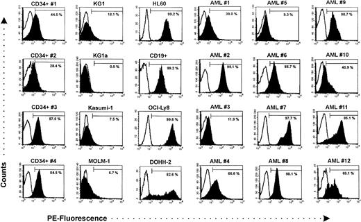
As shown in Fig 1, circulating CD34+ cells expressed significant amounts of CXCR-4. Analysis of hematopoietic progenitor cell subpopulations was performed using CD34+ cells after immunomagnetic cell separation. Coexpression of CXCR-4 and differentiation-related antigens (CD38, HLA-DR, CD11a, and CD45RA), which are low or absent on pluripotent progenitor and stem cells,19-22 was analyzed. In contrast, the CD34+/Thy-1+ subpopulation is enriched for more primitive progenitor cells.23 A representative analysis is shown in Fig 2. Expression of CXCR-4 was not related to differentiation, as demonstrated by coexpression analysis of CD38, Thy-1, HLA-DR, CD11a, and CD45RA. The percentage of CXCR-4+ cells in subpopulations enriched for primitive progenitors (77.2% ± 21.2% of the CD34+/CD38−cells, 75.7% ± 10.1% of the CD34+/Thy-1+cells, 62.8% ± 12.4% of the CD34+/HLA-DR− cells, 58.0% ± 15.0% of the CD34+/CD11a cells, and 60.9% ± 12.8% of the CD34+/CD45RA− cells) was approximately as great as the percentage of CXCR-4+ cells in the total CD34+ population (61.2% ± 14.7%, n = 4).
Analysis of CXCR-4 expression by flow cytometry. The results are shown as fluorescence histograms (solid, CXCR-4 expression; line, respective IgG control). CD34+ hematopoietic progenitor cells from four patients (CD34+ no. 1 through 4) were positive for CXCR-4, while CD34+ cell lines (KG1, KG1a, Kasumi-1, MOLM-1) expressed only low levels. In contrast, variable CXCR-4 expression was found in primary leukemic cells (AML #1 through #12). The cell line HL60, peripheral blood B lymphocytes, and B lymphoma cell lines (OCI-Ly8, DOHH-2) were brightly positive for CXCR-4.
Analysis of CXCR-4 expression by flow cytometry. The results are shown as fluorescence histograms (solid, CXCR-4 expression; line, respective IgG control). CD34+ hematopoietic progenitor cells from four patients (CD34+ no. 1 through 4) were positive for CXCR-4, while CD34+ cell lines (KG1, KG1a, Kasumi-1, MOLM-1) expressed only low levels. In contrast, variable CXCR-4 expression was found in primary leukemic cells (AML #1 through #12). The cell line HL60, peripheral blood B lymphocytes, and B lymphoma cell lines (OCI-Ly8, DOHH-2) were brightly positive for CXCR-4.
Coexpression analysis of CXCR-4 and differentiation-related antigens on purified, circulating CD34+ hematopoietic progenitor cells. Results from a representative three-color flow cytometry analysis are shown. After immunomagnetic separation and staining with CD34-PerCP and the respective FITC/PE-labeled antibodies, CD34+ cells were gated in a FSC/SSC and SSC/FL-3 dot plot. Only low SSC/CD34+ cells were further analyzed. Most of the cells of the more primitive subsets (CD34+/CD38−, CD34+/Thy-1+, CD34+/HLA-DR−, CD34+/CD45RA−, CD34+/CD11a−) coexpressed CXCR-4, as indicated by the shaded areas.
Coexpression analysis of CXCR-4 and differentiation-related antigens on purified, circulating CD34+ hematopoietic progenitor cells. Results from a representative three-color flow cytometry analysis are shown. After immunomagnetic separation and staining with CD34-PerCP and the respective FITC/PE-labeled antibodies, CD34+ cells were gated in a FSC/SSC and SSC/FL-3 dot plot. Only low SSC/CD34+ cells were further analyzed. Most of the cells of the more primitive subsets (CD34+/CD38−, CD34+/Thy-1+, CD34+/HLA-DR−, CD34+/CD45RA−, CD34+/CD11a−) coexpressed CXCR-4, as indicated by the shaded areas.
Variable amounts of CXCR-4 are expressed on primary AML blasts.
Primary leukemic blasts from the peripheral blood of patients with AML expressed variable amounts of CXCR-4 (Fig 1). The mean fluorescence intensity of CXCR-4 was more variable (range, 6 to 601) compared with normal CD34+ cells (range, 13 to 37), while the average percentage of CXCR-4 positive cells (66.7% ± 9.7%, n = 12) was comparable (normal CD34+ cells: 61.2% ± 14.7%, n = 4). Unexpectedly, expression of the SDF-1 receptor on the CD34+ leukemic cell lines KG1 (18.1%), KG1a (0.0%), Kasumi-1 (7.5%), and MOLM-1 cells (5.7%) was substantially lower than the average expression on the nonmalignant circulating CD34+ cells and AML leukemic blasts (Fig 1). In contrast to the CD34+ leukemic cell lines, there were cases of primary CD34+ AML cells, which were also brightly positive for CXCR-4. Interestingly, the CD34− leukemic cell line HL60 expressed high levels of CXCR-4. As a control, normal CD19+ lymphocytes were brightly positive for CXCR-4. The level of CXCR-4 expression was even higher in the B-lymphoma cell lines OCI-Ly8 and DOHH-2 (Fig 1). Surprisingly, expression of CXCR-4 mRNA was found in CD34+ progenitor cells, in all cell lines, and all AML blasts by RT-PCR (data not shown), including the cell line KG1a, which was negative for CXCR-4 by immunofluorecence. These results indicate that at least basal levels of CXCR-4 were produced by these cells.
SDF-1 supports transendothelial migration of progenitors and leukemic cells in vitro.
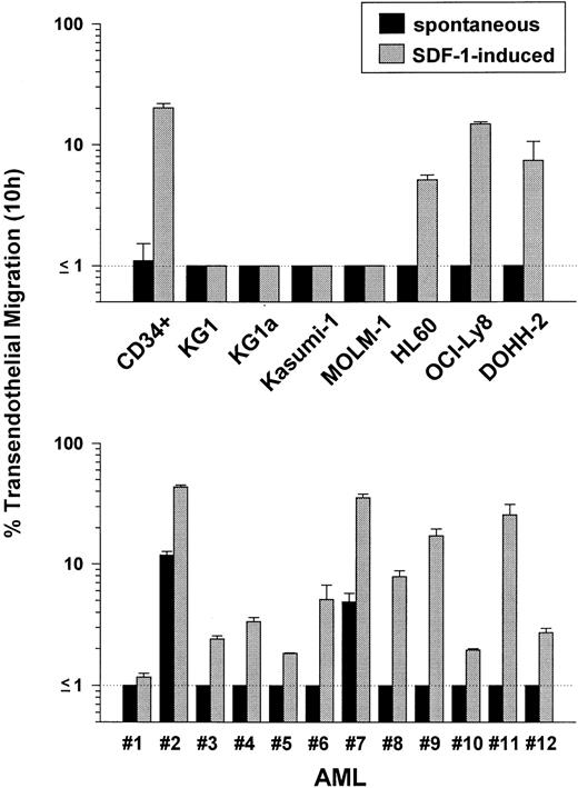
Transmigrated cells were recovered from the lower chamber of the transmigration system after 10 hours and enumerated. Circulating CD34+ cells efficiently transmigrated the bone marrow endothelium in vitro when SDF-1–containing conditioned medium was added to the lower chamber of the transmigration system (Fig 3, upper panel). In contrast, no migration was observed when the CD34+ leukemic cell lines KG1, KG1a, Kasumi-1, and MOLM-1 were added to the upper chamber of the transmigration system, independent of addition of SDF-1 containing conditioned medium to the lower chamber. The CD34 leukemic cell line HL60 and B-lymphoma cell lines efficiently migrated in response to SDF-1. The response of primary AML blasts to a transendothelial SDF-1 gradient was more heterogeneous (Fig 3, lower panel). Leukemic cells from some AML patients rapidly migrated across the bone marrow endothelial cell layer when SDF-1–containing conditioned medium was added to the lower chamber, while migration of cells from other samples was only weak. Interestingly, also significant spontaneous migration was observed in the samples with the greatest level of CXCR-4 expression (Fig 3, lower panel).
Transendothelial migration in vitro. A total of 5 × 10⁵ cells (purified PB CD34+ progenitors, different cell lines, or primary leukemic blasts) were added to the upper chamber of the transmigration system. After 10 hours, transmigrated cells recovered from the lower chamber were enumerated. Due to the detection limit of this assay, migration of >1% could be quantified reliably. Spontaneous migration (addition of control medium to the lower chamber) was compared with SDF-1–induced migration (addition of SDF-1–containing conditioned medium to the lower chember). CD34+ progenitors, CD34− leukemic (HL60), and lymphoma (OCI-Ly8, DOHH-2) cell lines showed significant migration, in contrast to CD34+ leukemic cell lines. SDF-1–induced migration of primary AML blasts was variable.
Transendothelial migration in vitro. A total of 5 × 10⁵ cells (purified PB CD34+ progenitors, different cell lines, or primary leukemic blasts) were added to the upper chamber of the transmigration system. After 10 hours, transmigrated cells recovered from the lower chamber were enumerated. Due to the detection limit of this assay, migration of >1% could be quantified reliably. Spontaneous migration (addition of control medium to the lower chamber) was compared with SDF-1–induced migration (addition of SDF-1–containing conditioned medium to the lower chember). CD34+ progenitors, CD34− leukemic (HL60), and lymphoma (OCI-Ly8, DOHH-2) cell lines showed significant migration, in contrast to CD34+ leukemic cell lines. SDF-1–induced migration of primary AML blasts was variable.
The chemotactic effect of MS-5–conditioned medium on AML blasts is due to SDF-1.
CXCR-4–positive primary AML blasts migrated across bone marrow endothelium in response to recombinant SDF-1 nearly as efficient as in response to the MS-5–conditioned medium (Fig 4). Addition of the antibody 12G5, which partially blocks CXCR-4,15 markedly reduced migration in response to both conditioned medium and recombinant SDF-1. These results demonstrate that SDF-1 contained in the MS-5–conditioned medium is the predominant chemotactic activity for AML blasts, which mediates its effects through the chemokine receptor CXCR-4.
Effect of a CXCR-4 blocking antibody on transendothelial migration of CXCR-4–positive primary AML blasts. Migration was expressed as relative transendothelial migration compared with migration in response to MS-5–conditioned medium (CM [MS-5] = 100%). The CXCR-4 antibody (mAb) 12G5 markedly reduced migration of CXCR-4–positive, primary AML blasts (AML no. 7, 8, 11, and 12) in response to SDF-1–containing conditioned medium (CM + mAb). Transendothelial migration induced by 500 ng/mL rhSDF-1β was nearly as efficient as migration induced by the conditioned medium. The chemotactic effect of both MS-5–conditioned medium and recombinant SDF-1 was reduced by the partially blocking CXCR-4 antibody to a similar extent.
Effect of a CXCR-4 blocking antibody on transendothelial migration of CXCR-4–positive primary AML blasts. Migration was expressed as relative transendothelial migration compared with migration in response to MS-5–conditioned medium (CM [MS-5] = 100%). The CXCR-4 antibody (mAb) 12G5 markedly reduced migration of CXCR-4–positive, primary AML blasts (AML no. 7, 8, 11, and 12) in response to SDF-1–containing conditioned medium (CM + mAb). Transendothelial migration induced by 500 ng/mL rhSDF-1β was nearly as efficient as migration induced by the conditioned medium. The chemotactic effect of both MS-5–conditioned medium and recombinant SDF-1 was reduced by the partially blocking CXCR-4 antibody to a similar extent.
The expression level of CXCR-4 correlates with transmigration in response to SDF-1.
A positive correlation (r = 0.97) was found between the expression density of CXCR-4 on the cell surface (as reflected by the mean fluorescence intensity) and the percentage of cells transmigrating in response to SDF-1–containing conditioned medium, indicating that the chemokine receptor CXCR-4 is functionally active in malignant AML blasts (Fig 5).
Correlation of CXCR-4 expression and SDF-1–induced transendothelial migration of primary AML blasts. A positive correlation (r = 0.97) was found between the expression level (mean fluorescence intensity) of CXCR-4 and the percentage of AML blasts (AML no. 1 through AML no. 12) transmigrating in response to SDF-1 (addition of SDF-1–containing conditioned medium to the lower chamber of the transmigration system).
Correlation of CXCR-4 expression and SDF-1–induced transendothelial migration of primary AML blasts. A positive correlation (r = 0.97) was found between the expression level (mean fluorescence intensity) of CXCR-4 and the percentage of AML blasts (AML no. 1 through AML no. 12) transmigrating in response to SDF-1 (addition of SDF-1–containing conditioned medium to the lower chamber of the transmigration system).
DISCUSSION
In this study, we show that significant levels of CXCR-4 are expressed on circulating normal CD34+ hematopoietic progenitor cells. The vast majority of the CD34+ cells consists of lineage committed progenitors, which are not capable of establishing long-term hematopoiesis.24 These progenitors might not even contribute to marrow recovery after high-dose therapy and progenitor cell transplantation.25 Therefore, we focused on the expression of CXCR-4 on more primitive progenitors. Previous studies have shown that coexpression of CD38, HLA-DR, CD11a, and CD45RA on CD34+ cells is related to differentiation and lineage commitment.19-22 Pluripotent progenitors including hematopoietic stem cells are mainly found in the subpopulations negative for these markers, while the CD34+/Thy-1+ subpopulation is enriched for more primitive progenitor cells.23 Because the proportion of progenitors coexpressing CXCR-4 was comparable or tended to be even greater in the more primitive subpopulations, we conclude that the expression of CXCR-4 on the cell surface is not related to differentiation and lineage commitment. Also primitive progenitors, including stem cells capable of long-term hematopoietic reconstitution after myeloablative therapy, might therefore express the chemokine receptor CXCR-4 and respond to a transendothelial SDF-1 gradient with enhanced migration. Dramatically reduced bone marrow hematopoiesis in SDF-1 −/− mice further supports the idea that the interaction of SDF-1 and CXCR-4 is critical for stem cell homing.11
We have previously shown that only a small proportion of CD34+ hematopoietic progenitor cells migrates spontaneously across bone marrow endothelium in vitro.17 However, only differentiated, CD34+/CD38++ cells are found among those spontaneously migrating progenitors. Particularly primitive progenitors may require additional stimulation for efficient migration across endothelium also in vivo. Given the fact that bone marrow stromal cells constitutively produce SDF-1, a transendothelial SDF-1 gradient could contribute to migration of more primitive progenitor cells during the process of stem cell homing in vivo.
In contrast to CD34+ hematopoietic progenitor cells, primary leukemic blasts from AML patients expressed variable amounts of the SDF-1 receptor. In malignant cells, a chemokine receptor might not be functionally active, even when expressed in large amounts on the cell surface. However, a CXCR-4 blocking antibody reduced migration of AML blasts in response to MS-5–conditioned medium as well as recombinant SDF-1, demonstrating that CXCR-4 is the functionally active SDF-1 receptor also in primary AML blasts. In addition, our results clearly show that the level of CXCR-4 expression is directly related to the ability of the leukemic cells to migrate across bone marrow endothelium in response to the ligand SDF-1. CXCR-4 expression was not related to positivity for CD34. Both, CD34+ AML blasts coexpressing high levels of CXCR-4 and CD34+ blasts virtually negative for CXCR-4 were observed.
Interestingly, significant spontaneous transendothelial migration was found in some primary AML blasts (AML no. 2, no. 7). Similar spontaneous migration across endothelium has been reported in mature monocytes.26 However, the mechanisms controlling leukocyte locomotion are only partially understood. The aquisition of spontaneous transendothelial migration might reflect differentiation of the AML blasts into the monocytic lineage.
Surprisingly, CXCR-4 was low or virtually absent in CD34+leukemic cell lines. At least basal levels of mRNA were produced by all cell lines, as suggested by the positive RT-PCR signals. In accordance with the flow cytometry data, the CD34+ leukemic cell lines did not transmigrate bone marrow endothelium, when SDF-1–containing conditioned medium was added to the lower chamber of the transmigration system. The larger size of the leukemic cell lines could not account for the absence of migration in vitro, as the cell line HL60, which consists of cells larger than KG1 or KG1a, showed significant migration in response to SDF-1. This CD34− cell line was brightly positive for CXCR-4, which demonstrated that expression of CXCR-4 was not downmodulated by culturing of the cells in vitro. Similary, B-lymphoma cell lines overexpressed CXCR-4 compared with normal B lymphocytes, associated with efficient transendothelial migration in response to SDF-1. However, a greater level of CXCR-4 expression on the larger malignant cell lines might be required for significant SDF-1–induced migration at least in the in vitro model using endothelium grown on 3-μm microporous membranes.
A recent study showed expression of CXCR-4 in CD34+hematopoietic progenitor cells by RT-PCR.27 However, as shown by the results of this report, a positive RT-PCR signal is not necessarily related to a functionally relevant cell surface expression of CXCR-4. Furthermore, even a few contaminating cells such as monocytes or lymphocytes may give rise to a positive PCR signal in separated CD34+ cells. Our results suggest that migration in response to SDF-1 is regulated by gradual changes in CXCR-4 expression rather than in an all-or-nothing manner.
It is still an open question whether hematopoietic stem cells can be infected by HIV-1.28-33 It has previously been reported that hematopoietic progenitor cells express low levels of CD4.34,35 RT-PCR data have also raised the possibility that CD34+ cells express both coreceptors required for entry of HIV-1, CXCR-4, and CKR-5.27 As far as CXCR-4 is concerned, we have shown that significant levels are expressed on hematopoietic progenitor cells including more primitive subsets. These results further confirm the hypothesis that also primitive, pluripotent hematopoietic progenitor cells can be targets for HIV-1 infection.
In conclusion, CXCR-4 is a cell surface antigen expressed in significant levels on CD34+ hematopoietic progenitor cells including primitive, pluripotent CD34+ subsets. Expression of this receptor is related to efficient SDF-1–induced transendothelial migration in vitro. It is therefore conceivable that the SDF-1/CXCR-4–mediated migration is involved in the homing of hematopoietic stem cells to the bone marrow. In contrast, primary AML blasts express more variable amounts of functionally active CXCR-4. Because migration is an important function related to the spreading of malignant diseases, further studies are required to determine whether CXCR-4 is a useful marker for the staging of hematopoietic malignancies, which might also be related to prognosis and treatment outcome.
ACKNOWLEDGMENT
We thank Alexandra Schüller and Petra Mayer for excellent technical assistance and Dr I.G. Schmidt-Wolf (University of Berlin, Berlin, Germany) for providing the OCI-Ly8 cell lines.
Supported by grants from Deutsche Forschungsgemeinschaft, Bonn, Germany (SFB 510) to R.M., F.B., W.B., and L.K.; by National Institutes of Health Grant NO. KO8-HL-02926, Dorothy Rodbell Cohen Foundation for Sarcoma Research, and The Rich Foundation (to S.R.); by The Gar Reichman Fund of the Cancer Research Institute and the Rosemary Breslin Fund (to M.A.S.M.).
Address reprint requests to Robert Möhle, MD, Department of Medicine II, University of Tübingen, Otfried-Müller-Str. 10, 72076 Tübingen, Germany.
The publication costs of this article were defrayed in part by page charge payment. This article must therefore be hereby marked "advertisement" is accordance with 18 U.S.C. section 1734 solely to indicate this fact.


![Fig. 4. Effect of a CXCR-4 blocking antibody on transendothelial migration of CXCR-4–positive primary AML blasts. Migration was expressed as relative transendothelial migration compared with migration in response to MS-5–conditioned medium (CM [MS-5] = 100%). The CXCR-4 antibody (mAb) 12G5 markedly reduced migration of CXCR-4–positive, primary AML blasts (AML no. 7, 8, 11, and 12) in response to SDF-1–containing conditioned medium (CM + mAb). Transendothelial migration induced by 500 ng/mL rhSDF-1β was nearly as efficient as migration induced by the conditioned medium. The chemotactic effect of both MS-5–conditioned medium and recombinant SDF-1 was reduced by the partially blocking CXCR-4 antibody to a similar extent.](https://ash.silverchair-cdn.com/ash/content_public/journal/blood/91/12/10.1182_blood.v91.12.4523/4/m_blod41204004x.jpeg?Expires=1766411680&Signature=qwJiHF5J7HaGGRQYnbPPSBacHjvO33l9aB155C-6y-xIY5pgcY6IhiwoiiijWCxvdWcDLXrOQdNkErBVLO9FSvp8HUPDUsT7b4xeqRsUdFkWoQAu9BKeAbQdjREW-i-4MNrsbguXLd3szTuS1wHGG1-KONyD8sUecl8pxRGgU9o~H~qsMiZcbDoT~rtLRGt8YVtZo2dhZozbwNVQhri8MV4Ma6-SGW1Ty2n0nSBTnNiV5ULxuU4VmHCNPCcN3kWG3qAnVWj2MRUV6fYKHWvIwYtUYBsXT~bJfgU48THMaMBAaT6u8kzDDyTY2DV8QlbtavtVlLkiY-XsnBltiIArfg__&Key-Pair-Id=APKAIE5G5CRDK6RD3PGA)

