Abstract
Cellular interactions between hematopoietic cells and stromal cells play important roles in the proliferation and differentiation of hematopoietic cells. The proliferation of a human erythroleukemia cell line, HEL cells, which can differentiate into macrophage- and megakaryocyte-like cells, and erythroid precursors was dramatically induced on coculture with a hematopoietic-supportive stromal cell line, HESS-5 cells, which can support long-term hematopoiesis in vitro without fetal bovine serum. HEL cells proliferated when they were cocultured with but not without direct cell contact. Because the coculture supernatants with direct cell contact and cytokines such as interleukins and growth factors did not exhibit growth-stimulating activity toward HEL cells, it was suggested that some molecule that has growth-stimulating activity exists on the surface of the cells. Extracellular matrix components such as fibronectin, laminin, vitronectin, and collagen did not affect the proliferation of HEL cells. An anti-CD18 monoclonal antibody, which recognizes the common β chain of the β2 integrin subfamily, induced dramatic proliferation of HEL cells. Moreover, the proliferation of HEL cells was inhibited by an antisense oligonucleotide of CD18 mRNA. As judged from these observations, the proliferation of HEL cells was mediated by CD18 molecules expressed on HEL cells. On the contrary, the common counter-receptor of the β2 integrin subfamily, intercellular adhesion molecule-1, which is expressed on CHO-K1 cells, did not stimulate the growth of HEL cells. It is known that other counter molecules of the β2 integrin subfamily, such as complement C3bi and fibrinogen, are not produced by stromal cells. These findings suggest that the proliferation of HEL cells may be induced through an interaction between a novel molecule of the β2 integrin subfamily on HEL cells and the counter-receptor on HESS-5 cells. The β2 integrin subfamily may regulate the growth of hematopoietic cells in hematopoiesis in vivo and/or cause the abnormal growth of leukemia cells.
DIRECT CELL INTERACTIONS are regulated by adhesion molecules, such as the integrin family, the cadherin family, the Ig superfamily, and the selectin family, and extracellular matrix components expressed on both hematopoietic cells and stromal cells.1-3 In vitro long-term coculture of hematopoietic cells and stromal cells revealed the important roles of direct cell contact in the maintenance, proliferation, and differentiation of hematopoietic stem/progenitor cells.4 5 Direct cell contact through a specific adhesion molecule leads not only to the maintenance and differentiation of hematopoietic cells, but also to cytokine production by the stromal cells. From these findings, it is well recognized that direct cell interactions play important roles in hematopoiesis in vivo.
Among the adhesion molecules, the functions of the integrin family in early hematopoiesis have been well analyzed.2,3,6-10 The integrin family members consist of two noncovalently associated subunits, ie, α and β chains. The integrins are classified into subfamilies in which a common β subunit is associated with a different number of α subunits.2 Integrin molecules are expressed on the surface of a wide variety of cell types such as hematopoietic stem cells, progenitor cells, and functional-terminal differentiated hematopoietic cells.3 In early hematopoiesis, VLA-4 (α4β1) and VLA-5 (α5β1) regulate the migration of hematopoietic stem cells underneath a stromal layer.11 Recently, it was demonstrated that a lack of the integrin β1 or α4 chain causes serious deficiencies in hematopoiesis and embryogenesis.8-10
It is also known that the β2 integrin subfamily has functions such as the adhesion, invasion, and chemotaxis of hematopoietic cells.12-15 The importance of the β2 integrin subfamily in the immune function in vivo was demonstrated by studies on the human genetic disease leukocyte adhesion deficiency (LAD) type I.14 However, little evidence has been reported that β2 integrin subfamily participates in the growth of hematopoietic cells.
In this study, we examined the adhesion-dependent proliferation of HEL cells on a stromal cell line, HESS-5 cells. The proliferation of HEL cells was not induced by the coculture supernatant of HEL and HESS-5 cells. Based on the results with monoclonal antibodies against integrin family members and a specific antisense oligonucleotide, we deduced that the proliferation of HEL cells induced by adhesion to stromal cells was mediated by the integrin β2 chain (CD18) expressed on HEL cells. It has not been previously reported that the CD18 molecule induces the proliferation of hematopoietic cells and cell lines that can differentiate into other lineages. The role of the CD18 molecule and the possibility of a novel member of the β2 integrin subfamily existing on HEL cells are also discussed.
MATERIALS AND METHODS
Monoclonal antibodies.
Unconjugated and fluorescein (FITC)-conjugated purified monoclonal antibodies (MoAbs) were purchased from Immunotech (Cedex, France) or the following vendors: anti-CD29 (clone Lia1/2, K20) and CD18 (clone 7E4, Lia3/2), Upstate Biotechnology Inc, Lake Placid, NY; CD61 (clone SZ21, PM6/13), YLEM, Rome, Italy; CD11a (clone 25.3.1, G43-25B), Pharmingen, San Diego, CA; DF1524, Sigma, St Louis, MO; B-B15, Diaclone Res, Besancon cedex, France; 38, Cymbus Biotechnology, UK; CD11b (clone BEAR 1, 44), Pharmingen, and LM2/1, Bender MedSystems, Austria; CD11c (clone BU15, B-Ly6), Pharmingen; and CD49b (clone Gi9), CD49c (clone M-KID 2), CD49d (clone HP2/1), CD49e (clone SAM1), and CD49f (clone Go H3) MoAbs and FITC-labeled anti-mouse IgG sheep F(AB′) fragment, Organon Teknika Corp, Durham, NC. Anti-CD11a (clone TS1/22) and CD18 (clone TS1/18) MoAbs were purified from the conditioned medium of their hybridomas.18 A mouse IgG fraction, as a control antibody, was purchased from Zymed Laboratories Inc (San Francisco, CA).
Cell lines.
The hematopoietic-supportive stromal cell line, HESS-5 cells, was established from murine bone marrow and spleen.16,17 HESS-5 cells were maintained in minimal essential medium α ([MEMα] GIBCO Laboratories, Grand Island, NY) supplemented with 10% (vol/vol) horse serum ([HS] Nichimen America, Los Angeles, CA) at 37°C under 5% CO2 in humidified air. The human erythroleukemia cell line, HEL cells, was provided by the Japan Cell Research Bank and maintained in RPMI 1640 medium (GIBCO Laboratories) supplemented with 10% (vol/vol) FCS.19 The hybridoma cell lines TS1/22.1.1.13 and TS1/18.1.2.11 were provided by the American Type Culture Collection and maintained in Dulbecco's modified Eagle's medium (GIBCO Laboratories) supplemented with 10% (vol/vol) FCS and 1 mmol/L sodium pyruvate.18
3H-thymidine incorporation assay of the cocultured leukemia cell line, HEL cells.
A confluent layer of the fibroblast or stromal cell line on a 96-well culture plate (Falcon, Lincoln Park, NJ) was irradiated with 9.0-Gy x-rays for prevention of incorporation of 3H-thymidine into the cell line. The cell lines were washed five times with RPMI 1640 medium supplemented with 0.1% (wt/vol) bovine serum albumin ([BSA] Sigma). HEL cells (5 × 10³) were cultured on irradiated confluent cell feeders in 200 μL RPMI 1640 medium supplemented with 0.1% (wt/vol) BSA. After 3 days in culture, proliferation of leukemia cells was measured as the incorporation of 3H-thymidine (Amersham, Buckinghamshire, England), added at a concentration of 0.1 μCi/well, during the last 6 hours of the incubation. The cells were harvested on glass fiber filters with a Cell Harvester (Packard, Meriden, CT), and then radioactivity was measured with a MATRIX 9600 counter (Packard).
Cell growth assay of HEL cells.
A confluent layer of HESS-5 cells on a six-well cell culture plate (Falcon) was irradiated with 9.0-Gy x-rays and then washed five times with RPMI 1640 medium supplemented with 0.1% BSA. Then, HEL cells (2 × 10⁵ cells/well) in 5 mL RPMI 1640 medium supplemented with 0.1% (wt/vol) BSA were cocultured with or without the irradiated HESS-5 cells in triplicate wells. To prevent direct cell-to-cell contact, the cells were separated by a microporous membrane, Cyclopore (pore size, 0.45 μm; Falcon). After 3 days in culture, viable HEL cells were recovered by gentle pipetting and then counted using the trypan blue exclusion method under a microscope with a hematocytometer. The recovered HEL cells were distinguished from HESS-5 cells by means of cell size and morphology.
For examination of the effects of extracellular matrix components on the proliferation of HEL cells, fibronectin-, laminin-, vitronectin-, and collagen-precoated dishes (Koken Inc, Tokyo, Japan) were used. Viable cells were counted by the method described.
Cell proliferation-stimulating activities toward HEL cells of culture supernatants, cytokines, and mAbs.
Culture supernatants were harvested from HESS-5 cells cocultured with or without HEL cells for 24 hours under the various culture conditions described. The growth-stimulating activities toward HEL cells in these culture supernatants were measured by means of a3H-thymidine incorporation assay involving HEL cells. HEL cells were seeded at a concentration of 5 × 10³cells/well in 96-well plates (Falcon) in a final 200 μL RPMI 1640 medium supplemented with 0.1% (wt/vol) BSA, and then serial dilutions of various conditioned media, cytokines, and mAbs were added to the HEL cells in triplicate. After 3 days' incubation, the proliferation of leukemia cells was measured as the incorporation of3H-thymidine as described.
Phenotypic analysis of the expression of adhesion molecules on HEL cells.
HEL cells (105 cells each) were incubated with 5 μg anti-CD29, CD18, CD61, CD49b, CD49c, CD49d, CD49e, CD49f, CD11a, CD11b, CD11c, CD41, or CD44 MoAbs in 100 μL of Ca2+- and 100 Mg2+-phosphate–buffered saline (PBS-) supplemented with 0.5% (wt/vol) BSA and 5 mmol/L EDTA for 30 minutes at 4°C. After incubation, the cells were washed twice with the same buffer and then stained with the FITC-conjugated anti-mouse IgG sheep F(ab′)2 fragment in the same buffer for 30 minutes at 4°C. After staining, the cells were washed twice and then analyzed by flow cytometry. For analysis of the expression of adhesion molecules on HEL cells, the specified gate for HEL cells was set in the plot of forward and side scatter.
Inhibition of CD18 molecule expression on HEL cells by an antisense oligonucleotide.
HEL cells (5 × 10⁵ cells) were plated in 2 mL RPMI 1640 medium supplemented with 0.1% (wt/vol) BSA and 5 μmol/L antisense (CD18/AS; positions 1 to 24), sense (CD18/S; positions 1 to 24), or random (CD18/RS) phosphorothionate oligodeoxynucleotide (PON) solubilized in PBS-. The PON sequences were as follows: CD18/AS, 5′-CAGTGGGGGGCGCAGGCCCAGCAT-3′; CD18/S, 5′-ATGCTGGGCCTGCGCCCCCCACTG-3′; and CD18/RS, 5′-CATCGAGCGAGGCGTCGAGCGGCG-3′.12After 24 hours' incubation, HEL cells were harvested and then treated with 0.2 mL 0.2% trypsin and 10 mg/mL proteinase K (GIBCO) at 37°C for 10 minutes for digestion of cell-surface CD18 molecules. After the proteinase treatment, HEL cells were cultured with anti-CD18 MoAb (clone 7E4) or cocultured on a confluent layer of HESS-5 cells at a concentration of 5 × 10³ cells/well of a 96-well cell culture plate in 200 μL RPMI 1640 medium supplemented with 0.1% BSA and 5 μmol/L PON. After 2 days in culture, the proliferation of leukemia cells was measured as the incorporation of3H-thymidine, added at a concentration of 0.5 μCi/well, during the last 6 hours of the incubation.
Effect of 3H-thymidine incorporation by HEL cells on coculture with human intercellular adhesion molecule-1–expressing CHO-K1 cells.
The human gene for ICAM-1 purchased from British Bio-technology Ltd (Oxon, Great Britain) and constructed in the pcDNA3.1 vector (Invitrogen, San Diego, CA). CHO-K1 cells were transfected by electroporation with hICAM-1–expressing vector, and stable transformants were selected by maintenance of the cells in RPMI 1640 medium supplemented with 10% FCS and 800 μg/mL geneticin (GIBCO). A single cell clone was isolated from the transformants by the limited dilution technique. After single cell isolation, a highly expressed clone of human ICAM-1 was selected by means of flow cytometric analysis. Expression of hICAM-1 on the cell surface was analyzed by the method already described with the anti-CD54 MoAb. The specified gate for CHO-K1 cells was set in the plot of forward and side scatter.
The method used to examine the growth-stimulating activity toward HEL cells of the stable transformant was the same as that for the coculture with HESS-5 cells already described.
RESULTS
Proliferation of leukemia cell lines on hematopoietic-supportive stromal cells.
We examined whether a human erythroleukemia cell line, HEL cells, proliferates on coculture on hematopoietic-supportive or -nonsupportive cell lines. Hematopoietic-supportive and -nonsupportive stromal cell lines were established from murine bone marrow and spleen as described previously.16
HEL cells did not proliferate alone in RPMI 1640 medium supplemented with 0.1% BSA. Proliferation of HEL cells was strongly induced on cocultivation with a hematopoietic-supportive stromal cell line, HESS-5 cells or SSXL CL.7 cells, but not a hematopoietic-nonsupportive fibroblast cell line, C3H/10T1/2 cells or hematopoietic-nonsupportive stromal cell lines (data not shown). This assay indicated proliferation of the leukemia cell lines, ie, entry into the S-phase of the cell cycle, but did not indicate escape from apoptosis, because the incorporation of 3H-thymidine into the nuclei of HEL cells was measured.
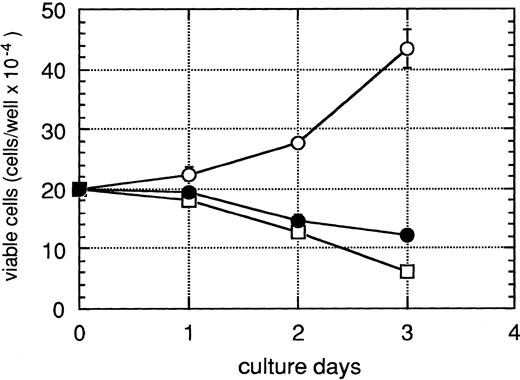
Figure 1 shows the number of viable HEL cells under various culture conditions with HESS-5 cells. After 3 days' culture in RPMI 1640 supplemented with 0.1% (wt/vol) BSA, the number of viable HEL cells was decreased. HEL cells cultured in RPMI 1640 supplemented with 0.1% (wt/vol) BSA died, exhibiting the morphologic and biochemical characteristics of apoptosis, which was induced by the deprivation of growth factors (data not shown). The number of viable HEL cells increased in the coculture with HESS-5 cells with direct cell contact. The increased number of the input HEL cells proliferated on HESS-5 cells in the same logarithmic ratio (data not shown). Although HESS-5 cells became thinner in serum-free medium, all HEL cells adhered to the HESS-5 cell layer, and some of the HEL cells proliferated on and underneath the layer of HESS-5 cells (data not shown).
Proliferation of HEL cells under various coculture conditions with a murine hematopoietic supportive stromal cell line, HESS-5 cells. HEL cells (2 × 10⁵ cells) were cultured without stromal cells (□), or cocultured with HESS-5 cells with direct cell contact (○) or without cell contact (•) in RPMI-1640 medium supplemented with 0.1% (wt/vol) BSA in triplicate wells. The cell contact between HEL cells and HESS-5 cells was prevented by a microporous membrane on the confluent cell layer of HESS-5 cells. The results are expressed as mean values ± SD of the mean.
Proliferation of HEL cells under various coculture conditions with a murine hematopoietic supportive stromal cell line, HESS-5 cells. HEL cells (2 × 10⁵ cells) were cultured without stromal cells (□), or cocultured with HESS-5 cells with direct cell contact (○) or without cell contact (•) in RPMI-1640 medium supplemented with 0.1% (wt/vol) BSA in triplicate wells. The cell contact between HEL cells and HESS-5 cells was prevented by a microporous membrane on the confluent cell layer of HESS-5 cells. The results are expressed as mean values ± SD of the mean.
On the other hand, HEL cells did not proliferate when they were cocultured with HESS-5 cells without direct cell contact using a microporous membrane filter, Cyclopore.
HEL cell growth-stimulating activities of various cytokines and culture supernatants of HESS-5 cells under various culture conditions.
It was examined whether various murine and human cytokines stimulate3H-thymidine incorporation by HEL cells, which were cultured in serum-free medium. HEL cells did not proliferate with murine interleukins, such as interleukin-1α (IL-1α), -1β, -3, -4, -5, -6, -7, -9, -10, and -11, at various concentrations. Murine colony-stimulating factors (CSFs) also did not induce3H-thymidine incorporation by HEL cells. The growth of HEL cells was not induced by stem cell factor (SCF), tumor necrosis factor α, or macrophage-inflammatory protein (MIP)-1α. Serum-related growth factors such as epidermal growth factor (EGF), basic fibroblast growth factor (basic FGF), and platelet-derived growth factor (PDGF) also did not induce proliferation of HEL cells. Only transferrin derived from human and bovine serum exhibited strong cell growth-stimulating activity toward HEL cells (data not shown). Although transferrin in bovine serum stimulates the proliferation of HEL cells, a neutralizing antibody against transferrin did not reduce the3H-thymidine incorporation or the proliferation of HEL cells cocultured with HESS-5 cells with direct cell contact (data not shown).
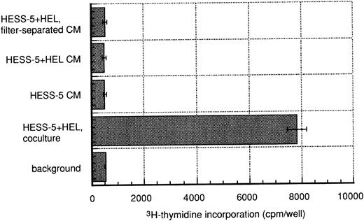
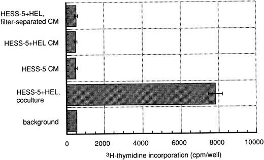
Next, we examined whether a coculture supernatant exhibited cell growth-stimulating activity toward HEL cells by means of the3H-thymidine incorporation assay (Fig2). In recent studies, some cytokines produced by stromal cell lines were found to be induced on the direct adhesion of hematopoietic cells.17 20-22 We also examined this possibility, ie, that some new cytokines were produced by HESS-5 cells on HEL cell adhesion that act on human cells.
Growth-stimulating activities toward HEL cells in various culture supernatants. Conditioned media (CM) were harvested from one day cocultures of HEL cells and HESS-5 cells. The supernatant of a culture of HESS-5 cells alone (HESS-5 CM), or a coculture of HEL cells and HESS-5 cells with direct cell contact (HESS-5 + HEL CM) or without cell contact, the latter being prevented with a microporous membrane (HESS-5 + HEL, filter-separated CM), was added to HEL cells (5 × 10³ cells each) at the final concentration of 25% (vol/vol) in triplicate wells. The assay used for3H-thymidine incorporation by HEL cells was described under Materials and Methods. Background indicates a culture of HEL cells alone in RPMI-1640 medium supplemented with 0.1% (wt/vol) BSA as a negative control. HESS-5 + HEL, coculture indicates a coculture of HEL cells and HESS-5 cells with direct cell contact as a positive control. The results are expressed as mean values ± SD of the mean.
Growth-stimulating activities toward HEL cells in various culture supernatants. Conditioned media (CM) were harvested from one day cocultures of HEL cells and HESS-5 cells. The supernatant of a culture of HESS-5 cells alone (HESS-5 CM), or a coculture of HEL cells and HESS-5 cells with direct cell contact (HESS-5 + HEL CM) or without cell contact, the latter being prevented with a microporous membrane (HESS-5 + HEL, filter-separated CM), was added to HEL cells (5 × 10³ cells each) at the final concentration of 25% (vol/vol) in triplicate wells. The assay used for3H-thymidine incorporation by HEL cells was described under Materials and Methods. Background indicates a culture of HEL cells alone in RPMI-1640 medium supplemented with 0.1% (wt/vol) BSA as a negative control. HESS-5 + HEL, coculture indicates a coculture of HEL cells and HESS-5 cells with direct cell contact as a positive control. The results are expressed as mean values ± SD of the mean.
3H-thymidine incorporation by HEL cells was induced on coculture with HESS-5 cells with direct cell contact. The culture supernatant of HESS-5 cells alone did not stimulate3H-thymidine incorporation by HEL cells cultured alone in RPMI 1640 supplemented with 0.1% (wt/vol) BSA. The coculture conditioned medium of HEL and HESS-5 cells with or without direct cell contact also did not induce 3H-thymidine incorporation by HEL cells. HEL cells cultured in these conditioned media died as in serum-free medium, as judged on microscopic observation (data not shown). These results indicate that the proliferation of HEL cells induced by adhesion to HESS-5 cells was not due to soluble factors produced by HESS-5 cells, but was produced by the direct cell interaction between HEL cells and HESS-5 cells.
Effects on the cell proliferation of HEL cells of extracellular matrix components.
It was investigated as to whether extracellular matrix components, which are possibly expressed on the cell surface membrane of HESS-5 cells, stimulate the proliferation of HEL cells. Extracellular matrix components, such as fibronectin, laminin, vitronectin, and collagen did not affect the proliferation of HEL cells in serum-free medium (data not shown). HEL cells also could not escape from apoptosis on the deprivation of growth factors in serum-free medium due to these extracellular matrix components.
Effects of MoAbs against cell adhesion molecules on the proliferation of HEL cells.
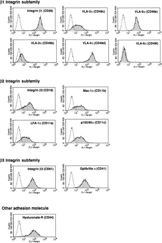
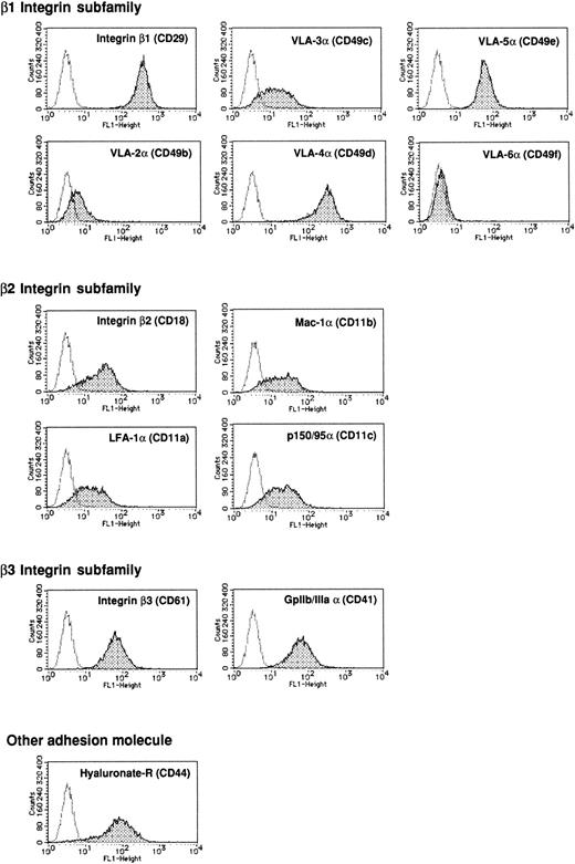
Next, we analyzed the expression of adhesion molecules on HEL cells by means of flow cytometry. We focused on the integrin family because the functions of this family in early hematopoiesis have been well analyzed as to adhesion molecules,3 and some integrins have signal transduction pathways.6 7 The expression patterns of the integrin β and α chains are shown in Fig3.
Phenotypic characterization of HEL cells stained with various anti-adhesion molecule antibodies. HEL cells (105cells each) were incubated with 5 μg of anti-CD29, CD18, CD61, CD49b, CD49c, CD49d, CD49e, CD49f, CD11a, CD11b, CD11c, CD41 or CD44 MoAb in PBS-supplemented 0.5% (wt/vol) BSA and 5 mmol/L EDTA for 30 minutes at 4°C. After incubation, the cells were washed twice with the same buffer and then stained with FITC-labeled anti-mouse IgG sheep F(ab′)2 fragment in the same buffer for 30 minutes. After staining, the cells were washed twice and then analyzed by flow cytometry. For analysis of the expression of adhesion molecules on HEL cells, the specified gate for HEL cells was set in the plot of forward and side scatter. The MoAb used is indicated at the top of each histogram. The shaded peak indicates the expression of the adhesion molecule on HEL cells stained with an anti-adhesion molecule MoAb. The dotted line indicates the background for HEL cells stained with only FITC-labeled anti-mouse IgG F(ab′)2 fragment.
Phenotypic characterization of HEL cells stained with various anti-adhesion molecule antibodies. HEL cells (105cells each) were incubated with 5 μg of anti-CD29, CD18, CD61, CD49b, CD49c, CD49d, CD49e, CD49f, CD11a, CD11b, CD11c, CD41 or CD44 MoAb in PBS-supplemented 0.5% (wt/vol) BSA and 5 mmol/L EDTA for 30 minutes at 4°C. After incubation, the cells were washed twice with the same buffer and then stained with FITC-labeled anti-mouse IgG sheep F(ab′)2 fragment in the same buffer for 30 minutes. After staining, the cells were washed twice and then analyzed by flow cytometry. For analysis of the expression of adhesion molecules on HEL cells, the specified gate for HEL cells was set in the plot of forward and side scatter. The MoAb used is indicated at the top of each histogram. The shaded peak indicates the expression of the adhesion molecule on HEL cells stained with an anti-adhesion molecule MoAb. The dotted line indicates the background for HEL cells stained with only FITC-labeled anti-mouse IgG F(ab′)2 fragment.
The integrin β1 chain (CD29), among members of the three different β chain subfamilies of the integrin family, was highly expressed on HEL cells. Although integrin β2 (CD18) and β3 (CD61) chains were expressed on HEL cells, their expression levels were lower than that of CD29. Of the integrin β1 subfamily, VLA-4α (CD49d) and -5α (CD49e) were expressed at relatively high levels on HEL cells, rather than another α chain of the β1 integrin subfamily. VLA-3α (CD49c) and -2α (CD49b) were expressed at low levels, and VLA-6α (CD49f) was not detected at all. Of the β2 integrin subfamily LFA-1, Mac-1, and p150/95, the α chains were expressed at the same level, which was relatively low compared with that of CD49d. The gpIIb/IIIa complex is a member of the β3 integrin subfamily. The expression level of the α chain of the GpIIb/IIIa complex (CD41) was intermediate but higher than that of the α chains of the β2 integrin subfamily. On the other hand, the hyaluronate receptor (CD44) is known to play a role in the rosette formation between bone marrow stromal cells and erythroid leukemic cells.23 The expression level of CD44 was intermediate, the same as that of CD61 and CD41.
We examined the effects of the MoAbs described, ie, MoAbs against cell adhesion molecules that can block cell adhesion or mitogenic activity, on the proliferation of HEL cells cocultured with HESS-5 cells with cell contact. However, none of the MoAbs blocked3H-thymidine incorporation by HEL cells induced by adhesion to HESS-5 cells (data not shown). Some MoAbs against integrins are known to stimulate the signal transduction pathway of target cells.7 Thus, we next examined whether the MoAbs against integrin β chains exhibit growth-stimulating activity toward HEL cells cultured alone in serum-free medium.
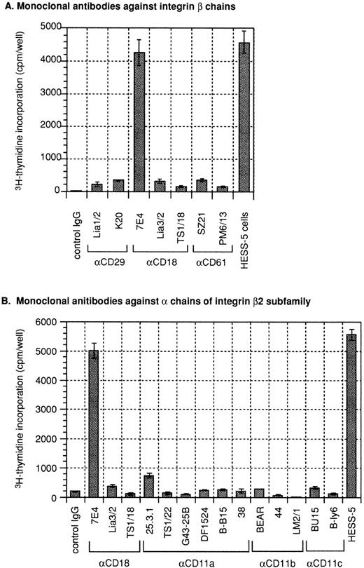
Surprisingly, an anti-CD18 MoAb (clone 7E4), which recognizes the integrin β2 chain, dramatically stimulated 3H-thymidine incorporation by HEL cells at the concentration of 10 μg/mL (Fig4A). The anti-CD18 MoAb (7E4) did not promote adhesion of HEL cells to the substrate, and HEL cells were able to proliferate in suspension without morphologic changes (data not shown). The activity was comparable to that on coculture with HESS-5 cells. The growth-stimulating activity toward HEL cells of the anti-CD18 MoAb (clone 7E4) was dose-dependent (data not shown). Other anti-CD18 MoAbs (clones Lia3/2 and TS1/18), which recognize a different epitope from that of clone 7E4 and inhibit the adhesion of T cells mediated by the CD11a/CD18 complex (LFA-1) and ICAM-1, did not exhibit such activity. The anti-CD29 MoAbs, which inhibit cell adhesion and thymocyte proliferation mediated by CD29, respectively, did not promote3H-thymidine incorporation by HEL cells. The anti-integrin α chain MoAbs CD49b, 49c, 49d, 49e, and 49f, which inhibit cell adhesion mediated by the β1 integrin subfamily, did not stimulate HEL cell proliferation (data not shown). The anti-CD29 MoAb stimulated the aggregation of HEL cells cultured alone in serum-free medium 12 to 24 hours after addition of the MoAb and anti-CD49d, and CD49e MoAbs stimulated the migration of HEL cells underneath HESS-5 cells (data not shown). These MoAbs, which exhibit agonistic activity toward a natural ligand, did not participate in the mechanism underlying the proliferation of HEL cells cocultured with HESS-5 cells with cell contact. The anti-CD61 MoAbs also did not promote3H-thymidine incorporation by HEL cells.
Growth-stimulating activities toward HEL cells of various anti-adhesion molecule MoAbs. Induction of3H-thymidine-incorporation of HEL cells by MoAbs against integrin β chains (A) and integrin β2 subfamily (B). Anti-adhesion molecule MoAbs were added at the final concentration of 10 μg/ml to 5 × 10³ cells/well of 96-well type culture plates of HEL cells in 200 μL of RPMI-1640 medium supplemented with 0.1% (wt/vol) BSA in triplicate. Growth-stimulating activity was measured by assaying3H-thymidine incorporation by HEL cells as described under Materials and Methods. The MoAbs used and their clone names are indicated below the bars in the figure. The results are expressed as mean values ± SD of the mean.
Growth-stimulating activities toward HEL cells of various anti-adhesion molecule MoAbs. Induction of3H-thymidine-incorporation of HEL cells by MoAbs against integrin β chains (A) and integrin β2 subfamily (B). Anti-adhesion molecule MoAbs were added at the final concentration of 10 μg/ml to 5 × 10³ cells/well of 96-well type culture plates of HEL cells in 200 μL of RPMI-1640 medium supplemented with 0.1% (wt/vol) BSA in triplicate. Growth-stimulating activity was measured by assaying3H-thymidine incorporation by HEL cells as described under Materials and Methods. The MoAbs used and their clone names are indicated below the bars in the figure. The results are expressed as mean values ± SD of the mean.
CD18 is a common β subunit of the β2 integrin subfamily comprising lymphocyte function-associated antigen-1 ([LFA-1] CD11a/CD18), Mac-1 (complement receptor type 3, CD11b/CD18), and p150/95 (complement receptor type 4, CD11c/CD18). Next, we investigated whether the MoAbs against the integrin α chains of the β2 integrin subfamily exhibit growth-stimulating activity toward HEL cells (Fig 4B). The anti-CD11a MoAbs (clone 25.3.1, TS1/22, G43-25B, and DF1524) recognize the LFA-1α chain and inhibit the adhesion between LFA-1 and ICAM-1. Although clone 25.3.1 of the anti-CD11a MoAb weakly stimulated3H-thymidine incorporation by HEL cells, the other MoAbs did not induce 3H-thymidine incorporation by HEL cells. Some clones of the anti-CD11b and CD11c MoAbs were examined as to growth-stimulating activity toward HEL cells, but they did not exhibit such activity.
Effect of an antisense oligonucleotide of CD18 on the proliferation of HEL cells.
To determine whether the growth-stimulating activity toward HEL cells on direct cell contact with stromal cells is mediated by the CD18 molecule, we next examined the inhibition of CD18 expression by antisense phosphorothionate oligonucleotides (PONs). For functional analysis of some molecules, antisense PONs were used for inhibition of expression of the molecules.24 25
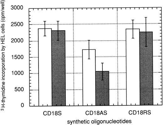
To block CD18 expression by HEL cells, we added a modified antisense PON complementary to positions 1 to 24 of the human CD18 coding region (CD18/AS) to the culture medium. The control PONs included a sense (positions 1 to 24; CD18/S) and a random (CD18/RS) sequence of CD18/AS. Because the CD18 molecule is expressed on the surface of HEL cells, trypsin, and proteinase K treatment was performed before coculture with HESS-5 cells to degrade the CD18 molecule that had already been expressed on the membrane at the time of PON treatment for 1 day. This procedure was important to block expression of the CD18 molecule on HEL cells. Then, the growth-stimulating activity toward HEL cells was examined by means of anti-CD18 MoAb stimulation or coculture with HESS-5 cells with supplementation of PONs at various concentrations. The results are shown in Fig 5. The growth of HEL cells under the coculture conditions with HESS-5 cells was inhibited in a dose-dependent manner only on the addition of CD18/AS, and the inhibition was significant at the final concentration of 5 μmol/L (P < .005; Fig 5). This result indicates that CD18 expressed on HEL cells mediates the proliferation signal into HEL cells when they are cocultured with HESS-5 cells.
Growth-stimulating activity toward HEL cells on coculture with HESS-5 cells with direct cell contact of the anti-sense PON of CD18 (integrin β2 chain). HEL cells were cultured in RPMI-1640 medium supplemented with 0.1% (wt/vol) BSA, and an antisense (CD18/AS; positions 1-24), sense (CD18/S; positions 1-24), or random (CD18/RS) PON at a final concentration of 2 μmol/L (□) or 5 μmol (▪). After 24 hours of incubation, the HEL cells were harvested and subjected to proteinase treatment for digestion of the cell surface CD18 molecules. After the treatment, the HEL cells were cocultured with a confluent layer of HESS-5 cells at the concentration of 5 × 10³ cells/well of a 96-well cell culture plate in 200 μL of RPMI-1640 medium supplemented with 0.1% BSA and 2 or 5 μmol/L PONs. After 2 days in culture, the 3H-thymidine incorporation assay described under Materials and Methods was performed. The results are expressed as mean values ± SD of the mean.
Growth-stimulating activity toward HEL cells on coculture with HESS-5 cells with direct cell contact of the anti-sense PON of CD18 (integrin β2 chain). HEL cells were cultured in RPMI-1640 medium supplemented with 0.1% (wt/vol) BSA, and an antisense (CD18/AS; positions 1-24), sense (CD18/S; positions 1-24), or random (CD18/RS) PON at a final concentration of 2 μmol/L (□) or 5 μmol (▪). After 24 hours of incubation, the HEL cells were harvested and subjected to proteinase treatment for digestion of the cell surface CD18 molecules. After the treatment, the HEL cells were cocultured with a confluent layer of HESS-5 cells at the concentration of 5 × 10³ cells/well of a 96-well cell culture plate in 200 μL of RPMI-1640 medium supplemented with 0.1% BSA and 2 or 5 μmol/L PONs. After 2 days in culture, the 3H-thymidine incorporation assay described under Materials and Methods was performed. The results are expressed as mean values ± SD of the mean.
Effect of coculture with human ICAM-1–expressing CHO-K1 cells on the proliferation of HEL cells.
ICAM-1 is thought to be the common counter-receptor of the β2 integrin subfamily.26-29 Thus, we investigated whether the ICAM-1 molecule stimulated the growth of HEL cells via the CD18 pathway.
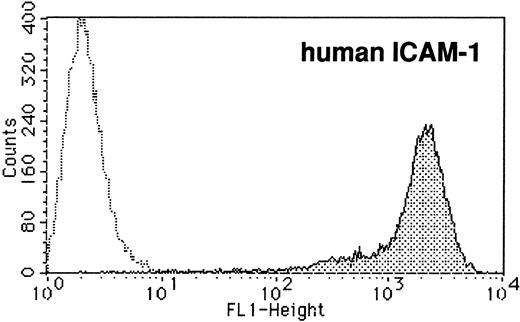
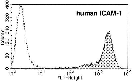
A stable transformant clone expressing a high level of human ICAM-1 (hICAM-1) was established by electroporation of the hICAM-1 expression vector into CHO-K1 cells and subsequent selection as to neomycin resistance. After the selection, stable single clones were isolated, and then high-expression clones of hICAM-1 were screened by means of flow cytometry. CD18 expression of the stable transformant is shown in Fig 6A. We performed a functional assay using PMA-activated lymphocytes of the binding of LFA-1 and ICAM-1 on CHO-K1 cells according to a previous report.30PMA-activated lymphocytes dramatically adhered to stable human ICAM-1–expressing CHO-K1 cells rather than mock-transfected CHO-K1 cells (data not shown). Moreover, this adhesion was inhibited by an anti-ICAM-1 antibody. These results confirmed that the expressed ICAM-1 molecule was active.
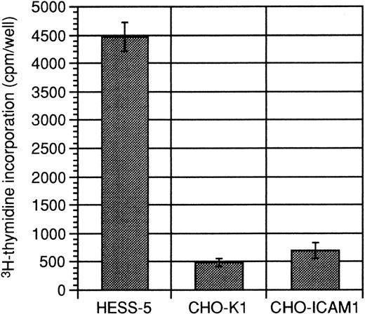
Growth-stimulating activity toward HEL cells on coculture with stable human ICAM-1 expressing CHO-K1 cells. (A) Phenotypic analysis of stable human ICAM-1-expressing CHO-K1 cells. Stable human ICAM-1–expressing CHO-K1 cells were prepared by the method described under Materials and Methods. The cells were stained with anti-human ICAM-1 MoAb and FITC-conjugated antimouse IgG F(AB′) fragment, successively, and then analyzed by flow cytometry. The shaded peak indicates the expression of ICAM-1 on the stable transformant and the dotted line indicates that on mock-transfected CHO-K1 cells. (B) Growth-stimulating activity toward HEL cells on coculture with stable ICAM-1–expressing CHO-K1 cells. HEL cells (5 × 10³cells) were seeded onto irradiated confluent layers of HESS-5 cells (HESS-5), CHO-K1 cells (CHO-K1), and stable human ICAM-1–expressing CHO-K1 cells (CHO-ICAM1) in 200 μL of RPMI-1640 medium supplemented with 0.1% BSA in triplicate. After 3 days of culture, the3H-thymidine incorporation by HEL cells was measured by the method described under Materials and Methods. The results are expressed as mean values ± SD of the mean.
Growth-stimulating activity toward HEL cells on coculture with stable human ICAM-1 expressing CHO-K1 cells. (A) Phenotypic analysis of stable human ICAM-1-expressing CHO-K1 cells. Stable human ICAM-1–expressing CHO-K1 cells were prepared by the method described under Materials and Methods. The cells were stained with anti-human ICAM-1 MoAb and FITC-conjugated antimouse IgG F(AB′) fragment, successively, and then analyzed by flow cytometry. The shaded peak indicates the expression of ICAM-1 on the stable transformant and the dotted line indicates that on mock-transfected CHO-K1 cells. (B) Growth-stimulating activity toward HEL cells on coculture with stable ICAM-1–expressing CHO-K1 cells. HEL cells (5 × 10³cells) were seeded onto irradiated confluent layers of HESS-5 cells (HESS-5), CHO-K1 cells (CHO-K1), and stable human ICAM-1–expressing CHO-K1 cells (CHO-ICAM1) in 200 μL of RPMI-1640 medium supplemented with 0.1% BSA in triplicate. After 3 days of culture, the3H-thymidine incorporation by HEL cells was measured by the method described under Materials and Methods. The results are expressed as mean values ± SD of the mean.
3H-thymidine incorporation by HEL cells cocultured with hICAM-1–expressing CHO-K1 cells is demonstrated in Fig 6B. Although cell contact with HESS-5 cells dramatically induced3H-thymidine incorporation by HEL cells, hICAM-1–expressing cells did not affect the growth of HEL cells. This clearly demonstrated that ICAM-1 is not the counter-receptor of the CD18 complex on HEL cells that exhibits growth-stimulating activity toward HEL cells.
DISCUSSION
In this study, we focused on the proliferation of hematopoietic cells on coculture with stromal cells via direct cell contact. We adopted an approach involving an in vitro xenogenic coculture system comprising a human multipotential erythroleukemia cell line, HEL cells,19,31 and a murine hematopoietic-supportive stromal cell line, HESS-5 cells, to avoid the effects of cytokines, because many murine cytokines cannot affect human cells.32,33Therefore, at least in the coculture of HEL cells and HESS-5 cells, some soluble factors did not affect the proliferation of HEL cells. This system is thought to be more useful for the identification of a novel molecule for hematopoietic cells than a syngeneic coculture system.32 33
The stage of differentiation of HEL cells was thought to be the early blast and/or erythroblast stage,19 and the cells can differentiate into macrophage-like cells, gpIIb/IIIa-positive megakaryocytic cells, on stimulation by 12-O-tetradecanoyl-phorbol 13-acetate or the erythrocyte precursor after exposure to hemin. Therefore, this leukemic cell line is thought to be a useful tool for studying the proliferation of hematopoietic cells, which mimic hematopoietic stem/progenitor cells, and the differentiation of a myeloid-erythroid lineage.19,31 HEL cells die, exhibiting the morphologic and biochemical characteristics of apoptosis induced by the deprivation of growth factors when they are cultured in a serum-free medium. In the coculture system, a hematopoietic-supportive stromal cell line, HESS-5 cells, but not -nonsupportive stromal cell lines, induced the proliferation of HEL cells without serum with direct contact but not without cell contact. The HEL cell growth-stimulating activity seemed to show a good correlation with the hematopoietic-supportive activity.16
Although, at first, we thought that some cytokines produced by HESS-5 cells in the steady state or induced by the adhesion of HEL cells affected the proliferation of HEL cells, the proliferation was not induced by various cytokines such as IL-1α, -1β, -3, -4, -5, -6, -7, -9, -10, or -11, GM-CSF, G-CSF, M-CSF, SCF, TNF-α, MIP-1α, EGF, basic FGF, or PDGF (data not shown), or the coculture supernatant of HESS-5 and HEL cells with direct cell contact. These results suggested that the signals for the proliferation of HEL cells are mediated by direct cell contact and some adhesion molecules expressed on both HEL cells and HESS-5 cells.
It is well known that the integrin family participates in hematopoiesis in vivo.2,3 Integrin molecules are expressed on the surface of a wide variety of cell types, such as hematopoietic stem cells, progenitor cells, and functional-terminal differentiated hematopoietic cells.3 In HEL cells, almost all of the examined integrin α and β chains were found to be expressed on the surface of the cells. The integrin β1 chain showed the highest expression among integrin β chains. In early hematopoiesis, VLA-4 (α4β1) and VLA-5 (α5β1) regulate the migration of hematopoietic stem cells underneath the stromal layer.11 Recently, it was demonstrated that α4 integrin was required for the early development of precursor cells for T and B lymphocytes, but not monocytes or natural killer cells.10 Moreover, β1 integrin was found to regulate the colonization of the fetal liver in early embryogenesis but not the formation or differentiation of hematopoietic stem cells into a different lineage using chimeric mice generated with β1 integrin–deficient embryonic stem cells.9
On the other hand, the integrin β2 and β3 chains were expressed at relatively medium levels on HEL cells. Surprisingly, only the proliferation induced by clone 7E4 of the anti-CD18 MoAb mimics the proliferation of HEL cells cocultured with HESS-5 cells with direct cell contact. The other MoAbs against CD29 (integrin β1), CD61 (integrin β3), CD49b (VLA-2α), CD49c (VLA-3α), CD49d (VLA-4α), CD49e (VLA-5α), CD49f (VLA-6α), CD11b (Mac-1α), CD11c (p150/95α), CD41 (gpIIb/IIIaα), and CD44 (hyaluronate receptor) did not induce the proliferation of HEL cells. Some MoAbs are known to exhibit agonistic activity, like binding to their own specific ligands. These results suggested that the proliferation induced by the anti-CD18 MoAb (7E4) mimics the proliferation of HEL cells cocultured with HESS-5 cells with direct cell contact. From the finding that the antisense PON of CD18 inhibited the growth-stimulating activity induced on coculture with HESS-5 cells with direct cell contact, we confirmed that the proliferation of HEL cells was mediated via the β2 integrin (CD18) subfamily on HEL cells.
Nortamo et al,34 focusing on the competition assay of anti-CD18 MoAbs that clone 7E4, which strongly inhibits cell adhesion, recognized epitope group Ib of the CD18 molecule. However, another blocking antibody against CD18 could not induce the proliferation of HEL cells. As judged from these results, the region of CD18 that participates in the signal transduction for HEL cell growth must be restricted to the short region of epitope group Ib of CD18.
On the other hand, CD18 is the common β chain of LFA-1, Mac-1, and p150/95. Although we examined some MoAbs against the α chains of the β2 integrin subfamily, they did not stimulate the proliferation of HEL cells. Previously, Kanner et al35 reported that CD18 is linked to tyrosine kinases that phosphorylate both phospholipase C-γ1 and the pp80 substrate, and that an anti-CD18 MoAb could induce the phosphorylation of these substrates. As judged from these observations, the signal transduction pathway for the β2 integrin subfamily may have been activated only by the agonistic MoAb against CD18 in the experiment involving MoAbs. Furthermore, the counterreceptors or ligands for the β2 integrin subfamily are known to be ICAM-1, ICAM-2, ICAM-3, complement C3bi, and fibrinogen.36-41 Of these ligands, only ICAM-1 is a common counterreceptor for the β2 integrin subfamily.26-29 Although the proliferation of HEL cells was weakly stimulated by the anti-CD11a (LFA-1α) MoAb, proliferation of the cells was not induced when they were cocultured with stable human ICAM-1–expressing CHO-K1 cells. Taken together, these results suggested that the known β2 integrin subfamily members do not participate in the proliferation of HEL cells. The ligands for Mac-1 and p150/95, such as fibrinolysis and coagulation factors, would not be produced by stromal cells. Therefore, a novel integrin molecule of the β2 integrin subfamily must exist on HEL cells. On the other hand, it is important to identify the counter-receptor, which stimulates the proliferation of HEL cells via the CD18 molecule, that exists on HESS-5 cells.
The CD18 molecule is known to be expressed on a wide variety of lineages of hematopoietic cells including hematopoietic stem/progenitor cells.42 The β2 integrin subfamily has functions such as the adhesion, invasion, and chemotaxis of hematopoietic cells.12-15 It was demonstrated by studies on LAD type I that the β2 integrin subfamily plays important roles in immune function.14 Granulocytes, monocytes, and lymphocytes isolated from LAD patients show profound defects such as binding to endothelial cells, phagocytosis, cell-mediated cytolysis, and responses to specific antigens. It was demonstrated that heterogeneous mutations in the CD18 common to the β2 integrin subfamily cause LAD type I.15 We speculate that CD18 regulates the cell growth and differentiation of some populations of hematopoietic cells. Moreover, the relationship between the acquisition of malignancy of leukemia cells and CD18 expression is interesting. Makrynikola and Bradstock43 reported that the expression of β2 integrins was noted in some cases of precursor-B acute lymphoblastic leukemia, as indicated by CD18 expression in 13 of 16 cases. It was also speculated that these leukemia cells, including HEL cells, undergo abnormal growth through overexpression and/or mutations in the CD18-mediated signaling pathway.
Further studies on the mechanism underlying the proliferation of HEL cells and identification of the member of the β2 integrin subfamily on HEL cells and the counterreceptor of HESS-5 cells would lead to clarification of the CD18-mediated proliferation of HEL cells and provide valuable information on the roles of β2 integrins in hematopoiesis.
ACKNOWLEDGMENT
We thank Dr Kazuhiro J. Mori of Niigata University for valuable suggestions and discussions.
Address reprint requests to Takashi Tsuji, PhD, Pharmaceutical Frontier Research Laboratories, JT Inc., Fukuura 1-13-2, Kanazawa-ku, Yokohama, Kanagawa 236, Japan.
The publication costs of this article were defrayed in part by page charge payment. This article must therefore be hereby marked “advertisement” in accordance with 18 U.S.C. section 1734 solely to indicate this fact.

