Abstract
Although a large body of data on mobilization have yielded valuable clues, the mechanism(s) dictating egress of stem/progenitor cells during baseline hematopoiesis and after their mobilization are poorly understood. We have previously provided functional in vivo evidence that cytoadhesion molecules, specifically the β1integrins, are involved in mobilization; however, the mechanism by which this was achieved was unclear. To provide further insights into the anti–very late antigen 4 (VLA4)/anti–vascular cell adhesion molecule 1 (VCAM-1)—induced mobilization, we used these antibodies to treat mutant mice with compromised growth factor receptor function. We found that mobilization by anti-VLA4 does not depend on a functional granulocyte colony-stimulating factor, interleukin-7 (IL-7), or IL-3α receptor. By contrast, the functional kit receptor is required, because W/Wv mice responded minimally, whereas Steel-Dickie (Sl/Sld) responded normally. Both Wv and Sl/Sld mice did not respond to anti–VCAM-1 treatment, in contrast to their +/+ littermates and despite normal levels of VCAM-1 expression in bone marrow cells. The defective response to anti–VCAM-1 in W/Wv mice was corrected after their transplantation with +/+ cells. mev/mev mice showed increased numbers of circulating progenitors before treatment and a heightened response after anti-VLA4 or anti–VCAM-1 treatment. Downmodulation of kit expression was detected in normal bone marrow cells after anti-VLA4 treatment. On the strength of the above findings we conclude that (1) anti–VLA4/VCAM-1—induced mobilization likely requires signaling for stimulation of cell migration; (2) this cooperative signaling involves the kit/kit ligand pathway, and provides a novel example of integrin/cytokine crosstalk; and (3) migration mediated through the kit/kit ligand pathway may be a common contributor to different mobilization stimuli. Dissection of the exact molecular pathways that lead to mobilization remains a future challenge.
HEMATOPOIETIC CELLS proliferate and differentiate within a unique bone marrow microenvironment consisting of several types of stromal cells and extracellular matrix.1-4 Terminally differentiated cells are then released into the circulation. In addition to circulating mature cells, peripheral blood contains a small number of ancestor cells, ie, stem cells and lineage-committed progenitor cells. Their existence in the peripheral blood has been functionally shown in previous transplantation experiments involving mice, dogs, and primates.5 Despite barely detectable levels under basal conditions, circulating stem/progenitor cells can be increased to significant levels after several treatment schemes, ie, after administration of pharmacological doses of hematopoietic cytokines/chemokines, alone and in combination, or during the recovery period from chemotherapy.6 Although information about circulating levels of stem progenitor cells has been available for several decades, the molecular mechanisms that lead both to their physiological release at basal hematopoiesis and to their enforced emigration remain poorly understood. A large number of studies regarding mobilization have been published in recent years, especially after the introduction of granulocyte colony-stimulating factor (G-CSF). However, many of these are concerned with the effectiveness of mobilization schemes, the spectrum and characterization of mobilized cells, and whether or not malignant cells are mobilized.6Although these questions are of clinical importance, the suggested schemes and observations have remained empirical and studies designed to explore mechanisms of mobilization have been limited.6
Within the bone marrow, hematopoietic stem/progenitor cells are located in extravascular spaces and to find their way to peripheral blood, they must move into sinusoids and transmigrate through the basement membrane and endothelial layer. If adhesive interactions are responsible for their anchoring in the first place, then these same interactions have to be severed, and the cells should acquire increased migratory properties. A wide repertoire of cytoadhesion molecules are present on hematopoietic cells, and their respective ligands are found on microenvironmental cells and matrix. Several of these molecules have been found in vitro to contribute to adhesive interactions between hematopoietic cells and surrogate populations of bone marrow stromal cells.7-12 However, it has not been clear to what extent this in vitro information was relevant in vivo.
We provided the first direct in vivo evidence that a certain class of cytoadhesion molecules, the β1 integrins, are involved in mobilization, by reporting that anti–very late antigen 4 (VLA4) treatment of primates and mice led to mobilization of stem/progenitor cells.13 In addition to anti-VLA4, we showed that antibodies to its ligand, vascular cell adhesion molecule 1 (VCAM-1), present on stromal cells, can also have the same effect,14implying that perturbations of either the hematopoietic cells themselves or marrow stromal cells can induce mobilization. Although we have found that the function-blocking property of the antibody was critical for response,15,16 the mechanism leading to mobilization after antibody treatment was not immediately apparent. Is anti-VLA4–induced mobilization the result of a simple biophysical event, ie, deadhesion, or are additional steps required? If so, what are the cooperating molecules? We addressed these questions by studying first the effects of combined treatments of anti-VLA4 and cytokine-induced mobilization. Coadministration of anti-VLA4 with cytokines (G-CSF, kit ligand (KL), and flt3 ligand), alone or in combination, led to enhancement of mobilization.16 Whether this enhancement involves accentuation of a single mechanism, ie, downregulation of VLA4, or the result of cooperation of more than one mechanism controlled by independent actions of cytokines or anti-VLA4, has not been clear.
To provide further insights, we have used function-blocking antibodies and mutant mice with compromised growth factor receptor function. We generated data which suggest that the anti-VLA4– or anti–VCAM-1—induced mobilization requires signaling, which is likely accomplished through the kit/kit ligand pathway, providing a novel example of integrin/cytokine interaction. Our data also raise the possibility that signaling by the kit/kit ligand pathway may be a common contributor to mobilization induced by other stimuli.
MATERIALS AND METHODS
Mice
WCB6F1 (Sl/Sld or Steel-Dickie) and WCB6F1 (+/+) littermates, WBB6F1(W/Wv) and WBB6F1 (+/+) littermates, C57B16 (mev/mev) and phenotypically normal littermates (either +/+ or mev/+), and A/J mice and (B6xA/J)F1 controls were purchased from Jackson Laboratories (Bar Harbor, ME). The G-CSF receptor (G-CSFR) (−/−) and (+/+) controls were generously provided by Dr D. Link (Washington University School of Medicine, St Louis, MO). The interleukin-7 receptor (IL-7R) (−/−) and (+/+) controls were provided by Dr Chris Clegg (Bristol-Meyers Squibb Pharmaceutical Research Institute, Seattle, WA), and were generated by Dr J.J. Peschon.17 All animals were housed under specific pathogen-free (SPF) conditions in a facility at the University of Washington approved by the American Association for the Accreditation of Laboratory Animal Care, or at the research facility of Bristol-Meyers Squibb.
Antibodies and Cytokines
Purified low endotoxin monoclonal rat antibodies to murine VLA4 (clone PS/2) and VCAM-1 (clone MK-2) were a gift from Dr R. Lobb (Biogen, Cambridge, MA). Antibodies were injected intravenously (i.v.) at 2 mg/kg body weight/d for 3 days. Cytokines used included: G-CSF, Filgrastim Neupogen (Amgen, Thousand Oaks, CA) administered at 100 μg/kg body weight intraperitoneally twice daily; Kit Ligand (KL, SCF, nonpegylated; a generous gift from Amgen) administered subcutaneously (sc) at 100 μg/kg body weight/d; and Flt-3 Ligand (FL, Chinese hamster ovary–derived; a generous gift from Immunex, Seattle, WA) administered sc at 400 μg/kg body weight/d for either 3 or 7 days.
Collection of Tissues
Tissue sampling was performed under anesthesia with Nembutal sodium or with Avertin. Peripheral blood was drawn from anesthetized animals either from the retro-orbital plexus of the eye or from the vena cava at the juncture with the portal vein. A measured volume of blood was washed with Dulbecco's phosphate-buffered saline (DPBS) and mononuclear cells were separated over a density gradient (Accudenz; Accurate Chemical & Scientific, NY), and resuspended in Iscove's Modified Dulbecco's medium (IMDM; HyClone, Logan, UT) + 0.1% bovine serum albumin (BSA) for culture. Bone marrow cells were obtained from donor animals anesthetized and killed by cervical dislocation. Femurs and tibias were removed and bone marrow cells were flushed aseptically in Hanks' Balanced Salt Solution (Hyclone) containing 0.1% BSA. Cells were dispersed into a single-cell suspension by repeated flushing and then allowed to settle for 1 minute to remove bone spicules.
Colony-Forming Unit–Culture (CFU-C) Assays
CFU-C assays were performed using a methylcellulose mixture consisting of 1.2% (wt/vol) methylcellulose (Fisher Scientific, Fairlawn, NJ), 30% fetal bovine serum (FBS; Intergen, Purchase, NY), 1% BSA, 0.1 mmol/L 2-mercaptoethanol, 5 U/mL recombinant erythropoietin (Genetics Institute, Cambridge, MA), 10% (vol/vol) Mouse IL-3 Culture Supplement (Collaborative Biomedical Products, Bedford, MA), 5% (vol/vol) pokeweed-mitogen-stimulated spleen-cell-conditioned medium, and 50 ng/mL recombinant murine stem cell factor (Peprotech, Rocky Hill, NJ), in IMDM. Cultures were incubated at 37°C in 5% CO2/95% air in a humidified chamber for 7 to 10 days. Mononuclear cells from 0.2 to 1.0 mL of peripheral blood per mouse and/or cells from appropriately diluted bone marrow suspensions were plated in replicate plates. Colonies were counted on the basis of morphological criteria using a dissecting microscope, and all colony types (burst-forming unit–erythroid and colony-forming unit–granulocyte-macrophage) were pooled for reporting total CFU-C.
Bone Marrow Transplantation
Bone marrow cell suspensions were obtained as described above from WBB6F1 +/+ animals and were diluted appropriately for i.v. injection into irradiated recipients using a volume of 0.5 to 1.0 mL per animal. W/Wv mice were used as recipients and irradiated at 400 cGy total from a dual source (Gammacell-40; Nordion International, Ontario, Canada) delivering between 129 and 132 cGy/min.
Fluorescence-Activated Cell Sorter (FACS) Analysis and Immunohistochemistry
BDF1 female mice were IV injected daily for 3 days with anti-mouse VLA4 (clone PS/2; Biogen, Cambridge, MA) 2 mg/kg body weight, and killed 8 hours after the final injection. Low density (<1.085 g/mL) bone marrow mononuclear cells were obtained from an Accudenz gradient, washed with PBS + 0.1% BSA, and stained with phycoerythrin (PE)-conjugated anti-CD117 (anti-c-kit, clone 2B8; Pharmingen, San Diego, CA). All staining was at 1 μg Ab/106 cells for 30 minutes on ice, followed by PBS washes. Cells were also incubated with or without PS/2 followed by fluorescein isothiocyanate (FITC) conjugated goat F(ab′)2 anti-rat IgG (Caltag, Burlingame, CA) to verify that the in vivo injections were at saturating levels. Analysis was performed on a FACSCalibur cytometer (Becton Dickinson Immunocytometry Systems, San Jose, CA).
Immunohistochemistry was performed on 6-μM frozen sections of femoral and tibial bone marrow plugs dehydrated in 3:1 acetone: methanol fixed in 0.2% formalin, then blocked sequentially with 10% FBS in DPBS and levamisole to suppress endogenous alkaline phosphatase expression. Alternate serial sections were stained with anti-mouse VCAM-1 (Clone MK-2; Biogen) or anti-human von Willebrand Factor (DAKO, Carpinteria, CA), followed by appropriate biotinylated secondary antibodies and streptavidin-alkaline phosphatase (Vector, Burlingame, CA). Color was developed with Vector alkaline phosphatase substrate kit and counterstained with hematoxylin.
RESULTS
Response of Mice With Hematopoietic Growth Factor Receptor Deficiencies
G-CSFR null mice.
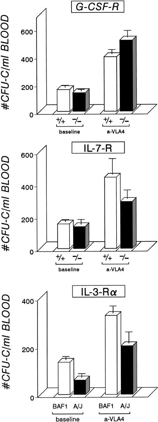
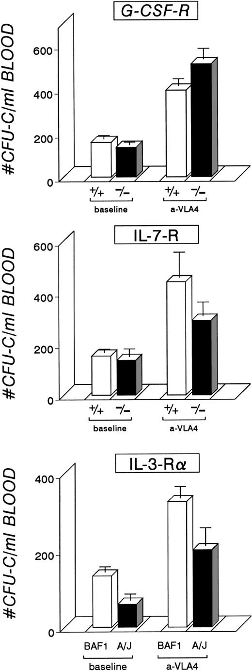
In previous experiments we have documented that coadministration of G-CSF and anti-VLA4 augments the mobilization response.16However, we could not determine whether there was exaggeration of a single mechanism or two different mechanisms. To resolve this issue we examined the anti-VLA4 mobilization response of mice that were null for the G-CSFR. Generation of these mice and description of their phenotype was previously published.18 G-CSFR null mice and controls were administered daily i.v. injections of anti-VLA4 for 3 days and killed the following day. The results (Fig1) show that G-CSF null and control mice respond similarly to the same dose of anti-VLA4 (control mice, 395 ± 37.2 CFU-C/mL blood; G-CSFR −/− mice, 514.2 ± 58.2 CFU-C/mL blood) and their responses were significant compared with their pretreatment values (P < .05). Therefore, the presence of an intact G-CSF receptor on hematopoietic cells is not required for anti-VLA4 response.
Five control mice and five receptor null mutant mice per group were treated by three IV injections of anti-VLA4 (see Materials and Methods). The mobilization response, measured as CFU-C/mL blood, is shown. All mice responded to anti-VLA4 treatment with a significant difference from their baseline CFU-C/mL levels.
Five control mice and five receptor null mutant mice per group were treated by three IV injections of anti-VLA4 (see Materials and Methods). The mobilization response, measured as CFU-C/mL blood, is shown. All mice responded to anti-VLA4 treatment with a significant difference from their baseline CFU-C/mL levels.
IL-7R null mice.
IL-7R deficient mice have impaired B and T lymphopoiesis.19It is also of interest that administration of IL-7 alone or in combination with G-CSF in normal mice has been shown to mobilize cells.20 IL-7R null mice and controls were injected with anti-VLA4 i.v. for 3 days and peripheral blood was drawn the fourth day to assess circulating levels of CFU-C (Fig 1). A significant mobilization was induced by anti-VLA4 in IL-7R −/− mice (P < .05) and their controls (P < .05), although some quantitative differences between the two sets of animals were noted (controls, 440.3 ± 103; IL-7R −/−, 289.2 ± 57).
A/J mice.
A/J mice have an impaired response to IL-3 because of a defect in the IL-3R α.21 The latter is not detectable on the surface of A/J mouse hematopoietic cells, but may be detected in the cytoplasm.22 The defect is not absolute because A/J mast cells in culture can generate IL-3R α in the presence of IL-3 and KL.22 A/J mice and BAF1 controls were treated with anti-VLA4. Again, a significant mobilization response (P < .05) was obtained (BAF1, 326 ± 30 CFU-C/mL of blood; A/J, 301 ± 48) compared with baseline levels from both types of animals (Fig 1). However, A/J mice were lower at baseline and after mobilization.
W/Wv and Sl/Sld mice.
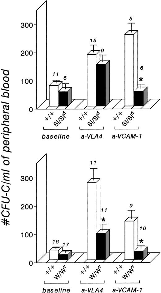
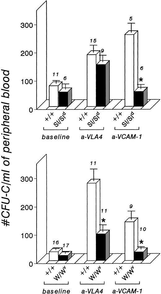
W/Wv mice have a defined defect in kit kinase activity, although the expression of kit protein on the cell surface and the binding to KL is normal.23 Control +/+ littermates responded to anti-VLA4–induced mobilization; however, W/Wvmice show a very blunted response (about 30% of normal), in contrast to the other mutant mice tested (11 WBB6F1 +/+ littermates, 273 ± 41 CFU-C/mL blood; 11 W/Wv mice, 93 ± 23) (Fig 2). We were intrigued by this muted response and proceeded to test Sl/Sld mice. Sl/Sld mice lack expression of membrane-bound KL,24 have a similar clinical phenotype to Wvmice, but have normal kit receptor expression and function in their hematopoietic cells, which can correct, by bone marrow transplantation, the defect in W/Wv mice.25 When Sl/Sld mice were treated with anti-VLA4, they responded to treatment similarly to their +/+ littermate controls (15 WCB6F1 +/+, 183 ± 27; 9 Sl/Sld mice, 148 ± 25), in contrast to W/Wv mice (Fig 2).
CFU-C/mL blood from control +/+ littermate mice and from Sl/Sld and W/Wv mice before and after anti-VLA4 and anti–VCAM-1 treatment (see Materials and Methods for details). Sl/Sld mice respond to anti-VLA4 similarly to controls (P = .35), in contrast to W/Wv mice which showed a blunted response (P < .0016). Both types of mutant mice did not respond to anti–VCAM-1 treatment, in contrast to their +/+ littermates. *Indicates significant differences from control responses (P < .05) and numbers above each column indicate the number of mice treated.
CFU-C/mL blood from control +/+ littermate mice and from Sl/Sld and W/Wv mice before and after anti-VLA4 and anti–VCAM-1 treatment (see Materials and Methods for details). Sl/Sld mice respond to anti-VLA4 similarly to controls (P = .35), in contrast to W/Wv mice which showed a blunted response (P < .0016). Both types of mutant mice did not respond to anti–VCAM-1 treatment, in contrast to their +/+ littermates. *Indicates significant differences from control responses (P < .05) and numbers above each column indicate the number of mice treated.
The expression of VLA4 in bone marrow cells, either lineage-positive or lineage-negative cells, was similar in W/Wv and Sl/Sld mice and did not differ from their +/+ littermate controls (data not shown). Furthermore, the data could not be explained by differences in the CFU-C content per femur, because Sl/Sld mice, if anything, had less CFU-C per femur and are more anemic than W/Wv mice (Table1). We also tested whether these mice differed in their response to anti-VCAM-1, the VLA4 ligand. Littermate +/+ control mice of either the Sl/Sld or the W/Wv series responded to VCAM-1. However, to our surprise, both Sl/Sld and W/Wv mice showed no response to anti–VCAM-1 treatment compared with their pretreatment values (5 WCB6F1 +/+ mice, 254 ± 33; 6 Sl/Sld mice, 53 ± 16 CFU-C/mL blood; 9 WBB6F1 +/+ mice, 136 ± 31; 10 W/Wv mice, 30 ± 9 CFU-C/mL blood) (Fig2). Constitutive expression of VCAM-1 was shown on immunohistochemistry of marrow sections, in sinusoidal endothelial cells of both mutant strains similar to their control littermates (data not shown).
It is known that the Steel (Sl) microenvironment is defective in the expression of membrane-bound KL and the maintenance of normal hematopoiesis.24, 25 Because of the abnormal microenvironment, one might have anticipated that the Sl mice might not respond to anti–VCAM-1. This result was totally unexpected for W/Wv mice because these mice have a functionally normal hematopoietic stroma.25 One may argue that W/Wvmice may respond subnormally to all mobilization treatments including anti-VLA4 or anti–VCAM-1. However, there was no difference in response to G-CSF between Sl/Sld and W/Wv mice (see data in following sections). Although the G-CSF response was subnormal in both types of mice (about 50% of control responses), both strains did mobilize progenitors to a level significantly different from baseline level (Fig 2), unlike the almost complete lack of mobilization with VCAM-1 treatment (W/Wv, 30 ± 9; Sl/Sld, 53 ± 16). To explain these data, we hypothesized that for either anti-VLA4 or anti-VCAM mobilization in W/Wv mice, signaling is required but does not occur because of unresponsive hematopoietic cells.
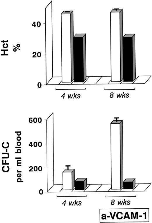
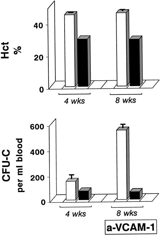
If our reasoning was correct, then by providing normal cells in the W/Wv mice we should correct the mobilization defect. For this reason we transplanted bone marrow cells from +/+ littermate controls into sublethally-irradiated W/Wv recipients (see Materials and Methods). Four and 8 weeks after transplantation the hematocrits in these mice had returned to control levels (Fig3), as would have been expected after engraftment of normal +/+ stem/progenitor cells. W/Wvrecipients were also transplanted with W/Wv donor cells and served as transplantation control animals. The posttransplant hematocrit (Hct) in these mice at 4 and 8 weeks after transplantation was at 29% and circulating progenitor cells at 4 weeks posttransplant were much lower than those observed in the recipients of +/+ donor cells (Fig 3). Eight weeks after transplantation all transplanted animals were treated with anti–VCAM-1 to test mobilization response. As seen in Figure 3, a significant response to anti–VCAM-1 is noted after transplantation in the recipients of +/+ donor cells. This response was similar to or better than the response of +/+ mice after anti–VCAM-1 treatment (545.7 ± 30.5 posttransplant in W/Wv recipients of +/+ cells; 136.2 ± 30.7 in +/+ controls post—anti–VCAM-1) (Fig 2). By contrast, no response was observed in the recipients of W/Wv donor cells. A second group of five W/Wv recipients transplanted with +/+ donor cells also had a normal Hct and responded well to anti–VCAM-1 treatments 8 weeks after transplantation (Hct 44.8 ± 1.15; post—anti–VCAM-1, 257.5 ± 12.6 CFU-C/mL blood). The data from these experiments suggested that the absence of the response in W/Wv, when administered anti–VCAM-1, is a result of the absence of signal transmission to defective W/Wv cells. If normal cells are administered within the same W/Wvmicroenvironment, then anti–VCAM-1 treatment becomes effective.
Transplantation of W/Wv mice with +/+ bone marrow cells (□) or W/Wv (▪) bone marrow cells. Eight weeks posttransplant W/Wv mice administered +/+ donor cells had normal Hct levels and responded significantly to anti–VCAM-1, in contrast to W/Wv recipients of W/Wv donor cells.
Transplantation of W/Wv mice with +/+ bone marrow cells (□) or W/Wv (▪) bone marrow cells. Eight weeks posttransplant W/Wv mice administered +/+ donor cells had normal Hct levels and responded significantly to anti–VCAM-1, in contrast to W/Wv recipients of W/Wv donor cells.
mev/mev mice.
The above data suggested that a functional kit receptor is important in the mobilization process. Mice homozygous for me locus have a severe phenotype and die at about 20 days, but the biology of the disease has been studied in the viable mouse variant (mev) which die much later, at about 9 weeks.26 Mutant me mice exhibit remarkable hematopoietic defects including autoimmunity, massive expansion of myeloid cells, and splenomegaly with increased number of CFU-E and hypersensitivity to erythropoietin.26 The me locus encodes Shp1, and cells from these mice are expected to exhibit an augmented or prolonged activation of kit autophosphorylation, because Shp1 is a downstream negative effector of kit signaling in vivo.27 Thus, we tested the responses of mev/mev mice to both anti-VLA4 and anti–VCAM-1 (Fig 4). Baseline circulating levels of CFU-C/mL are significantly higher in mev/mevmice (650 ± 15.8), versus either mev/+, or +/+ controls (104.7 ± 26.8). Bone marrow mononuclear cells (BM-MC) and CFU-C/femur are similar in mev/mev and +/+ controls. (BM-MCs in +/+ mice, 13.02 ± 1.74 × 10⁶per femur; mev/mev mice, 10.23 ± 0.61 × 10⁶/femur; CFU-C/femur in +/+ mice, 25,066 ± 2,296/femur; mev/mev, 25,936 ± 2,027/femur). Our data suggest that the unexplained splenomegaly in this model26 could be caused by an increased migratory capacity and splenic redistribution of stem/progenitor cells in mev mice. The opposite result is observed in W/Wv mice (Fig 2), which have very low basal numbers of circulating CFU-C (18 ± 3.4/mL blood). There is an exaggerated response to both anti-VLA4 and anti–VCAM-1 in mev mice compared with controls (post–anti-VLA4: mev/mev, 3,655 ± 970; controls, 863 ± 333; post—anti–VCAM-1: mev/mev, 1,942 ± 855; controls, 171.5 ± 57). Thus, constitutive activation of kit signaling results in increased migration of cells into the periphery and a heightened response to other mobilizing agents. The data provide additional evidence consistent with involvement of kit signaling in mobilization.
Bone marrow mononuclear cells were tested for kit and VLA4 expression after three daily injections of anti-VLA4. Labeling of cells with 2° antibody only, or anti-VLA4 and 2° antibody, gave superimposable profiles (not shown) indicating that cells were saturated by anti-VLA4 in vivo. Scattergram on the left panel was identical when untreated or treated animals were tested and the insert depicts cells in blast window analyzed for both kit expression (using directly conjugated anti-kit) and VLA4 expression. Note that after anti-VLA4 treatment both kit and VLA4 expression are downmodulated (middle and right panel). Downmodulation of kit was similar in two other experiments comparing untreated and treated bone marrow cells.
Bone marrow mononuclear cells were tested for kit and VLA4 expression after three daily injections of anti-VLA4. Labeling of cells with 2° antibody only, or anti-VLA4 and 2° antibody, gave superimposable profiles (not shown) indicating that cells were saturated by anti-VLA4 in vivo. Scattergram on the left panel was identical when untreated or treated animals were tested and the insert depicts cells in blast window analyzed for both kit expression (using directly conjugated anti-kit) and VLA4 expression. Note that after anti-VLA4 treatment both kit and VLA4 expression are downmodulated (middle and right panel). Downmodulation of kit was similar in two other experiments comparing untreated and treated bone marrow cells.
Response of W/Wv mice to cytokines.
To study the importance of normal kit signaling in mobilization by other cytokines we tested the response of W/Wv mice to a number of mobilization regimens. We confirmed published data28 that the response to G-CSF is approximately 50% of controls (W/Wv mice, 181.8 ± 19.9; +/+ controls, 388.6 ± 36; Sl/Sld, 199.7 ± 29.6%; +/+ controls, 416.8 ± 40). Reduced responses were also observed with treatments involving other cytokines or cytokine combinations administered for 3 days (post–G + FL for 3 days: W/Wv mice, 1,714 ± 344 CFU-C/mL; +/+ controls, 3,535 ± 346; post-FL treatment for 3 days: W/Wv, 83.7 ± 9.7; +/+ controls, 333.3 ± 26.3; post–FL + KL for 3 days: W/Wv, 146.3 ± 29.3; +/+ controls, 569 ± 79). Although there was a significant difference after 3 days of FL treatment between +/+ and W/Wv mice, after 7 days of FL treatment there was excellent response in W/Wv mice, with no significant differences from controls (post-FL treatment for 7 days: W/Wv, 5,965.3 ± 1,300; +/+ controls, 3,511 ± 172). The FL mobilization response after day 7 in W/Wv mice was accompanied by a marked proliferative response within the bone marrow (CFU-C/femur in W/Wv, 607,300 ± 56,910; +/+ controls, 210,322 ± 19,473; P < .05).
The normal responses of W/Wv mice to FL at day 7 stand in contrast to the subnormal responses previously observed with G-CSF or combinations of G + FL or KL + FL or FL alone for 3 days. The data with FL at 7 days would suggest that either W/Wv mice may not respond early on as readily as control mice, or that the FL response is an exception to the responses with other cytokines. A subnormal response was noted by other investigators with different doses of G-CSF28; however, very high doses have not been tested. If with high doses the subnormal response persists, one may suggest that the response to FL is qualitatively different from that of other cytokines. FL signals via binding to its tyrosine kinase receptor, flt3, present on hematopoietic stem/progenitor cells. Thus, it is theoretically possible that the mobilizing defect in the kit tyrosine kinase receptor of W/Wv mice may be compensated for by signaling through the flt3 alternative tyrosine kinase.
Downmodulation of c-kit expression after anti-VLA4 treatment.
To test whether any changes could be detected in kit expression after anti-VLA4 treatment, we treated normal B6D2F1 mice for 3 days with anti-VLA4 (2 mg/kg/d). Eight hours after the last injection femurs were removed for preparation of bone marrow cell suspensions. Bone marrow mononuclear cells were labeled either with goat anti-rat FITC or with anti-VLA4 (rat anti-mouse α4) followed by secondary antibody (goat anti-rat FITC). The FACS histograms of these two populations were virtually superimposable (data not shown), indicating that the cells were saturated in vivo by anti-VLA4 antibody. Cells labeled with anti-VLA4 plus 2° antibody (as above) were also labeled with anti-kit PE antibody. A separate bone marrow aliquot was also labeled with anti-kit PE only and all samples analyzed by FACS. Control bone marrow cells from mice that were not treated with anti-VLA4 were processed and labeled similarly, then run concurrently with the other samples. Before anti-VLA4 treatment most of the population of cells within the blast window (Fig 5, left panel)were doubly labeled with anti-kit and anti-VLA4 (Fig 5, middle panel). After in vivo anti-VLA4 treatment (Fig 5, right panel), a significant proportion of kitlo or kitneg cells was observed, along with reduction of anti-VLA4 brightly labeled cells. The data suggest that bone marrow cells display downmodulation of both kit and VLA4 after anti-VLA4 treatment.
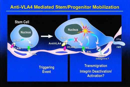
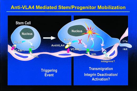
A model to explain anti–VLA4/VCAM-1—induced mobilization. Normally there is strong adhesion of hematopoietic cells to stromal endothelial cells through the VLA4/VCAM-1 pathway, which overrides any positive effects of kit on migration of stem/progenitor cells expressing kit. Consequent to antifunctional antibody treatment, there is deadhesion, which, either indirectly (by relieving the negative pressure on kit) or directly through communicating molecules, activates kit with ensuing stimulation of migration. During this process cells that are ready to egress downmodulate both α4 and kit. It is likely that increased migration via a kit-dependent pathway is achieved by activation/deactivation of another integrin to provide mechanistic support of cell movement. This effect may be strengthened by alleviation of a transdominant effect that VLA4 may exert on other integrins.
A model to explain anti–VLA4/VCAM-1—induced mobilization. Normally there is strong adhesion of hematopoietic cells to stromal endothelial cells through the VLA4/VCAM-1 pathway, which overrides any positive effects of kit on migration of stem/progenitor cells expressing kit. Consequent to antifunctional antibody treatment, there is deadhesion, which, either indirectly (by relieving the negative pressure on kit) or directly through communicating molecules, activates kit with ensuing stimulation of migration. During this process cells that are ready to egress downmodulate both α4 and kit. It is likely that increased migration via a kit-dependent pathway is achieved by activation/deactivation of another integrin to provide mechanistic support of cell movement. This effect may be strengthened by alleviation of a transdominant effect that VLA4 may exert on other integrins.
DISCUSSION
Mechanisms of Anti–VLA4/VCAM-1—Induced Mobilization
Adhesion/migration phenomena of mature leukocytes and the sequence of events from their activation to their extravasation and final recruitment in tissue sites of inflammation have been studied extensively and described in detail, especially using in vitro experimental models.29 Largely on the basis of in vitro information, several in vivo studies have been conducted in animal inflammation models using either function blocking antibodies or anti-adhesive peptides or small molecules.30 Detailed analyses of the antibodies' mechanism of action in vivo, their specific tissue requirements, or their cooperative molecules have not been described and extrapolations have been made from in vitro data. However, in vivo the situation may be very complex, with a coexistent network of negative and positive mediators and a cascade of events triggered after the initial stimulus, so that the in vivo setting cannot be faithfully mimicked with in vitro modeling. Consequently, the in vivo behavior of specific molecules may not be predicted from in vitro data. For example, although anti-β2 integrin antibodies inhibit adhesive interactions of CD34+ cells in vitro,10 in vivo administration of anti-β2has not been effective in mobilizing CD34+ cells, in contrast to anti-α4 antibody and despite the similar effects of these antibodies in vitro.13
In the case of anti-VLA4 administration in vivo, a simplistic view is that it can cause disruption of adhesive phenomena within the bone marrow, similar to its mode of action in vitro in long-term bone marrow cultures.9 Weakening of the adhesion between bone marrow cells and their microenvironment provides a biophysical mechanism whereby physiologically important connections of stem cells with their microenvironment are now severed. For mobilization to occur, hematopoietic cells need to migrate to intravascular spaces through basement membrane and endothelial cells, and such a migration is a prerequisite for their egress from the bone marrow to the circulation. Although changes in integrin-mediated adhesion per se can promote directed migration, adhesion-based mechanisms modulating cell locomotion are particularly complex and are usually driven by a multistep cascade of reactions involving several adhesion molecules.31-34 Events leading to mature leukocyte transmigration involve activation of several classes of adhesion molecules by ligands, activating antibodies, or peptide mimetic molecules.29 Our adhesion-blocking antibody abrogates α4-mediated adhesion in vitro, but is not known to behave like anti-β1 activating antibodies initiating signaling. One then would ask: Is a single deadhesion step in vivo sufficient to initiate subsequent events leading to mobilization, or are signaling and additional cooperative molecules required for mobilization? β1 integrins are considered as “locomotive” adhesion receptors and it is possible that antibody treatment per se can influence cell motility. Recently an antibody was described which inhibits neutrophil adhesion and increases motility.35Although this mode of action may be a theoretical possibility in the anti-VLA4–induced mobilization process, the data presented herein support alternative possibilities.
One can propose that the initial deadhesion step leads to engagement of several other intracellular signaling molecules, some of which are interconnected with growth factor signaling pathways. In hematopoietic stem/progenitor cells, the functional association between kit and integrins is of extreme interest. c-kit occupies a unique place in hematopoiesis because it affects proliferation, differentiation, and migration of multiple cell lineages, including hematopoietic stem/progenitor cells.23 Because migration is dependent on adhesive interactions with extracellular matrix, c-kit could mediate some of its function through inside-out signaling and modulation of integrins.36-40 Mast cells and eosinophils exhibit rapid induction to fibronectin adherence with KL stimulation, dependent on kit kinase activity.37, 39 Cells from hematopoietic cell lines with a progenitor cell phenotype also show a transient modulation of integrin avidity after KL stimulation.40 In addition to c-kit, another tyrosine kinase receptor, the platelet-derived growth factor receptor, has also been reported to influence integrin-mediated adhesive phenomena.41 However, the relevance of the short-lived in vitro effects of kit ligand to its delayed in vivo mobilization effect is unclear.
In the present study we provide evidence that kit receptor kinase activity modulates the effectiveness of anti-α4 integrin–induced mobilization. W/Wv cells with compromised kit kinase activity have a blunted mobilization, whereas cells from Sl/Sld mice with a similar clinical phenotype but normal kit kinase activity in stem/progenitor cells respond like normal control mice to anti-α4–induced mobilization. Because W/Wv mice do not respond appropriately to other mobilizing stimuli,28 one could argue that hyporesponsiveness is a general phenomenon for W/Wv and as such may not be directly associated with kit function. However, hyporesponsiveness to cytokines, specifically G-CSF, is not a unique feature of W/Wv because it was observed in Sl mice.27 On the other hand, other receptor mutant mice, like the G-CSF null mice, IL-7R null mice, or mice with compromised IL-3 function (A/J mice) respond normally to anti-α4 mobilization. Although testing of additional mutant mice (ie, Mpl −/− mice) would have been of interest, the data thus far do suggest that the association between kit function and anti-α4 mobilization may be causative and may be involved in changes in cell motility. Furthermore, both W/Wv and Sl mice did not respond to anti–VCAM-1—induced mobilization, in contrast to their +/+ littermates and despite normal expression of VCAM-1 in bone marrow. Collectively, the data would imply that signals initiated by anti–VCAM-1 cannot be transmitted to hematopoietic stem/progenitor cells, either because their kit function is compromised, as in W/Wv mice, or because of the absence of membrane-bound KL in the microenvironment of Sl mice. The finding that W/Wvmice 8 weeks posttransplant of +/+ donor bone marrow cells mobilized normally after anti–VCAM-1 treatment supports this view. Bone marrow mononuclear cells after anti-VLA4 treatment display (by FACS analysis) downmodulation of kit, which may be consequent to their prior activation, and is similar to what has been reported for human cells.42 The increased mobilization in mev/mev mice with constitutive activation of kit is also compatible with this hypothesis. The increased spleen size and increased CFU-C content in mev mice noted previously26 may result from a heightened migratory capacity of stem/progenitor cells leading to high levels of ongoing mobilization and spleenic seeding.
Mobilization: How Many Mechanisms? Any Emerging Theme?
Because several cytokines with diverse actions and different cognate receptor distribution mobilize a wide spectrum of stem/progenitor cells, one may suggest that some of the events triggered by cytokine administration may be common. We would like to propose that a major common pathway involved in mobilization, integrating signaling through integrins and initiating cell migration, is the kit/kit ligand pathway.
Merging information from the present study and the previously published studies,6 the following general mechanistic model for mobilization is proposed. Mobilization is a multistep process, usually accomplished in two phases: (1) the initiation phase, which encompasses the initial triggering event, and (2) the amplification phase, which includes several steps implemented through integrin/cytokine crosstalk or integrin/other adhesion molecules cross-influence and proliferative effects. The triggering event could be applied to cells themselves or components of their bone marrow microenvironment (stromal cells, extracellular matrix components). For example, anti-VLA4 or anti–VCAM-1 treatment are examples of effects on hematopoietic stem/progenitor cells or microenvironment cells, respectively. Such triggering events could act alone, leading to a short-lived mobilization (ie, post–IL-8,43 Mip1α,44 IL-1, or early postendotoxin6). Activation of signaling pathways by subsequent steps could lead to sustained and more efficient mobilization. Cooperative signaling could be mediated through growth factor receptors with tyrosine kinase activity (kit, flt3) and/or through integrin cross-talk.45 In the case of hematopoietic growth factors, this effect is also compounded by an increase in proliferation of cells within the bone marrow because of growth factor synergy affecting proliferation.
Do all the different growth factors have their own triggering or initiating event? Growth factors like G-CSF, which also affect end stage cells, could initiate that event in neutrophils, so that if neutrophils are functionally incompetent or not present, they do not respond and no mobilization ensues (ie, as in G-CSF −/− mice,17 or in chronic granulomatous disease patients.46) With KL or FL, the situation is even more speculative. The receptors for these ligands are present mostly on early cells and on certain cells within the microenvironment. Thus, endothelial cells expressing kit, or macrophages and/or dendritic cells with flt3 receptors could be affected in addition to primitive hematopoietic cells. Downmodulation of kit on cells may provide a stimulus for migration. Profound proliferative effects within the bone marrow resulting from many cytokines could impact existing pools of cells within bone marrow spaces or have secondary effects on blood flow.47 A delayed response for KL or FL would be compatible with the notion that proliferative events precede mobilization. However, proliferative effects themselves may not necessarily initiate mobilization.17
It has recently been reported that G-CSFR–deficient mice do not respond to IL-8 or cyclophosphamide-induced mobilization, suggesting that these stimuli act indirectly through G-CSF signalling. W/Wv mice with deficient kit signaling and mpl-deficient mice respond subnormally to G-CSF.28 Given the fact that many signaling molecules are shared by these hematopoietic growth factor receptors, it is conceivable that if signaling is required for mobilization, and if in these mice suboptimal stimuli are generated, mobilization may not be optimal. The net effect of mobilization is also dependent on proliferation events. Absence of synergistic actions between cytokines compound the magnitude of mobilization. In addition, one may predict that incompetence of cells to respond to the triggering event, either through receptor deficiency or deficiency of several relevant signaling proteins, may cause defects in mobilization. Thus, one can test whether mice deficient in signaling molecules (ie, Lck) are capable of responding to known mobilizing stimuli. These and other murine models should help further dissect the pathways responsible for mobilization.
ACKNOWLEDGMENT
We are grateful to Dr D. Link for allowing us to study his G-CSF null mice and to Drs J.J. Peschon and Chris Clegg for the IL-7R null mice. The generous gift of anti-VLA4 and anti–VCAM-1 antibodies by Dr R. Lobb (Biogen) and flt3 ligand by Dr S. Lyman (Immunex) is greatly appreciated. We also thank Dr J. Harlan for helpful discussions and critical reading of the manuscript and Sherri Brenner for her skillful secretarial assistance.
Address reprint requests to Thalia Papayannopoulou, Hematology, Box 357710, University of Washington, Seattle, WA 98195.
The publication costs of this article were defrayed in part by page charge payment. This article must therefore be hereby marked "advertisement" is accordance with 18 U.S.C. section 1734 solely to indicate this fact.

