Abstract
T-cell precursors develop within the thymus in contact with multiple supportive elements, among which thymic epithelial cells (TEC) are known to exert a dominant role in their homing, survival, and functional differentiation. All these functions are supported by cell-cell contacts and cytokine release. Signaling events triggered in lymphoid cells by adhesion to TEC are well characterized, but little is known about the opposite phenomenon. To address this issue, we derived cultures of TEC from human normal thymus. TEC monolayers were cocultured with thymocytes and immunostained with monoclonal antibodies (MoAbs) to integrin (2, 3, 4, and 6) and β (β1 and β4) chains. Optical and confocal analysis showed that integrins were polarized on TEC at discrete surface locations: 6β4 lined the basal surface of TEC monolayers, whereas 3β1 was found mostly at TEC-TEC contacts; it is noteworthy that both 3β1 and 6β4 became highly enriched also at the boundaries with adherent thymocytes. Functional studies performed with MoAbs anti-β1 and -β4 integrins showed that β1, and, to a much lower extent, β4 heterodimers are involved in the TEC-thymocyte adhesion. Thymocyte contact or MoAb-mediated ligation of 3, 6, β1, and β4 integrins was investigated as a potential inducer of intracellular signaling in TEC. Thymocyte adhesion or cross-linking of MoAbs bound to integrins clustered at the TEC/thymocyte contact sites led to activation of interleukin-6 (IL-6) gene transcription factors, namely NF-IL6 serine phosphorylation and NF-κB nuclear targeting, as well as to increased IL-6 secretion. We propose that integrin clustering occurring during TEC-thymocyte contacts modulates in TEC the gene expression of a cytokine involved in thymocyte growth and functional differentiation.
THE THYMIC EPITHELIUM is composed of multiple cellular subsets of ectodermal or endodermal embryonic origin, characterized by tonofilaments, surrounded by extracellular matrix, and interconnected by desmosomes to form an intralobular meshwork filled with developing thymocytes. Although the discrete functions of the various subsets have yet to be elucidated in humans, it is known that thymic epithelial cells (TEC) exert pivotal roles in the homing, intrathymic migration, and differentiation of lymphoid precursors through the release of cytokines (ie, interleukin-1α [IL-1α] and IL-1β, IL-3, IL-6, IL-8, and transforming growth factor β3 [TGFβ3]),1-3 the secretion of extracellular matrix proteins, and the establishment of adhesive interactions. Thymocytes adhesion seems also effective at modulating functions and differentiation process of TEC. Evidence in mouse models demonstrated that thymocyte contact is required by thymic epithelium to properly develop during embryogenesis.4 In addition, observations obtained with human TEC derived in vitro showed that thymocyte adhesion results in tyrosine phosphorylation of cytoplasmic proteins, increased IL-6 mRNA transcription, and/or cytokine production.4-6 The regulation of IL-6 gene expression in TEC is of particular relevance within the thymic microenvironment as regards its activities on lymphoid activation and cytotoxic differentiation of thymic precursors as well as its antiapoptotic functions in epithelial and lymphoid cells.7-10 The IL-6 gene expression is mainly regulated at a transcriptional level, although mechanisms of posttranscriptional control have been also described.3,11,12 To date, two major regulatory factors, namely NF-κB and NF-IL6, have been described to be involved in IL-6 transcription in different cell lineages.11 It is noteworthy that transfection studies in murine carcinoma cell lines have shown that overexpression of NF-IL6 and the p65 subunit of NF-κB synergistically activates an IL-6 promoter-reporter construct, indicating that these two factors are sufficient to sustain the activation of IL-6 gene.13 NF-IL6, a member of the CCAAT/enhancer binding (C/EBP) protein-related family, is a DNA binding protein recognizing similar motifs in liver-specific, acute-phase reaction, and cytokine genes.11,14 Its transactivation potential is regulated at translational and posttranslational levels. Different usage of initiation codons accounts for expression of isoforms, which differ in the N-terminal half of the protein containing the activation domain and hence in transcriptional activity.15,16 Posttranslational modifications, consisting in serine/threonine phosphorylation of specific amino acids within the functional domains, further regulate both the transcriptional and the DNA binding activity, as demonstrated in epithelial and fibrosarcoma cell lines.17,18 NF-κB is an ubiquitous dimeric factor composed of p50 and p65 (RelA) subunits sequestered inactive in the cytosol bound to a family of inhibitors, IκBs, that mask its nuclear localization sequence.19 Activation relies on serine hyperphosphorylation and proteosomal degradation20 or on tyrosine posphorylation of inhibitors,21 either one of which is sufficient to disrupt the IκB-NF-κB complexes, thus allowing NF-κB to move into the nucleus.
Our previous data demonstrated that binding of lymphoid cells to TEC increases the DNA binding activity of their endogenous NF-κB as well as their IL-6 production,22 prompting us to investigate whether IL-6 gene expression in TEC could be regulated at a transcriptional level by signals delivered from adhesive interactions with thymocytes. Several molecules have been identified that act as a receptor or counter-receptor in TEC adhesion to thymocytes such as CD6-100kD ligand, galectin, LFA3, intercellular adhesion molecule-1 (ICAM-1), vascular cell adhesion molecule (VCAM), cadherin, and integrins.23-25 The latter are of particular interest because of their signal transduction properties. Integrin ligation with natural ligands or monoclonal antibodies (MoAbs) initiates a variety of events including induction of Calcium transients, changes in cAMP levels, activation of Na+/H+ antiporter, or protein tyrosine phosphorylation, which can ultimately lead to the expression of several genes, including those of cytokines.26
In this report, we show that binding of normal thymocytes to TEC is followed by the activation of IL-6 gene transcription factors, namely NF-κB nuclear translocation and serine phosphorylation of the 36- and 43-kD NF-IL6 isoforms, and is associated with increased IL-6 production. Moreover, we show that both activation of IL-6 transcription factors and increased IL-6 secretion are induced by cross-linking of α3β1 and α6β4 integrin heterodimers, which cluster at the TEC/thymocyte contact sites and are functionally involved in their binding.
Previous reports have shown that TGF-β and epidermal growth factor (EGF) induce IL-6 production in TEC through posttranscriptional control mechanisms.3 We herewith propose that TEC IL-6 production can be regulated also at the transcriptional level through signals delivered by integrin recruitment during their adhesion to thymocytes.
MATERIALS AND METHODS
Cell cultures.
TEC primary cultures were derived from normal thymus of patients (<5 years of age) undergoing corrective cardiac surgery as previously described.27 Briefly, thymus specimens were minced and trypsinized (0.05% trypsin/0.01% EDTA) at 37°C for 3 hours. Cells were collected every 30 minutes, pooled, plated onto lethally irradiated 3T3-J2 feeder-layer cells (a gift of Prof H. Green, Harvard Medical School, Boston, MA) at 2.5 × 10⁴/cm2and cultured at 5% CO2 in growth medium composed as follows: Dulbecco’s modified Eagle’s medium (DMEM) and Ham’s F12 media (3:1 mixture), 10% fetal calf serum (FCS), insulin (5 μg/mL), transferrin (5 μg/mL), adenine (0.18 μmol/L), hydrocortisone (0.4 μg/mL), cholera toxin (0.1 nmol/L), triiodothyronine (2 nmol/L), EGF (10 ng/mL), glutamine (4 mmol/L), and penicillin-streptomycin (50 IU/mL). Confluent TEC primary cultures were detached by trypsin-EDTA, plated on NIH-J2 feeder layer, and expanded to confluent secondary cultures in growth medium. TEC obtained from secondary cultures already devoid of NIH-J2 cells were replated without the feeder layer at 1.2 × 10⁴/cm2 cell density, cultured in growth medium for 24 hours and maintained before use for 48 hours in modified growth medium containing one third of the concentration of insulin, transferrin, adenine, hydrocortisone, cholera toxin, triiodothyronine, and EGF. Media were purchased from Seromed (Berlin, Germany) and supplements were purchased from Sigma-Aldrich (Milano, Italy). EGF was from Austral Biological (San Ramon, CA).
Human thymocytes were prepared by mechanical disruption of fresh thymus specimens. At least 95% viable cells were isolated from the cell suspension by Ficoll-Hypaque gradient centrifugation, washed, and used immediately after preparation.
Antibodies.
The following antibodies were used for immunostaining, cell treatment, Western blotting, electromobility shift assay (EMSA), and Ca2+ mobilization assays: MoAbs anti-CD2, -CD3, -CD4, -CD8, and -CD16 and fluorescein isothiocyanate (FITC)-labeled F(ab)2 goat antimouse Ig from Becton Dickinson (Mountain View, CA); anti-CD1a, anti-CD18, anti–ICAM-1, anti-α3 (CD49c), and anti-a2 (Gi9) from Immunotech International (Marseille, France); MoAbs B9-12 (anti-MCH class I) and D1-12 (anti-MHC class II) provided by Dr R.S. Accolla (University of Pavia, Varese, Italy); MoAb 10.1.2 (gift of Dr G. Corte, CBA Genova, Genova, Italy); MoAb 3E1 (anti-β4; gift of Dr E. Engvall, The Burnham Institute, La Jolla, CA); MoAb MAR 4 (anti-β1) and MAR 6 (anti-α6; kindly provided by Dr S. Ménard, Istituto Nazionale Tumori, Milano, Italy); MoAb Lam89 (anti-laminin 1) from Sigma; MoAb GB3 (anti-laminin 5; gift of Dr Patrick Verrando, University of Nice, Nice, France); MoAb anti-VCAM (British Biotechnology Laboratories Ltd, Oxford, UK) through the courtesy of Dr R. Giavazzi (Istitut M. Negri, Bergamo, Italy); F(ab)2 goat antimouse Ig from Pierce (Pierce, Oud Beijerland, The Netherlands); swine antirabbit or rabbit antimouse IgG for immunoistochemistry from Dako (Glostrup, Denmark); antiserum anti-p105 recognizing both the p50 precursor (p105) and the p50 mature form (kindly provided by Dr A. Israel, Institut Pasteur, Paris, France); rabbit antiserum anticingulin (gift of Dr S. Citi, University of Padova, Padova, Italy); antisera anti–NF-IL6 (C19) and anti–NF-κB p65 (A) from Santa Cruz Biotechnology (Santa Cruz, CA); MoAb antiphosphoserine in agarose-coupled or uncoupled form, horseradish peroxidase-labeled rabbit antimouse, goat antirabbit antisera, and nonspecific mouse Igs from Sigma. Flow cytometry was performed in a FacScan flow cytometer (Becton Dickinson).
Adhesion assays and MoAb treatment.
As detailed in the previous paragraph, morphological and functional studies were performed with TEC grown to monolayers without the NIH-J2 cell support and adapted to low concentration of supplements in the culture medium. Thymo-epithelial cocultures destined to immunohistochemical studies were performed with TEC plated at 1.2 × 10⁴/cm2 on round glass coverlips fitting the 24-well Costar (Acton, MA) plates and grown to confluence. Freshly isolated thymocytes were added at a 5:1 thymocyte:TEC ratio and removed 12 hours later by gentle washing. Cocultures destined to transcription factor studies were performed in 6-well Costar plates at same cell ratio. Thymocytes were removed after 1, 3, or 5 hours of contact by extensive vigorous washings. TEC were detached by trysin-EDTA, scored for contamination with residual thymocytes by immunostaining with MoAbs anti-CD1a and -CD2, and used as a source of cytoplasmic and nuclear extracts.
TEC destined to binding assays with thymoctes were plated 24 hours before use in 24-well Costar plates at high density (5 × 10⁵/well), so that their extremely tight confluence could prevent the direct attachment to the plastic of thymocytes.
Cocultures used for IL-6 production assays were performed for 12 hours. After thymocyte removal, TEC were recovered by trypsin-EDTA, analyzed for the absence of contaminants as described above, and plated onto new plates. Cell supernatants were collected 24 and 72 hours from plating. TEC destined to Ca2+ mobilization analysis were grown to confluency on square glass coverslips fitting the spectrofluorimeter cuvette. MoAbs anti-α3, -α6, -β1, and -β4 chains, anti-CD71, and MHC class I were used purified at 5 μg/mL, previously identified as the saturating concentration for all antibodies by flow cytometry. Primary antibodies were cross-linked, if required, after 1 hour of incubation, by 2 additional hours of incubation with F(ab)2goat antimouse Ig at 10 μg/mL.
Thymo-epithelial coculture immunostaining.
Cocultures fixed in 3% paraformaldehyde (electron microscope grade), 2% sucrose in phosphate-buffered saline (PBS), pH 7.6, for 5 minutes at room temperature were further treated as previously reported.28 Briefly, fixed monolayers were permeabilized (3 minutes at 4°C) in HEPES-Triton X-100 buffer (20 mmol/L HEPES, 300 mmol/L sucrose, 50 mmol/L NaCl, 3 mmol/L MgCl2, 0.5% Triton X-100, pH 7.4). Staining for F-actin was performed with fluorescein-labeled phalloidin (F-PHD; Sigma; 200 nmol/L for 20 minutes at 37°C in the dark). Adhesion molecules were detected with the relevant MoAbs (see above), followed by rhodamine-tagged swine antirabbit or rabbit antimouse IgG. Primary antibodies were replaced by mouse IgG or preimmune rabbit sera in control samples. Coverslips were incubated with the appropriate rhodamine-tagged secondary antibodies routinely supplemented with 200 nmol/L F-PHD. After a final thorough washing, coverslips were mounted in Mowiol 4-88 (Hoechst, Frankfurt am Main, Germany) and observed in a Zeiss (Oberkochen, Germany) Axiophot microscope equipped for epifluorescence and a 63× planapochromatic lens. Stained coverslips were photographed with Kodak T-MAX 400 films (Eastman Kodak, Rochester, NY) exposed at 1000 ISO and developed at 1600 ISO in T-MAX developer for 10 minutes at 20°C. The same coverslips were analyzed in parallel with a confocal laser scanning microscope (CLSM Bio-Rad 1024; Bio-Rad, Hercules, CA). Image files were recorded on different channels and digitally reconstructed to provide z-axis views. Files obtained from confocal microscopy were assembled and printed with Adobe Photoshop 3.5 (Adobe Systems Inc, Mountain View, CA).
TEC-thymocyte binding assay.
Binding assays were performed with freshly isolated, unfractionated normal thymocyte prestained with the PK H26-GL red fluorescent dye (Sigma). Dye loading was achieved according to the manufacturer’s recommendations. Thymocytes were added to TEC monolayer grown to confluence at 5:1 thymocyte/TEC ratio and allowed to adhere for 1 hour at 37°C in humidified atmosphere of 5% CO2 in binding medium composed of incomplete medium and 3% bovine serum albumin (BSA; Sigma). At the end of the coculture, the nonadherent thymocyte were gently removed by three subsequent washings performed with binding medium. TEC monolayer and bound thymocytes where then detached with trypsin-EDTA treatment, washed, vigorously resuspended to disrupt aggregates, and analyzed by flow cytometry. TEC monolayers and PKH 26 GL-labeled thymocytes were separately incubated with the various MoAbs or with the aspecific mouse Ig (all used at 5 μg/mL) for 30 minutes at 37°C before binding. Assays in the absence of divalent cations were performed in PBS Ca2+Mg2+-free buffer at 3% BSA. TEC and thymocytes were washed twice with the buffer before binding. The results of each experiment were expressed as the ratio of TEC and thymocytes recovered and as the percentage of the ratio of the untreated control.
EMSA.
Nuclear and cytoplasmic extracts were prepared from TEC cells as previously described,30 with minor modifications. Protein concentration was assessed by Coomassie protein assay reagent (Pierce, Rockford, IL). DNA binding activity was determined by using a 32P γ-ATP end-labeled double-strand oligonucleotide encompassing the NF-κB binding site located in the IL-6 promoter region as probe. Cell extracts (6 μg) were incubated for 30 minutes at room temperature with the probe (1 × 10⁵ cpms/sample) in 20 μL of binding buffer (20 mmol/L Tris-HCl, pH 7.5, 0.1 mol/L NaCl, 1 mmol/L dithiothreitol [DTT], 1 mmol/L EDTA, 1 mg/mL BSA, 0.1% NP-40, 4% glycerol) containing 1 μg of Poly(dI-dC) (Pharmacia, Uppsala, Sweden). Cytoplasmic extracts were pretreated with 0.2% deoxycholic acid in binding buffer for 10 minutes at room temperature before probing. Antisera anti–NF-κB subunits were preincubated with nuclear extracts for 30 minutes on ice before probing. Nucleoprotein complexes were electrophoresed on a 5% polyacrylamide (30:1.2) gel in 0.05 mol/L Tris-borate/1 mmol/L EDTA at 150 V. Gels were dried and exposed to Amersham Hyperfilm films (Amersham, Little Chalfont, UK). Film densitometry was performed with an UltroScan Densitometer and the built-in software (LKB, Bromma, Sweden).
Cell labeling, immunoprecipitation, and Western blotting.
TEC monolayers were labeled overnight with 25 μC/mL of14C-leucine (324.9 mC/mmol; NEN Research Dupont, Boston, MA) in incomplete growth medium. Cell lysates were prepared as previously described for EMSA. Twenty micrograms of cytoplasmic or nuclear extracts was precleared with 20 μL of swollen protein A-Sepharose beads (Pharmacia) in lysis buffer (20 mmol/L Tris-HCl, pH 7.2, 0.5 mmol/L DTT, 1 mmol/L EDTA, 0.5 mmol/L phenylmethylsulfonyl fluoride [PMSF] adjusted to 0.15 mol/L NaCl) for 1 hour at room temperature and incubated with the anti–NF-IL6 antiserum at 1:103 dilution for an additional 2 hours on ice. Immunocomplexes were precipitated with 20 μL of protein A-Sepharose beads for 1 hour at room temperature and extensively washed in 0.05 mol/L Tris-HCl, pH 7.2, 0.15 mol/L NaCl, 0.1% NP40. Cell lysates (20 μg/sample) or immunoprecipitates (loaded at equal amount of counts) were fractionated by 12% sodium dodecyl sulfate-polyacrylamide gel electrophoresis (SDS-PAGE), electroblotted to nitrocellulose membrane (Bio-Rad, Milano, Italy), and probed with antiphosphoserine MoAbs in TBST 1.5% wt/vol powdered low fat dry milk. Cross-immunoblotting was performed by immunoprecipitation of 20 μg of precleared cell extracts with 30 μL of antiphosphoserine MoAb agarose-coupled (Sigma) for 1 hour on ice probed with the anti–NF-IL6 antiserum at 1:5,000 dilution. Membranes were washed with TBST buffer and specific bands were shown by enhanced chemiluminescence system (ECL; Amersham).
Intracellular calcium assays.
Free cytosolic Ca2+ concentration was determined by using Indo-1 acetoxymethyl ester (Indo-1-AM; Molecular Probes Europe BV, Leiden, The Netherlands).31 Optimal cell loading was achieved with 3 μmol/L Indo-1-AM in DMEM at 1% FCS without phenol red for 40 minutes at 37°C. Coverslips washed in fresh buffer were immobilized in the test cuvette in working buffer consisting of 140 mmol/L NaCl, 5 mmol/L KCl, 1 mmol/L MgCl2, 1 mmol/L NaH2PO4, 20 mmol/L HEPES, 5 mmol/L glucose, 0.5 mmol/L EGTA, or 1 mmol/L CaCl2, when indicated, at pH 7.4. MoAbs were used at 5 μg/mL. Relative fluorescence intensity was measured on a Hitachi F-2000 spectrofluorimeter (Hitachi, Ltd, Tokyo, Japan) at excitation and emission wavelengths of 340 and 400 nm, respectively, with slits of 20 nm. The [Ca2+]in was calculated according to the following formula (Grynkiewicz et al31): [Ca2+]in = kd (F − Fmin)/(Fmax − Fmin), where kd, the Ca2+-Indo-1 dissociation constant, was assumed to be 250 nmol/L; Fmax is fluorescence intensity obtained upon addition of 0.05% Triton X-100 in the presence of 1 mmol/L CaCl2; and Fmin is the level of fluorescence obtained after the addition of 1 mmol/L MnCl2. Because we observed that TEC treated with 100 μmol/L ATP increased intracellular Ca2+ independently from extracellular Ca2+, this responsiveness was assumed to be a criterion of cell suitability for analysis and, hence, was evaluated in all MoAb-unresponsive samples.
IL-6 production.
Cell supernatants were assayed for IL-6 production using an enzyme-linked immunosorbent assay (ELISA) kit according to the manufacturer’s instructions (CLB, Amsterdam, The Netherlands). Optical density (OD) values were plotted on the standard curve and expressed as nanograms per 106 cells recovered.
RESULTS
Thymo-epithelial cocultures.
Activation of IL-6 transcription factors in TEC after contact with thymocyte was investigated by adhesion assays with time-course. Cocultures were performed by using TEC monolayers at the third passage of culture grown to confluence and unfractionated thymocytes. Flow cytometry of seven cultures derived from five independent donors (Table1) consistently showed that greater than 90% of TEC expressed a high amount of MHC-class I, β1, and α3 molecules and moderate amounts of α5; 75% to 80% expressed α6 and β4 chains; ICAM-1 ranged between 9.5% and 20%; all cultures lacked CD18, CD16, and MHC-class II molecules (not shown); VCAM, recently described as a marker of cortical epithelium in vivo,32 was also undetectable in our TEC since the primary culture, whereas greater than 90% of TEC of all cultures were stained by the 10.1.2 MoAb, previously shown to recognize an α3 variant expressed by medullary epithelium in vivo.29
TEC monolayers were maintained in modified growth medium (see Materials and Methods) and in the absence of feeder layer for at least 48 hours before the adhesion assays to minimize the strong stimulating activity of the variety of growth factors required to raise and expand the primary cultures. Culturing TEC in modified growth medium did not affect the quantitative or qualitative features of their phenotype, the polarization of integrins, or the extent of thymocyte binding (see below). Freshly isolated, unfractionated thymocytes were used at 5:1 thymocyte/TEC ratio, allowed to adhere to TEC for 1, 3, and 5 hours at 37°C in humidified atmosphere of CO2, and ultimately removed by extensive and vigorous washings with medium. Control TEC were washed in the same way.
Thymocyte adhesion induces NF-κB nuclear translocation and NF-IL6 phosphorylation in TEC.
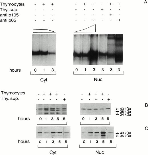
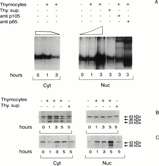
NF-κB binding activity was assayed by EMSA in cytoplasmic and nuclear extracts prepared from TEC previously scored negative for residual CD1+ or CD2+ cells by flow cytometry. As shown in Fig 1A, the constitutive nuclear NF-κB binding activity found in untreated TEC was increased more than 3.5-fold after 3 hours of thymocyte contact. This increase was consistent with the two thirds decrease of NF-κB activity observed in the cytoplasm at the same time point, indirectly indicating that hyperphosphorylation and subsequent proteolysis of NF-κB inhibitors had occurred.20 The subunit composition of translocated complexes was investigated by binding assays performed in the presence of antisera that recognize the p65 or the p50 proteins. As evidenced by the changes of NF-κB gel retardation pattern specifically induced by each antiserum, the translocated complexes found to be increased after 3 hours of thymocyte contact contained both p65 and p50 subunits, which have been previously shown to constitute transcriptionally active heterodimers.19 TEC extracts were next examined for the presence of serine-phosphorylated NF-IL6 isoforms, because previous findings have demonstrated that NF-IL6 protein and DNA interactions are positively regulated by serine/treonine phosphorylation of its functional domains.11,17 18 We therefore performed Western blot analysis of (1) cytoplasmic and nuclear lysates immunoblotted with an anti–C-terminal NF-IL6 antiserum recognizing all protein isoforms (Fig 1B) and (2) NF-IL6 immunoprecipitates probed with antiphosphoserine MoAbs (Fig 1C). Cell extracts were prepared from14C-metabolically labeled cells so that immunoprecipitates could be loaded at equal amounts of counts. As shown in Fig 1B, the cytoplasmic NF-IL6 pool was composed of 24-, 36-, and 43-kD isoforms, whereas the nuclear pool was mainly constituted of the 43-kD form. The cytoplasmic pool was increased after 1 hour of thymocyte contact, whereas the nuclear 43-kD isoform remained quantitatively unchanged in the same experimental conditions. By contrast, as shown in Fig 1C, increasing amounts of 43- and 36-kD phosphorylated proteins were immunoprecipitated from cytoplasmic (2-fold over control) and, more importantly, from nuclear extracts after 3 and 5 hours of adhesion with thymocytes (more than 10-fold over control). The latter results were further confirmed by cross-immunoblotting of antiphosphoserine MoAb immunoprecipitates probed with anti–NF-IL6 antiserum (not shown). Supernatants of thymocytes cocultured with TEC for 12 hours failed to induce NF-κB and NF-IL6 activation (Fig 1A, B, and C). These results demonstrated that posttranslational regulation of NF-κB and NF-IL6 transcription factors had occurred in TEC after signals delivered by thymocyte adhesion rather than by the release of soluble factors during the contact and led us to investigate whether they were associated with topographical rearrangements of adhesion molecules at cell contact sites.
NF-κB binding activity and NF-IL6 phosphorylation in TEC after adhesion to normal thymocytes. (A) NF-κB binding activity of cytoplasmic (Cyt) and nuclear (Nuc) extracts of TEC untreated, cocultured with freshly isolated thymocyte for 1 and 3 hours or incubated with thymocyte supernatant (Thy.sup) for 3 hours. Thymocyte supernatant was obtained from thymocytes cocultured with TEC for 12 hours. EMSA were performed with 6 μg of cell extracts probed with the32P-labeled IL6-κB oligonucleotide. Cytoplasmic proteins were treated with 0.2% deoxycholic acid before binding to dissociate NF-κB/IκB inhibitors. Nuclear complexes contained p50 and p65 subunits, as assessed by the band supershifting obtained with anti-p105 or anti-p65 antisera. DNA binding activity was quantitated by gel densitometry and expressed as a percentage of untreated controls (top of the gels). (B and C) PAGE analysis of NF-IL6 phosphorylation and isoform composition of cytoplasmic and nuclear extracts prepared from14C-leucine–labeled TEC untreated and cocultured with thymocytes for 1, 3, and 5 hours or incubated with thymocyte supernatants for 5 hours. (B) Immunoblotting of cytoplasmic and nuclear lysate (20 μg) probed with the anti–NF-IL6 antiserum. (C) Immunoblotting of cytoplasmic and nuclear NF-IL6-immunoprecipitates (103 cpm/lane) probed with antiphosphoserine MoAbs. Arrowheads indicate the positions of proteins with apparent molecular weights of 24, 36, and 43 kD. Sections of gels are shown. The results are representative of at least three experiments performed independently by using TEC and thymocytes derived from different donors.
NF-κB binding activity and NF-IL6 phosphorylation in TEC after adhesion to normal thymocytes. (A) NF-κB binding activity of cytoplasmic (Cyt) and nuclear (Nuc) extracts of TEC untreated, cocultured with freshly isolated thymocyte for 1 and 3 hours or incubated with thymocyte supernatant (Thy.sup) for 3 hours. Thymocyte supernatant was obtained from thymocytes cocultured with TEC for 12 hours. EMSA were performed with 6 μg of cell extracts probed with the32P-labeled IL6-κB oligonucleotide. Cytoplasmic proteins were treated with 0.2% deoxycholic acid before binding to dissociate NF-κB/IκB inhibitors. Nuclear complexes contained p50 and p65 subunits, as assessed by the band supershifting obtained with anti-p105 or anti-p65 antisera. DNA binding activity was quantitated by gel densitometry and expressed as a percentage of untreated controls (top of the gels). (B and C) PAGE analysis of NF-IL6 phosphorylation and isoform composition of cytoplasmic and nuclear extracts prepared from14C-leucine–labeled TEC untreated and cocultured with thymocytes for 1, 3, and 5 hours or incubated with thymocyte supernatants for 5 hours. (B) Immunoblotting of cytoplasmic and nuclear lysate (20 μg) probed with the anti–NF-IL6 antiserum. (C) Immunoblotting of cytoplasmic and nuclear NF-IL6-immunoprecipitates (103 cpm/lane) probed with antiphosphoserine MoAbs. Arrowheads indicate the positions of proteins with apparent molecular weights of 24, 36, and 43 kD. Sections of gels are shown. The results are representative of at least three experiments performed independently by using TEC and thymocytes derived from different donors.
Thymocyte adhesion to TEC monolayers induces repolarization of α3β1 and α6β4 heterodimers.
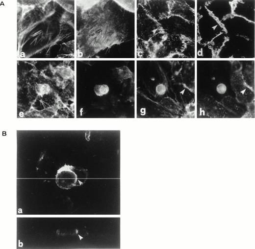
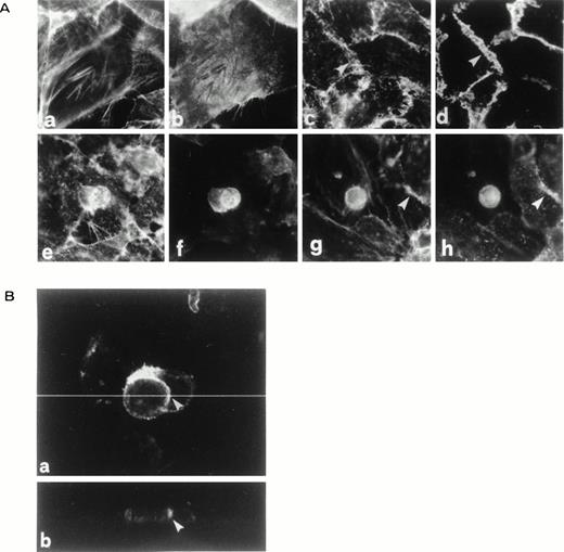
The immunofluorescence analysis of TEC monolayers or TEC-thymocyte cocultures performed for 12 hours at 37°C is shown in Fig2A. Analysis of TEC monolayers (Fig 2A, a through d) demonstrated that both α3β1 and α2β1 (the latter is not shown) lined the TEC intercellular boundaries (c shows F-actin and d shows the corresponding pattern of α3), whereas α6β4 was localized at the adhesion surface of TEC to the plastic (a and b show F-actin and β4, respectively). It is noteworthy that, as previously described for keratinocytes,28 spread and attached TEC displayed a submembraneous F-actin feltwork and few organized microfilament bundles (a) that exclude α6β4 (compare a and b), whereas α3β1 codistributed with the microfilamentous feltwork (compare c and d) at intercellular boundaries indicated by arrowheads. As a positive control of TEC cell-to-cell junctions, we used an antibody to cingulin as a marker of tight junctions (not shown). Immunofluorescence analysis of TEC-thymocyte cocultures showed that thymocyte adhesion partially redistributed α3β1 (h obtained with MoAb to α3 and g showing the corresponding F-actin pattern) and α6β4 (f obtained by an MoAb to β4 and e showing the corresponding F-actin pattern) at the TEC-to-thymocyte contact sites (e through h). In fact, α6β4 (f) and α3β1 (h), but not α2β1 (not shown), became enriched at the interface between TEC and engulfed thymocytes. Note that the focus of frames (e) through (h) is removed from the substratum adhesion to highlight TEC-to-thymocyte interface.
Immunostaining of thymo-epithelial cocultures. All the experiments were performed on cells cultured at 37°C under standard incubation conditions as described in Materials and Methods. (A) a through d: immunostaining of TEC monolayers for F-actin (a and c), β4 (b), and 3 (d). β4 and 3 stainings are shown coupled to their F-actin stainings (a and c, respectively). Frames a and b were focused at the basal surface of TEC monolayers to show the complementary distribution of F-actin with β4, showing that β4 is excluded from F-actin–rich areas. Frames c and d were focused slightly above the plane of adhesion to show intercellular boundaries. e through l: immunostaining of TEC thymocytes cocultures for F-actin (e and g), β4 (f), and 3 (h). Frames e through h were all focused above the basal surface of TEC monolayers to show the aggregation of β4 (f) and 3 (h) at TEC-thymocyte interface. Arrowheads in g and h show a TEC intercellular boundary that is still in focus. (B) Confocal analysis of β4 enrichment at TEC-thymocyte interface (a). Digital reconstruction and rotation at the dotted line (a) to show the z axis (b) confirm β4 enrichment at the TEC-thymocyte interface. Arrowheads in a and b indicate the same position before and after rotation.
Immunostaining of thymo-epithelial cocultures. All the experiments were performed on cells cultured at 37°C under standard incubation conditions as described in Materials and Methods. (A) a through d: immunostaining of TEC monolayers for F-actin (a and c), β4 (b), and 3 (d). β4 and 3 stainings are shown coupled to their F-actin stainings (a and c, respectively). Frames a and b were focused at the basal surface of TEC monolayers to show the complementary distribution of F-actin with β4, showing that β4 is excluded from F-actin–rich areas. Frames c and d were focused slightly above the plane of adhesion to show intercellular boundaries. e through l: immunostaining of TEC thymocytes cocultures for F-actin (e and g), β4 (f), and 3 (h). Frames e through h were all focused above the basal surface of TEC monolayers to show the aggregation of β4 (f) and 3 (h) at TEC-thymocyte interface. Arrowheads in g and h show a TEC intercellular boundary that is still in focus. (B) Confocal analysis of β4 enrichment at TEC-thymocyte interface (a). Digital reconstruction and rotation at the dotted line (a) to show the z axis (b) confirm β4 enrichment at the TEC-thymocyte interface. Arrowheads in a and b indicate the same position before and after rotation.
This finding prompted the search for laminin 1 and 5, both substrates for either integrin heterodimers, in between TEC and thymocytes. Both laminin types were expressed at TEC attachment surface (not shown), whereas neither was demonstrated at the TEC-thymocyte interface. The same applied for fibronectin and collagen type IV.
This very unusual localization of α6β4 at TEC-thymocyte contact sites was further investigated by confocal analysis (Fig 2B, a and b). Upon the removal of background fluorescence by optical sectioning, it was confirmed that α6β4 was specifically enriched at the boundary between thymocytes and the engulfing TEC. Because the latter are the only source of α6β4 in our cellular system, we conclude that the contact of thymocytes induced clustering of α6β4 within the TEC membrane just where thymocytes came in touch. The α3β1 confocal pattern was identical (not shown).
These observations indicated that α3β1 and α6β4, among the considered surface antigens, were selectively induced to cluster at the TEC-thymocyte contact sites by an unknown mechanism that apparently excluded any known bridging component of the extracellular matrix. The next step was to investigate whether these integrins were also functionally involved in the TEC adhesion to thymocytes, in the assumption that they might specifically provide signals leading to activation of IL-6 transcription factors.
TEC adhesion to thymocytes is inhibited by MoAbs anti-β1 and, to a lesser exent, anti-β4 integrins.
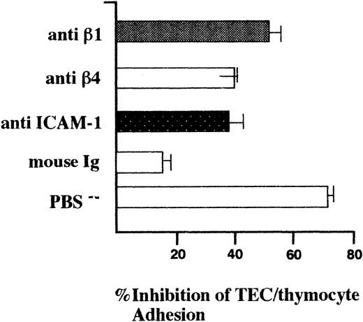
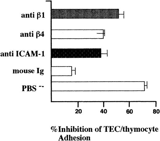
The involvement of β1 and β4 heterodimers in the TEC-thymocytes adhesion was evaluated by inhibition assays performed using MoAbs recognizing their extracellular domain. Assays were performed for 1 hour at 37°C in humidified atmosphere of 5% CO2 between TEC monolayers grown to tight confluence to prevent the direct attachment of thymocyte to the plastic and freshly isolated thymocytes previously labeled with the PKH26-GL red fluorochrome dye. As shown in Fig 3, the absence of divalent cations (PBS−) in the binding medium yielded 70% inhibition (70 ± 3.3 SE), indicating that integrin functions were largely involved in TEC-thymocyte adhesion. Precoating both TEC and thymocytes with MoAbs anti-β1 or anti-β4 reduced the thymocyte binding (58% ± 3% SD and 41% ± 3.5% SD, respectively) when compared with the untreated controls. A considerable inhibition (39.5% ± 12% SD) was exerted by MoAbs anti–ICAM-1, whereas a 15% ± 2% SD inhibition was observed in the presence of nonspecific mouse Ig.
Inhibition of TEC-thymocyte binding by MoAbs anti-β1 and anti-β4 integrins. Binding assays were performed between TEC monolayers plated and grown to tight confluence and PKH26-GL dye-labeled thymocytes (1:5 TEC/thymocyte ratio) for 1 hour at 37°C in humidified atmosphere of 5% CO2. Purified antibodies were separately incubated with TEC and thymocytes for 30 minutes before binding at 5 μg/mL and maintained during the test. PBS−indicates assays performed in PBS Ca2+Mg2+-free. Nonadherent thymocytes were removed by gentle washings. TEC and bound thymocytes were detached by trypsin-EDTA, washed, vortexed to disrupt aggregates, and counted by flow cytometry. Results were expressed as the ratio of TEC:thymocytes recovered and as a percentage of the ratio of the untreated controls. Shown are the mean values ± SD of four independent experiments performed with TEC and thymocytes obtained from different donors.
Inhibition of TEC-thymocyte binding by MoAbs anti-β1 and anti-β4 integrins. Binding assays were performed between TEC monolayers plated and grown to tight confluence and PKH26-GL dye-labeled thymocytes (1:5 TEC/thymocyte ratio) for 1 hour at 37°C in humidified atmosphere of 5% CO2. Purified antibodies were separately incubated with TEC and thymocytes for 30 minutes before binding at 5 μg/mL and maintained during the test. PBS−indicates assays performed in PBS Ca2+Mg2+-free. Nonadherent thymocytes were removed by gentle washings. TEC and bound thymocytes were detached by trypsin-EDTA, washed, vortexed to disrupt aggregates, and counted by flow cytometry. Results were expressed as the ratio of TEC:thymocytes recovered and as a percentage of the ratio of the untreated controls. Shown are the mean values ± SD of four independent experiments performed with TEC and thymocytes obtained from different donors.
Based on the finding that integrins clustered at the TEC-thymocyte contact sites were also functionally involved in their adhesion, we investigated whether MoAb-mediated ligation and/or aggregation of α3, α6, β1, and β4 could signal to NF-κB and NF-IL6 activation. To this purpose, all MoAbs were initially examined as regards their ability to functionally interact with these integrins on TEC monolayers by measuring [Ca2+]in fluctuation elicited by their binding. As summarized in Table2, an increase in intracellular calcium was elicited by binding of MoAbs to both α and β subunits. This increase required extracellular Ca2+, but not MoAbs cross-linking (not shown), thereby indicating that signals accross the membrane were delivered by the divalent binding of the MoAbs to the α3, α6, β1, and β4 extracellular domains.
Cross-linking of MoAbs bound to α3, α6, β1, and β4 integrins is required for activation of both NF-κB and NF-IL6 transcription factors.
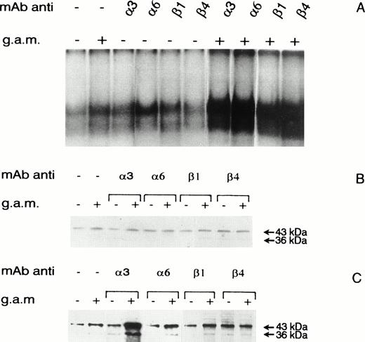
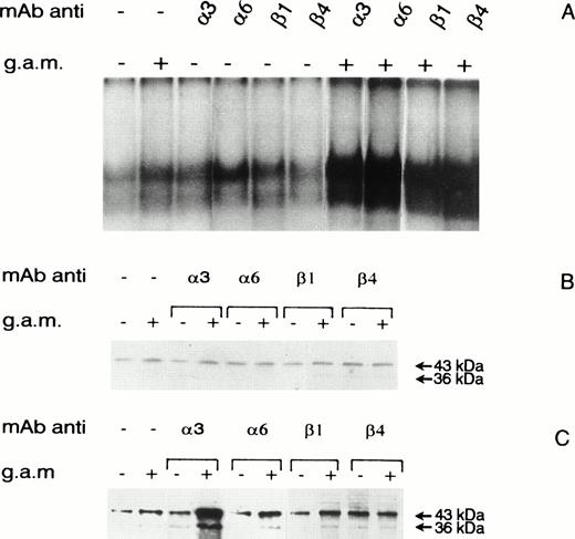
Integrin ligation and cross-linking were then investigated as inducers of signals leading to NF-κB nuclear translocation (Fig4A) and NF-IL6 phosphorylation (Fig 4B and C). Nuclear extracts were prepared from TEC incubated at 37°C with anti-α3, -α6, -β1, or -β4 MoAbs for 3 hours with or without the subsequent addition of F(ab)2 goat antimouse Ig (GAM) and washed. Control TEC were washed in the same way. NF-κB binding activity and NF-IL6 phosphorylation were evaluated as described for Fig1. As shown in Fig 4A, considerable increases (>2.8-fold the GAM-treated controls) of NF-κB binding activity were induced by cross-linking of both anti-α or anti-β MoAbs, whereas the activity observed after their divalent binding was comparable to that observed in the presence of the GAM reagent alone. MoAbs cross-linking was also required to induce NF-IL6 serine phosphorylation (3- to 8-fold higher than the GAM-treated controls, depending on the MoAbs used; Fig 4C), with the exception of the anti-β4, whose divalent binding was followed by a threefold increase of phosphorylated 36- and 43-kD isoforms. As previously observed in cell extracts prepared from TEC stimulated by thymocyte adhesion, the amounts of NF-IL6 proteins in the nucleus remained unmodified after binding and/or cross-linking of the various MoAbs (Fig 4B).
NF-κB binding activity and NF-IL6 phosphorylation in TEC untreated and treated with anti-3, -6, -β1, and -β4 MoAbs in the presence or absence of cross-linking. (A) NF-κB binding activity of nuclear extracts (6 μg) prepared from TEC untreated or treated for 3 hours with the various MoAbs at 5 μg/mL. When indicated, bound MoAbs were cross-linked by the addition of F(ab)2 GAM at 10 μg/mL for 2 hours. (B and C) PAGE analysis of NF-IL6 phosphorylation and isoform composition in nuclear extracts prepared from 14C-leucine–labeled TEC untreated or treated as previously mentioned. (B) Immunoblotting of nuclear lysate (20 μg) probed with anti–NF-IL6 antiserum. (C) Immunoblotting of nuclear NF-IL6 immunoprecipitates (103 cpm/lane) probed with antiphosphoserine MoAbs. Arrowheads indicate the positions of proteins with apparent molecular weight of 36 and 43 kD. Sections of gels are shown. Results shown are representative of three experiments performed independently by using TEC and thymocytes derived from different donors.
NF-κB binding activity and NF-IL6 phosphorylation in TEC untreated and treated with anti-3, -6, -β1, and -β4 MoAbs in the presence or absence of cross-linking. (A) NF-κB binding activity of nuclear extracts (6 μg) prepared from TEC untreated or treated for 3 hours with the various MoAbs at 5 μg/mL. When indicated, bound MoAbs were cross-linked by the addition of F(ab)2 GAM at 10 μg/mL for 2 hours. (B and C) PAGE analysis of NF-IL6 phosphorylation and isoform composition in nuclear extracts prepared from 14C-leucine–labeled TEC untreated or treated as previously mentioned. (B) Immunoblotting of nuclear lysate (20 μg) probed with anti–NF-IL6 antiserum. (C) Immunoblotting of nuclear NF-IL6 immunoprecipitates (103 cpm/lane) probed with antiphosphoserine MoAbs. Arrowheads indicate the positions of proteins with apparent molecular weight of 36 and 43 kD. Sections of gels are shown. Results shown are representative of three experiments performed independently by using TEC and thymocytes derived from different donors.
IL-6 production by TEC is enhanced after adhesion of thymocytes or cross-linking of MoAbs bound to α3, α6, β1, and β4 integrins.
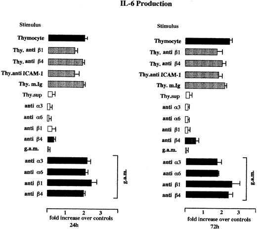
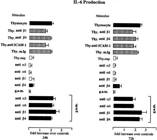
IL-6 production by TEC challenged with thymocyte adhesion was consistent with the observed functional activation of IL-6 gene transcription factors (Fig 5). Thymocyte contact increased TEC IL-6 secretion, as assessed in the culture supernatants collected 24 hours after the removal of the cell stimulus (1.96- ± 0.4-fold SE over controls), whereas supernatants of thymocytes cocultured with TEC or cultured in TEC medium for 12 hours exerted negligible activity. The increase of IL-6 production induced by thymocyte contact was impaired by the incubation of TEC and thymocytes before adhesion with MoAbs anti-β1 or anti–ICAM-1 (30% ± 8% SE and 25% ± 15%, respectively), whereas β4 activity seemed very close to the effect of nonspecific mouse Ig (8% ± 6% and 9% ± 4%, respectively).
IL-6 production of TEC after coculture with unfractionated thymocytes or treatment with MoAbs anti-3, -6, -β1, and -β4 integrins in the presence or absence of cross-linking. Cocultures were performed for 12 hours at 1:5 cell TEC-thymocyte ratio. Thymocyte supernatants were incubated for 12 hours. Treatment with the various MoAbs during the thymocyte adhesion was performed as described in Fig 3. Direct treatment with the same MoAbs of TEC monolayers was performed as described in Fig 4. Cross-linking with GAM was performed overnight. IL-6 production was measured by ELISA in culture supernatants withdrawn 24 and 72 hours after the removal of stimuli and the replating. Because of the variability of the basal level of IL-6 production by the different TEC cultures (range, 2 to 20 ng/106 cell/d; mean, 8.4 ± 2.6 SE), IL-6 production was expressed as the ratio of sample/untreated control. Shown are the means ± SE of the ratios observed in two independent experiments performed with TEC obtained from different donors.
IL-6 production of TEC after coculture with unfractionated thymocytes or treatment with MoAbs anti-3, -6, -β1, and -β4 integrins in the presence or absence of cross-linking. Cocultures were performed for 12 hours at 1:5 cell TEC-thymocyte ratio. Thymocyte supernatants were incubated for 12 hours. Treatment with the various MoAbs during the thymocyte adhesion was performed as described in Fig 3. Direct treatment with the same MoAbs of TEC monolayers was performed as described in Fig 4. Cross-linking with GAM was performed overnight. IL-6 production was measured by ELISA in culture supernatants withdrawn 24 and 72 hours after the removal of stimuli and the replating. Because of the variability of the basal level of IL-6 production by the different TEC cultures (range, 2 to 20 ng/106 cell/d; mean, 8.4 ± 2.6 SE), IL-6 production was expressed as the ratio of sample/untreated control. Shown are the means ± SE of the ratios observed in two independent experiments performed with TEC obtained from different donors.
IL-6 production by TEC was also increased by cross-linking of MoAbs bound to α and β integrins (≥2-fold over the GAM-treated controls), whereas the divalent binding was ineffective with the exception of anti-β4, followed by a 0.5-fold increase at 72 hours. The analysis of IL-6 accumulation at 72 hours confirmed the enhancing activity of thymocyte contact, the impairment exerted by the presence of MoAbs anti-β1 (28% ± 12% SE) and anti–ICAM-1 (23% ± 9% SE), the similarity between the effect of the presence of MoAbs anti-β4 (15% ± 7%) and nonspecific mIg (14% ± 4% SE), and highlighted the stronger activity exerted by β rather than α molecules cross-linking. These results indicate that the activation of NF-κB and NF-IL6 transcription factors induced by thymocyte adhesion was associated with induced IL-6 gene expression. Moreover, thymocyte induction could be impaired by MoAbs anti-β1 or -β4 integrins and mimicked by clustering of α or β subunits of α3β1 and α6β4 heterodimers.
DISCUSSION
A great deal of interest has arisen on intracellular signaling and the mechanisms of gene expression induced by cell-cell and cell-extracellular matrix adhesion.33 Cell-cell adhesion was investigated here in a coculture system of TEC and thymocytes obtained from normal donors, aimed at mimicking in vitro some of the thymo-epithelial interactions occurring physiologically within the thymus. With our culture procedure, we reproducibly obtained the development of organized monolayers of TEC that display a homogeneous morphology, retain their surface phenotype throughout the various culture passages, and produce their own matrix. Their lack of VCAM, together with the uniform expression of the α3 integrin recognized by the 10.1.2 MoAb, is suggestive of a medullary, rather than cortical, origin.28,31 TEC cultures maintained in vitro the constitutive production of variable amounts of IL-6. It has been reported that cytokines and growth factors induce IL-6 production in TEC through mechanisms most likely affecting the messenger stability.3,4 In the present report, we suggest that IL-6 production can be upregulated at the transcriptional level by signals delivered by integrin recruitment after thymocyte adhesion. In fact, adhesion to thymocytes triggered in TEC the intracellular cascade leading to the simultaneous presence in the nucleus of activated NF-κB and NF-IL-6 transcription factors, which are known to synergize in the inducible IL-6 gene expression.13,35 It is noteworthy that the nuclear pool of phosphorylated NF-IL6 was mainly constituted by the 43- and 36-kD isoforms previously shown to be endowed with transcriptional activity in humans.15 Additionally, NF-κB and NF-IL6 activation was followed by increased cytokine secretion, thus indicating that the two phenomena were associated.
The finding that thymocyte supernatants failed to activate NF-κB or NF-IL6 transcription factors ruled out the functional involvement of cytokines released by thymocytes in the activation of IL-6 gene expression and led us to investigate the inducing activity of adhesion molecules. Among them, we focused on α3β1 and α6β4 integrins because of (1) their repolarization at the TEC-thymocytes cell boundaries in the cocultures and (2) the involvement of their β subunits in the TEC-thymocyte binding.
TEC grown up to organized monolayers displayed, in fact, two sets of integrins polarized at discrete locations, α2β1 and α3β1 heterodimers, mostly located at the TEC-TEC boundaries, and the laminin receptor α6β4, sharply polarized at the basal domain of the monolayer and strictly facing the basement membrane. This pattern was remodeled by thymocyte contact so that α3β1 and α6β4, but not α2β1 heterodimers, clustered around individual thymocytes, therefore indicating that the latter trigger the local aggregation of otherwise located molecules without any evidence of simultaneous local enrichment of laminin 5, their natural common ligand.36 The functions of α3β1 and α6β4 heterodimer in TEC-thymocyte binding could not be directly investigated (ie, by blocking experiments with inhibiting peptides), because both heterodimers recognize conformational structures,37,38 and it was hence evaluated by MoAbs recognizing their β extracellular domains. The strongest inhibition, reaching almost a 60% reduction of the untreated controls, was exerted by MoAbs anti-β1 integrins. The inhibition observed in the presence of anti-β4 MoAbs was also considerable, particularly if compared with the activity of MoAbs anti–ICAM-1 molecules, which are well known components of the molecular pairs mediating the lympho-epithelial adhesions.33,39 Consistent with these results, the selective ligation of β1 and ICAM-1 receptors during the thymocyte adhesion impaired the overall adhesion potential of the TEC-thymocyte interaction, hence decreasing the TEC IL-6 production induced by thymocyte contact. Anti-β4 MoAbs seemed uneffective in the same experimental context. However, it has to be considered that any inhibitory effect due to β4 ligation would be counteracted by the peculiar signaling properties of this integrin (see below). The thymocyte ligands that may recognize α3β1 and α6β4 heterodimers are still unknown. It has been reported that α3β1 heterodimers can interact homotypically in keratinocytes.40 If a similar interaction occurs between TEC and thymocytes, the predominant expression of this integrin on very immature (double negative) and mature (single positive) thymocytes41 could affect the intrathymic migration and hence the differentiation of discrete thymocyte subsets. On the other hand, the inhibition exerted by MoAbs anti-β4 chains, together with the lack of detectable laminins interposed at the cell boundaries, raise the hypothesis of a novel laminin-like thymocyte ligand, possibly expressed by a restricted thymocyte subset. Further studies aimed at investigating these points are required.
Functional studies performed with the same MoAbs also demonstrated that recruitment of α3β1 and α6β4 heterodimers at the TEC surface can lead to activation of IL-6 transcription. Cross-linking of α or β subunits in fact yielded both NF-κB translocation and NF-IL6 phosphorylation. By contrast, with the exception of β4, which induced NF-IL6 phosphorylation and a detectable increase of IL-6 production, the divalent ligation of α3, α6, and β1 was ineffective. This occurred despite the fact that all MoAbs used for the analysis were functionally active, as demonstrated by the calcium transients elicited by their binding. The peculiar behavior of β4 might be possibly related to the properties of its large cytodomain, endowed with many potential docking sites for kinase activity42 and indirectly connecting the extracellular domain with the cytoskeleton.43 Our observations disagree with a recent report showing NF-κB translocation in monocytic cells upon β1 ligation with F(ab)2 MoAbs.44 However, the different cell lineages used in our and in the reported experiments may possibly explain this discrepancy.
Consistent with the requirement of cross-linking for inducing NF-κB and NF-IL6 activation, integrin cross-linking was also required for induction of IL-6 production. The finding that single subunits of the heterodimers may signal in a similar or even greater way than thymocytes could seem paradoxical. Rather, it has to be considered that MoAb cross-linking can theoretically recruit all molecules expressed at the TEC surface, whereas TEC interaction with thymocyte ligands can be affected by ligand(s) expression and concentration, the number of cells expressing the molecule(s) at the surface, the affinity-avidity state of integrin receptors, and the adhesive/deadhesive functions of proximal receptors within the membrane environment.
Although we did not investigate the signaling pathway(s) linking integrin recruitment at the TEC surface with posttranslational regulation of IL-6 gene transcription factors, current evidences indicate that thymocytes can trigger α3β1 and α6β4 recruitment and/or aggregation at the TEC surface. This phenomenon, among many others occurring at the same time, may, in turn, initiate the intracellular cascade leading to IL-6 gene expression.
Because IL-6 receptors are shared by both thymocytes and TEC45 and IL-6 is implicated in TEC growth4 and EGF-mediated differentiation,46 our results imply that thymocytes can regulate the expression of a cytokine exerting autocrine and paracrine activities within their microenvironment. Moreover, our findings may be also of help to investigate the molecular mechanisms regulating IL-6 gene expression in autoimmune diseases such asmiasthenia gravis,6 where the IL-6 overproduction has been related to the thymic morphological and functional abnormalities.
ACKNOWLEDGMENT
The authors gratefully acknowledge Dr M. Merola and Prof M. Palmieri (Institute of Biological Chemistry, University of Verona, Verona, Italy) for discussion and helpful suggestions concerning IL-6 transcription factor experiments.
Supported by Grants ISS-AIDS No. 9306.34 (1995) to G.T. and 9404-20 (1996) to P.C.M. P.C.M. was also supported by grants from CNR target project “Applicazioni Cliniche della Ricerca Oncologica” (ACRO), from AIRC (Associazione Italiana per la Ricerca sul Cancro, Milano), and from Telethon (Grant No. 762).
The publication costs of this article were defrayed in part by page charge payment. This article must therefore be hereby marked “advertisement” in accordance with 18 U.S.C. section 1734 solely to indicate this fact.
REFERENCES
Author notes
Address reprint requests to Dunia Ramarli, MD, Istituto di Immunologia e Malattie Infettive, Policlinico Borgo Roma, Via delle Menegone, 37134 Verona, Italy; e-mail: dunia@borgoroma.univr.it>.

