Abstract
We have studied the reconstitution of the T-cell compartment after bone marrow transplantation (BMT) in five patients who received a graft-versus-host disease (GVHD) prophylaxis consisting of methotrexate, cyclosporin, and 10 daily injections (day −4 to day +5) of Campath-1G. This treatment eliminated virtually all T cells (7 ± 8 T cells/μL at day 14) which facilitated the analysis of the thymus-dependent and independent pathways of T-cell regeneration. During the first 6 months, the peripheral T-cell pool was repopulated exclusively through expansion of residual T cells with all CD4+ T cells expressing the CD45RO-memory marker. In two patients, the expansion was extensive and within 2 months, the total number of T cells (CD8>>CD4) exceeded 1,000/μL. In the other three patients, T cells remained low (87 ± 64 T cells/μL at 6 months) and remained below normal values during the 2 years of the study. In all patients, the first CD4+CD45RA+RO− T cells appeared after 6 months and accumulated thereafter. In the youngest patient (age 13), the increase was relatively fast and naive CD4+ T cells reached normal levels (600 T cells/μL) 1 year later. In the four adult patients (age 25 ± 5), the levels reached at that time-point were significantly lower (71 ± 50 T cells/μL). In all patients, the T-cell repertoire that had been very limited, diversified with the advent of the CD4+CD45RA+RO− T cells. Cell sorting experiments showed that this could be attributed to the complexity of the T-cell repertoire of the CD4+CD45RA+RO− T cells that was comparable to that of a normal individual and that, therefore, it is likely that these cells are thymic emigrants. We conclude that after BMT, the thymus is essential for the restoration of the T-cell repertoire. Because the thymic activity is restored with a lag time of approximately 6 months, this might explain why, in particular in recipients of a T-cell–depleted graft, immune recovery is delayed.
FOR MANY PATIENTS with leukemia, allogeneic stem cell transplantation is the only curative therapy available.1-3 Its efficacy is due to the combination of high-dose chemotherapy, which can be administered despite the myeloablative effect, the intense transplant conditioning, and the antileukemic effect of the allogeneic T cells in the graft. After transplantation, most hematological lineages regain their normal function rapidly. Unfortunately, this is not the case for the T-cell lineage and patients can be subject to a variety of infections for prolonged periods of time.4-9
The reason why T-cell immunity remains impaired, notwithstanding the fact the peripheral T-cell pool is rapidly repopulated, has been the subject of a number of studies over the past years. Early reports already showed the existence of severe imbalances in T-cell subpopulations.10-12 Furthermore, a high number of T cells after transplantation appears to be anomalous, expressing numerous activation or memory markers.13-15 More recently, it has been shown that after the elimination of the peripheral T-cell pool, T cells can be regenerated through two different pathways.16-19 One is thymus dependent and might be considered as a recapitulation of ontogeny. In addition, the T-cell compartment can be repopulated through peripheral expansion of mature T cells cotransfused with the bone marrow (BM) graft. The relative importance of the second pathway is dependent on the activity of the thymus.16-18 20 Peripheral expansion will only be significant when the function of the thymus is impaired and its contribution in young euthymic mice is negligible.
In humans, the same correlation between an insufficient thymic function and the prevalence of peripheral expansion as a mechanism to restore the T-cell compartment exists. Therefore, it is likely that in most patients after BM transplantation (BMT), peripheral expansion is the primary pathway. Although radiographic imaging of the thymus has shown that after intense chemotherapy the thymic pathway is reactivated,18,21 this thymic rebound might become insufficient with increasing age. This has been shown in other studies that have analyzed the thymus rebound indirectly by measuring the production of naive CD4+lymphocytes.15,18 22-25
Whether a unique phenotype exists for naive T cells is still controversial and, therefore, their identification is not straightforward. The different splicing forms of CD45 are the surface markers most widely used to discriminate between naive and memory cells. Until primed by antigen, naive cells keep the CD45RA+RO− phenotype of thymic emigrants. Upon activation, T cells become CD45RA−RO+, but may re-express CD45RA thereafter. Memory CD8+ T cells may lose the expression of CD45RO completely,15,26 but this does not seem the case for CD4+ lymphocytes that remain CD45RARO double-positive.27 Therefore, the CD4+lymphocytes with a CD45RA+RO− phenotype that repopulate a patient after eradication of the peripheral T-cell pool most likely represent thymic emigrants. This is further supported by the inverse correlation found between the reappearance of CD4+CD45RA+RO− T cells and age,18 plus the fact that these cells remain absent in the thymectomized host.28
The contribution of the thymus to the reconstitution of the T-cell compartment after stem cell transplantation is not precisely known. Although the production of naive T cells can be taken as representative for the activity of the thymus, the mere presence of CD4+CD45RA+RO− T cells cannot. In particular, when the graft contains large quantities of T cells, the percentage of CD4+CD45RA+RO− T cells might just reflect the number of naive donor cells that have remained quiescent. Therefore, thymic activity will be most easily monitored in recipients of T-cell–depleted stem cell grafts. In this report, we have studied five patients who received the monoclonal antibody (MoAb) Campath-1G29 30 in vivo. Besides eliminating cotransfused donor T cells, the in vivo treatment also lyses residual T cells of recipient origin so that graft-versus-host disease (GVHD) as well as graft rejection are avoided. After this therapy, very few mature T cells were detected in the peripheral blood of the patient, which allowed a precise analysis of the thymus-dependent and -independent pathways of T-cell regeneration.
PATIENTS, MATERIALS, AND METHODS
Patients.
The clinical characteristics, conditioning regimen, and posttransplantation immunosuppressive treatments of the five patients are shown in Table 1. All were transplanted with the marrow of an unrelated donor molecularly matched for HLA-A, -B, -C, -DR, and DQα. GVHD prophylaxis consisted of Campath-1G (10 mg/d), from day −4 to day +5. Methylprednisolone was administered on days −4, −3, and −2, at doses of 750, 500, and 250 mg, respectively. From day −1 onward, patients received a 12-hour infusion of cyclosporin A. The dose was adjusted to attain a trough level of 200 ng/mL. From around day 30, patients received oral cyclosporin as indicated in Table 1. Methotrexate was administered at a dose of 15 mg/m2 (day +1) and 10 mg/m2 (day +3, +6, +11).
Blood samples and cell sorting.
Peripheral blood cells were collected at various times after transplantation. Mononuclear cells were obtained after Ficoll-Hypaque (Pharmacia, Uppsala, Sweden) density gradient centrifugation. Cells were fractionated into CD3+CD4+, CD3+CD8+, CD4+CD45RA+RO− and CD4+CD45RA−RO+ cell populations by fluorescence-activated cell sorting (FACS) (FACSTAR PLUS, Becton Dickinson, Mountain View, CA) after being stained with anti-CD3 (mouse-IgG2b-biotinylated) and/or anti-CD4 (mouse-IgG2a or mouse-IgG2b-biotinylated) and/or anti-CD8 (mouse-IgG1) and/or anti-CD45RA-fluorescein isothiocyanate (FITC) (mouse-IgG1) and/or anti-CD45RO (mouse-IgG2a). Where needed, the MoAbs were stained with either subclass specific antisera (Southern Biotechnologies, Birmingham, AL) or streptavidin-red 670 (Life Technologies, Inc, Gaithersburg, MD). Purity of the cell sorted populations was always more than 97%.
T-cell receptor (TCR)-spectratyping.
Total RNA was purified from 500 to 8,000 T cells using the RNeasy kits from Qiagen (Qiagen, Hilden, Germany). Two micrograms ofEscherichia coli rRNA was added to each sample as carrier. RNA was eluted with 45 μL water and lyophilized to reduce the volume down to 8 μL. First-strand cDNA synthesis was performed with 0.5 mmol/L dNTPs, 250 ng oligo-dT, and 100 U reverse transcriptase (Life Technologies, Basel, Switzerland) in a final volume of 15 μL. The samples were incubated for 30 minutes at 37°C, 30 minutes at 42°C, and denatured for 5 minutes at 95°C. Polymerase chain reaction (PCR) amplification of the cDNA was performed using a radiolabeled constant primer and two primers corresponding to the variable region of the TCR β-chain, based on the procedure described by Maslanka et al.31 The following primers were used: BC:5′-AGATCTCTGCTTCTGATGGCT-3′, Mix 1-long: BV1, 5′-CAGTTCCCTGACTTGCACTC-3′, -short: BV5.1, 5′-CTCGGCCCTTTATCTTTGCG-3′, Mix 2-long: BV12, 5′-CAAAGACAGAGGATTTCCTCC-3′, -short: BV2, 5′-GCTTCTACATCTGCAGTGC-3′, Mix 3-long: BV3, 5′-GAGAGAAGAAGGAGCGCTTC-3′, -short: BV13, 5′-GTCGGCTGCTCCCTCCC-3′, Mix 4-long: BV6.1, 5′-GATCCAGCGCACACAGC-3′, -short: BV4, 5′-GCAGCATATATCTCTGCAGC-3′, Mix 5-long: BV7, 5′-CCTGAATGCCCCAACAGC-3′, -short: BV8, 5′-CCAGCCCTCAGAACCAG-3′, Mix 6-long: BV14, 5′-GTCTCTCGAAAAGAGAAGAGG-3′, -short: BV9, 5′-GGAGCTTGGTGACTCTGCTG-3′, Mix 7-long: BV20, 5′-CACACCCCAGGACCGGCAG-3′, -short: BV11, 5′-CAGGCCCTCACATACCTCTCA-3′, Mix 8-long: BV15, 5′-GTCTCTCGACAGGCACAGGC-3′, -short: BV17, 5′-CCAAAAGAACCCGACAGCTTT-3′, Mix 9-long: BV21, 5′-GGCTCAAAGGAGTAGACTCC-3′, -short: BV16, 5′-GAACTGGAGGATTCTGGAGTT-3′, Mix 10-long: BV5.3, 5′-CCCTAACTATAGCTCTGAGC-3′, -short: BV18, 5′-GTGCGAGGAGATTCGGCAGC-3′, Mix 11-long: BV22, 5′-GTTGAAAGGCCTGATGGATC-3′, -short: BV24, 5′-GGGGACGCAGCCATGTACC-3′. The PCR reactions were performed in 20 μL in presence of: 1 μL cDNA, 50 mmol/L KCl, 10 mmol/L Tris-HCl pH 8.8, 1 mmol/L MgCl2, 100 μg/mL bovine serum albumin, 0.2 mmol/L dNTPs, 30 ng BC primer, 3 ng (γ-32P) adenosine triphosphate–labeled BC primer, 30 ng each BV primers, and 0.5 U Taq DNA polymerase (Life Technologies, Basel, Switzerland). Thirty-seven cycles of 30 seconds at 94°C, 30 seconds at 59°C, and 60 seconds at 72°C were followed by a final extension of 5 minutes at 72°C. Three microliters of the PCR samples were mixed with an equal volume of formamide/dye loading buffer, heated at 90°C for 2 minutes and separated on 6% polyacrylamide/urea gels for 4 hours. The gels were dried and exposed with one intensifying screen for 3 to 12 hours.
RESULTS
Patients, conditioning, and GVHD prophylaxis.
The characteristics of the five patients enrolled in this study who received a BM graft from an HLA-A, -B, -C,-DR,-DQ identical donor as a treatment for chronic or acute leukemia are depicted in Table 1. The patients received a conditioning regimen based on total body irradiation and cyclophosphamide. GVHD prophylaxis consisted of methotrexate, cyclosporin, and the MoAb Campath-1G.29 30All patients engrafted (polymorphonuclear neutrophils > 0.5 × 10⁹/L), with a median time of 21 days (range, 11 to 30) and remained in complete remission during the time of the study. Patients 1, 2, and 4 developed acute GVHD stage I-II, which was successfully treated with systemic steroids. Chronic GVHD occurred in patient 2 and 3, whereas cytomegalovirus (CMV) viremia was only observed in patient 1.
Restoration of the T-cell compartment.
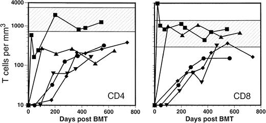
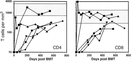
Because of the very efficient T-cell depletion by the 10 daily injections (day −4 to day +5) of Campath-1G, the number of T cells was very low during the first month after transplantation (7 ± 8 T cells/μL at day 14). Thereafter, two distinct repopulation patterns were observed. In patients 1 and 2, who suffered from GVHD II, the total number of T cells exceeded 1,000/μL already in the second month (Fig 1). In patient 2, the vast majority of the cells were CD8+, which caused the CD4/CD8 ratio to be reversed, a phenomenon that has been observed by others.10,14 32 In the other three patients with GVHD grade 0-I, expansion was limited. T-cell numbers were approximately 10-fold lower (87 ± 64 T cells/μL at 6 months) and remained below normal values during the 2 years of the study.
Time course of the reconstitution of CD4+and CD8+ T cells in five patients after BMT. (▪), P1; (▴), P2; (⧫), P3; (•), P4; (▾), P5. Shaded area represents range found in normal controls.
Time course of the reconstitution of CD4+and CD8+ T cells in five patients after BMT. (▪), P1; (▴), P2; (⧫), P3; (•), P4; (▾), P5. Shaded area represents range found in normal controls.
Limited T-cell repertoire diversity during the first 6 months after BMT.
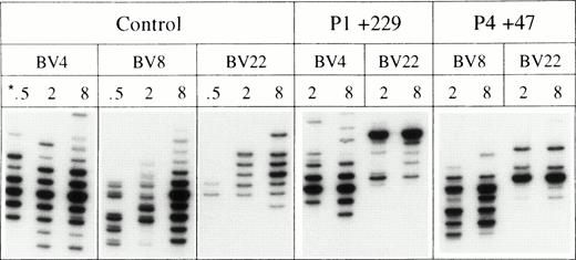
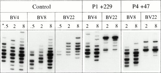
During the first months after transplantation, the T-cell compartment is repopulated mainly through peripheral expansion of mature T cells that are cotransfused with the graft.19,33,34 Therefore, T-cell repertoire complexity in recipients of a T-cell–depleted graft is initially lower than in recipients of an unmanipulated marrow.34,35 To measure the effect of the T-cell depletion with the MoAb Campath-1G, we determined the diversity of the T-cell repertoire at different time points after transplantation. Using the spectratype method,36 we compared the number of TCRs with a different CDR3 length of the variable region of the β-chain (BV) in different patients. By testing 21 BVs in two or three samples containing only low numbers (<104) of T cells, we obtained results that enabled us to compare the T-cell diversity at different time points after transplantation. The first panel in Fig 2 shows the result of three BVs representative for a polyclonal repertoire of a normal control. It shows that in a sample of as few as 500 T cells, the number of cells expressing a BV family with a high frequency such as BV 4 is sufficiently high to generate seven bands of different CDR3 size. Because CDR3s of an average size (27 to 33 nucleotides) are much more abundant than extremely short or long ones,37 the three most central bands were of higher intensity than the others, indicating that these bands represented more than one TCR. When a higher number of T cells was tested (2.103 to 8.103), the distribution of bands became more Gaussian and with 12 bands discernible at 8.103 T cells, the heterogeneity of the CDR3s was almost maximal. BV-families with lower frequencies yielded different results. For instance, for BV 8, the seven central bands were present only upon testing 8.103 T cells whereas this was not yet the case for a BV with a low frequency such as BV 22.
Comparison of spectratypes of a normal control and two patients 229 and 47 days after transplantation. *The respective lanes show the bands generated by 500, 2,000, and 8,000 (CD3+) T cells after PCR amplification with the BV-specific primers.
Comparison of spectratypes of a normal control and two patients 229 and 47 days after transplantation. *The respective lanes show the bands generated by 500, 2,000, and 8,000 (CD3+) T cells after PCR amplification with the BV-specific primers.
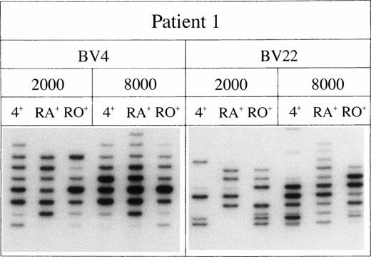
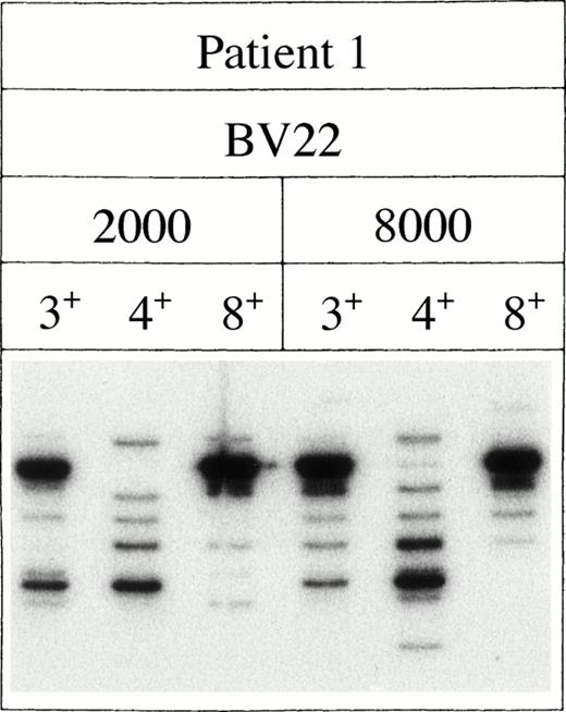
This method appeared to be extremely useful to compare the diversity of the T-cell repertoire in normal individuals with that of patients after transplantation. Oligoclonal repertoires in patients were readily discerned by the fact that the number of bands in a BV family was lower than in the corresponding family of the normal control. More importantly, a characteristic feature of a restricted repertoire was that the spectratypes of some BV families were quite similar in different samples, notwithstanding the fact that they contained higher numbers of T cells. This is depicted in panels 2 and 3 of Fig 2, which shows the heterogeneity of BV4/8/22 of two patients after transplantation. Clearly, the diversity of the T-cell repertoire of both patients was severely limited. Although the phenomenon was less pronounced for the BV4 family in patient 1, for the other families shown, the number of bands generated by 8.103 T cells was approximately the same as generated by 2.103 cells. This was particularly striking for BV22 that seemed to consist of very few clones only. The latter was confirmed by a separate analysis of the BV22+CD4+ and BV22+CD8+T cells in patient 1 (Fig 3). Because the major bands of the BV22 family are found either in the CD8+or CD4+ FACS-sorted fraction, it is indeed very likely that these bands represent single clones.
The dominant bands represent single CD4+ or CD8+ T-cell clones. The data represent the number of bands generated by either 2,000 or 8,000 cells FACS-sorted on the basis of the expression of CD3, CD4, or CD8.
The dominant bands represent single CD4+ or CD8+ T-cell clones. The data represent the number of bands generated by either 2,000 or 8,000 cells FACS-sorted on the basis of the expression of CD3, CD4, or CD8.
Table 2 shows the analysis of the repertoire of the CD4+ and CD8+ T cells in all five patients. To facilitate the comparison of different populations we expressed the diversity of the respective T-cell repertoires as the number of BV families of which the seven central bands were detectable. On the basis of results obtained from normal controls, we estimated that this was true for BV families of which at least 50 to 100 T cells in the sample tested used TCRs in a random fashion. The data show that in the patient samples, very few BV families showed this diversity. Furthermore, the complexity of the repertoire seemed to reflect the extent of expansion: TCR diversity was lower in CD8+ T cells than in CD4+ cells while the repertoire of patients 1 and 2, in whom T-cell expansion had been more extensive, seemed to be more restricted than that of patients 3 through 5.
The T-cell repertoire is restored by CD4+CD45RA+thymic emigrants.
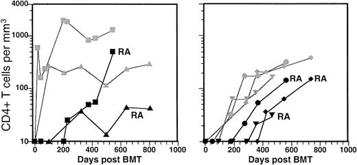
After transplantation, mature T cells expand and reconstitute the peripheral T-cell pool. During this expansion, these cells express a number of activation markers10,13,14 and acquire the CD45RO+-phenotype of memory cells. Because not all T cells expand, the number of T cells that express a CD45RA+-phenotype, ie, the cells that have remained quiescent, will be proportional to the number of T cells present before expansion. Therefore, the percentage of CD45RA+ T cells is relatively high after peripheral blood stem cell (PBSC) transplantation,38,39 low after BMT,14 39 and zero when the initial number of T cells has been reduced by T-cell depletion. During the first 200 days after transplantation, no CD4+CD45RA+RO− T cells were detected in any of the five patients (Fig4). Although the number of T cells gradually increased, all T cells expressed memory markers, indicating that peripheral expansion was still the only pathway through which the peripheral T-cell pool was repopulated. As a result, no changes in the T-cell repertoire were observed during this period. Spectratypes remained remarkably constant with the same bands dominating the respective BV families and without any noticeable diversification (data not shown).
Time course of the reconstitution of CD4+CD45RA+RO− (RA) T cells in the two patients with the fast T-cell reconstitution (left) and the three with the slow reconstitution (right). (▪), P1; (▴), P2; (⧫), P3; (•), P4; (▾), P5. The gray lines/symbols represent the reconstitution of the CD4+ T cells already shown in Fig1.
Time course of the reconstitution of CD4+CD45RA+RO− (RA) T cells in the two patients with the fast T-cell reconstitution (left) and the three with the slow reconstitution (right). (▪), P1; (▴), P2; (⧫), P3; (•), P4; (▾), P5. The gray lines/symbols represent the reconstitution of the CD4+ T cells already shown in Fig1.
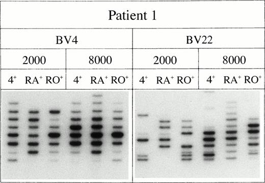
After approximately 200 days, the first CD4+CD45RA+RO− T cells appeared. In patient 1 (age 13), these cells rapidly accumulated, while in the adults (patients 2 through 5, age 25 ± 5), the increase was much slower. At the same time the T-cell repertoire diversified. The diversification was due to the high complexity of the T-cell repertoire of the CD4+CD45RA+RO− T cells (Fig 5). In all patients, T cells sorted on basis of their CD4+CD45RA+RO−phenotype generated significantly more bands than their CD4+CD45RA−RO+ counterparts (Table 3). Clearly, the diversity of the T-cell repertoire of the emerging T cells with a naive phenotype was comparable to that of normal controls, while the repertoire of the CD4+CD45RO+ T cells remained very limited.
The repertoire of the CD4+CD45RA+RO− T cells is diverse. The respective lanes show the bands generated by 2,000 or 8,000 T cells sorted on the basis of their CD4+CD45RA+RO− or CD4+CD45RA−RO+ phenotype.
The repertoire of the CD4+CD45RA+RO− T cells is diverse. The respective lanes show the bands generated by 2,000 or 8,000 T cells sorted on the basis of their CD4+CD45RA+RO− or CD4+CD45RA−RO+ phenotype.
Based on these significant differences in T-cell repertoire, we believe strongly that the CD4+CD45RA+RO− T cells are a distinct population of thymic emigrants and that, therefore, the role of the thymus must be considered as essential for the restoration of the T-cell immunity after BMT.
DISCUSSION
Malfunctioning of the T-cell compartment causes a significant part of the life-threatening immunodeficiencies after BMT. Although other hematopoietic lineages start to function sufficiently already in the weeks after transplant, T-cell immunity as well as T-cell–dependent B-cell responses might remain impaired for years.5 T-cell immunity is restored with greater difficulty than other components of the immune system while the little T-cell memory cotransfused with the graft is usually not sufficient to substitute the memory of the recipient that has been destroyed by the chemotherapy and the transplant conditioning. Indeed, posttransplant T-cell repertoires appear to be of limited diversity,34,36,40-42 even when large quantities of donor T cells are cotransfused with the BM.35 43
The distortion of the T-cell repertoire is caused by the fact that after eradication by chemotherapy, the peripheral T-cell pool will be initially repopulated through expansion of mature T cells.19 Because T cells that encounter antigen expand faster,33,44 a significant part of the final repertoire might be comprised of limited numbers of dominant T-cell clones directed against viruses such as CMV,45 against the histocompatibility antigens of the host,46,47 or possibly against residual leukemic cells.48,49 As a result, most T cells express activation markers during the first months after transplantation,13-15 while the transfused T cells with a naive phenotype, which have remained quiescent, will be diluted out and become a minority.
T cells can also be generated from T-cell progenitors through thymopoiesis. However, in particular in adult recipients of hematopoietic stem cell grafts, the function of the thymus might not be sufficient to generate a complete new pool of naive T cells. Moreover, the residual thymic activity might be further reduced by the combination of irradiation, chemotherapy, and GVHD that causes severe damage to the thymic microenvironment.50-52
After eradication of the peripheral T-cell pool, restoration of full immune competence might therefore depend on the capacity of the thymus to generate a new T-cell repertoire. This has not yet been shown directly, because monitoring thymus activity in humans is cumbersome. Recently, several studies have shown that a change in expression of the different CD45-isoforms on CD4+ T cells can be taken as evidence of a thymic rebound.17,18,20,24 Because the CD45RA+RO− phenotype of naive T cells is lost upon activation,53 thymic emigrants are distinct from the CD45RO+ memory cells generated through peripheral expansion. Because only CD8+ memory cells may revert to a CD45RA+RO− phenotype,26,54the increase of CD4+CD45RA+RO− T cells in the blood of recipients of hematopoietic stem cell grafts must be proportional to the residual thymic activity of the host. This is further supported by the correlation between the number of naive CD4+ T cells and the age of the recipient18,23,24 and by a case report that shows that naive CD4+ T cells do not reappear in a recipient who had been thymectomized before transplantation.28
In this report we have studied the reconstitution of the T-cell compartment in 5 patients who received the MoAb Campath-1G as GVHD-prophylaxis. These type of patients are well suited to discriminate the thymus-independent from the thymus-dependent pathway of T-cell repopulation. Because the treatment eliminates virtually all T cells, 1 month after transplantation, the number of T cells with a naive phenotype is negligible when compared with the number of T cells generated through peripheral expansion. Therefore, any CD4+CD45RA+RO− T cells detected thereafter must be generated by the thymus, and the onset of the thymic rebound can be monitored more precisely than in the presence of pretransplant CD4+CD45RA+RO− T cells, which might expand without acquisition of memory markers.55-57 We found that during the first 6 months after transplantation, all peripheral T cells were generated through peripheral expansion. In two of five patients, the expansion was fast and (supra)normal numbers were reached within weeks, while in the other three, the number of T cells increased only slowly. It is noticeable that the two patients with the rapid increase suffered from GVHD II and that a significant overshoot of CD8+ T cells was found in the patient with a CMV infection. The differences in T-cell reconstitution between the patients were very significant and one could argue that, while these type of correlations have been suggested by others,58-61they are most evident when patients are vigorously T-cell depleted.
In all patients, CD4+CD45RA+RO− T cells started to appear from the sixth month on. This delay of the thymic rebound is significantly longer than that is observed in patients after chemotherapy.18 The difference might be explained in several ways. Firstly, the conditioning received by patients transplanted with allogeneic stem cells is more intense than standard chemotherapy. Moreover, the additional immune suppression given after transplantation might interfere with early intra-thymic T-cell development.62-64 Secondly, allogeneic stem cell transplantation could be associated with a slower T-cell reconstitution61,65 while even mild forms of GVHD can damage the thymic epithelium.51 52 However, also in a larger group studied (Roux E, Dumont-Girard F, Helg C, Chapuis B, Roosnek E; manuscript in preparation), direct correlations between T-cell reconstitution at one hand and immunosuppression or the occurrence of GVHD on the other were hard to unveil. Because the age of the patient was by far the most important parameter correlated with thymic activity, and GVHD and its treatment were in general more severe in the older patients, these parameters could not be analyzed separately. CD4+CD45RA+RO− T cells emerged always faster in young patients who received cyclosporin than in patients over 30 years old who were already off immunosuppression. Therefore, larger studies in which patients on immune suppression can be compared with their age-matched controls are needed to determine to which extent immune suppression hinders the reconstitution of the T-cell compartment.
The thymic rebound was essential for the restoration of the T-cell repertoire. Until the appearance of the CD4+CD45RA+RO− T cells, the repertoire remained extremely limited. By measuring the number of bands representing different CDR3-lengths of the variable region of the TCR β-chain in very low numbers of T cells, we were able to monitor the diversification of the repertoire very precisely. We chose the lowest cell number (2.103) such that in normal controls BV families with a frequency higher than 5%, ie, the BVs expressed by more than 100 cells in the sample, generated Gaussian curves of which the 7 most central bands were always present (Fig 2). In contrast, BV families with a frequency between 1% and 5% were incomplete, with the BVs with the lowest frequencies generating one or two bands only. At the highest cell number (8.103), the seven most central bands were present in an average of 16 of the 21 BV families (Table 3), a number that, even when a much higher number (105) of cells was tested, was never reached in the patient samples before the advent of the CD4+CD45RA+RO−T cells. The fact that the number of bands generated by as many as 105 T cells in a patient was still lower than that generated by 8.103 T cells of which the TCR’s are randomly distributed, shows that the diversity in these patients is only a fraction of that in a normal individual. Therefore, it is not surprising that their immune reactivity during the first year after transplantation is severely diminished and that, for the restoration of the immune response against most antigens, a patient may depend on the generation of a new T-cell repertoire.
The data shown in this report are characteristic for patients who are vigorously T-cell depleted and who represent probably the most extreme example of an immune system with a restricted T-cell repertoire. However, although TCR diversity might be 2- to 3 logs higher in recipients of unmanipulated grafts,35 TCR diversity also remains limited in these patients.36,41,43Although such a repertoire might be sufficient to respond to some antigens, it is not unlikely that the capacity to respond to others is lost. Therefore, also in these patients, an effective immune response might depend on the completion of the repertoire by thymic emigrants. This is suggested by the fact that immunizations to recall antigens are more efficient in the second year after transplantation, and more recently, by studies reporting correlations between immune reactivity, age, and thymic activity.23,25 If so, the administration of cytokines that could rejuvenate the thymus after transplant might have a significant therapeutical potential.19,23,66 67
F.D.-G. and E.R. contributed equally to this work.
Supported by a grant of the Swiss National Science Foundation (32-043622.95).
The publication costs of this article were defrayed in part by page charge payment. This article must therefore be hereby marked “advertisement” in accordance with 18 U.S.C. section 1734 solely to indicate this fact.
REFERENCES
Author notes
Address reprint requests to Eddy Roosnek, PhD, Unitéd’Immunologie de Transplantation, Hôpital Cantonal Universitaire de Genève, 24 rue Micheli-du-Crest, CH-1211 Genève 14, Switzerland; e-mail: edro@hcuge.ch.

