Unlike in other lymphoproliferative diseases such as acute lymphocytic leukemia (ALL) and non-Hodgkin malignant lymphomas, central nervous system (CNS) and leptomeningeal involvement are extremely rare in chronic lymphocytic leukemia (CLL), so much so that no mention of this complication is to be found in the most recent and authoritative textbooks of hematology. Although a large autopsy series has revealed that invasion of the meninges by B-cell CLL occurs in 20% of cases,1 clinical syndromes are exceedingly rare.2 Two reviews have been published recently, one of them collating 21 cases3 and the other, 13 cases.4 In the first review, however, 6 cases are reported to pertain to a subset of cases with T-cell phenotype, and in the second there were 4 cases with the prolymphocytic leukemia (PLL) and/or prolymphoid phenotype, which has been considered as possibly predisposing to meningeal invasion.5 In most cases this complication arose in long-standing disease, but in a small minority it appeared in the early stages.3 In one case there was a syndrome of inappropriate secretion of antidiuretic hormone (SIADH).6Most patients were treated with combinations of RT and intrathecal MTX, and in 12 patients there was improvement, with most of them showing complete resolution.4
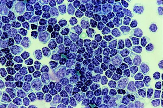
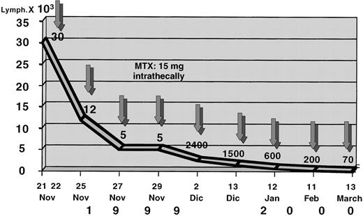
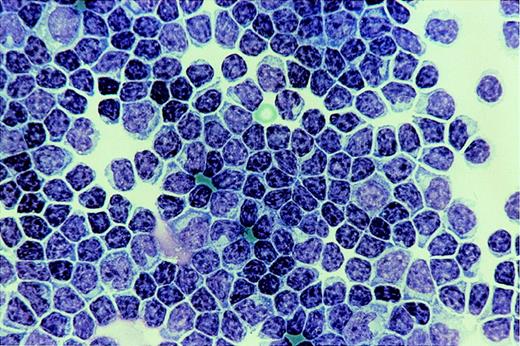
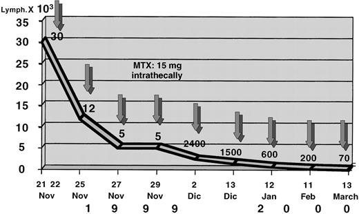
A 70-year-old (at the time of this writing) male was diagnosed with B-CLL in Rai/Binet stages 0/A in 1987. A nondiffuse (mixed nodular and interstitial) histologic pattern was found in the bone marrow (BM) biopsy specimen and did not change significantly in the following annual examinations. The lymphocyte doubling time was initially slow (longer than 12 months) but grew increasingly shorter. Therapy was given whenever his lymphocyte count exceeded 50 × 104 cells/μL. For 10 consecutive years he was treated with 11 courses of chlorambucil, a total of 2.15 g. Anemia was controlled by repeated courses of rh-erythropoietin (rh-EPO), at 30 000 U/wk for 6 consecutive weeks. In 1997 he received one 1 Gy irradiation per week for 6 weeks on the (nonenlarged) spleen, with good control of the lymphocyte level. Between 1998 and 1999 he received 2 courses of fludarabine, with little effect on the lymphocyte count. Because of an adverse cardiovascular event that occurred during the second fludarabine course, cytotoxic therapy was temporarily discontinued, and the lymphocyte count reached 120 × 104/μL, with some liver and spleen enlargement. On December 15, 1999, he complained of fatigue, confusion, and “leg pains.” On December 22, 1999, he was hospitalized at the Montallegro Clinic in Genoa, Italy. There were severe cognitive disturbances but no classical meningeal symptoms. An immediate lumbar puncture, however, disclosed an almost milky cerebrospinal fluid (CSF) with 3 × 10⁴ cells/ μL, all of them typical CLL lymphocytes with extremely few prolymphocytes or otherwise atypical lymphocytes. A cytospin preparation is shown in Figure 1. The immunophenotype was CD23, CD22, CD5, k+, identical to the circulating lymphocytes. Glucose was 12 mg/dL, and protein, 500 mg/dL. Intrathecal MTX, 15 mg, was administered 5 times on alternate days and then on increasingly longer intervals. Timing of therapy and CSF clearing are shown in Figure 2. The last lumbar puncture, on March 13, 2000, showed an otherwise normal CSF with 70 lymphocytes/μL. Magnetic resonance imaging performed after the first 2 intrathecal MTX injections showed abnormal tissue in the upper cervical peridural space, with many diffuse paravertebral enlarged lymph nodes. There was no discernible leptomeningeal involvement. Cervical irradiation (22.50 Gy in 15 fractions) was performed after the intensive intrathecal MTX treatment had been completed, and on January 26, 2000, total resolution of the peridural infiltration was ascertained. Because of the patient's acquired refractoriness to CT (chlorambucil, fludarabine, cyclophosphamide/fludarabine), the patient has now resumed weekly 1 Gy irradiations on the spleen. After 4 such irradiations, the lymphocyte count is 12 × 104 /μL, and his Karnofsky score is 100.
Much progress is being made toward a clearer definition of patient population and in efforts to improve the complete remission (CR) rate in CLL.7 But even without achieving CR, a significant prolongation both of quantity and quality of life can currently be obtained with an adequate control of the disease, which notoriously affects older patients. Meningeal leukemia is admittedly a rare complication, although certainly underdiagnosed,2-4and the present case, where the CSF lymphocyte count reached a peak that has not yet been reported, shows that a complete resolution is attainable with no adverse neurologic sequelae.


