Abstract
The herpes simplex virus thymidine kinase gene type 1 (HSV-Tk) ganciclovir (GCV) system is a novel therapeutic strategy for the modulation of graft-versus-host disease (GVHD), a major complication of allogeneic stem cell transplantation (allo-SCT). Retroviral-mediated gene transfer of the HSV-Tk gene into donor T lymphocytes before allo-SCT may allow their in vivo selective depletion after treatment with GCV. The expression of theHSV-Tk gene was analyzed in vitro in CEM cells, a human lymphoblastoid cell line, transduced with 2 different vectors, each containing the HSV-Tk gene and a selectable marker gene. GCV-resistant clones were identified within the clones expressing the marker gene. Characterization of the molecular events leading to this resistance revealed a 227-bp deletion in the HSV-Tk gene due to the presence of cryptic splice donor and acceptor sites within the HSV-Tk gene sequence. Furthermore, it was confirmed that this deletion was present in human primary T cells transduced with either vector and in 12 patients who received transduced donor T cells, together with a T-cell–depleted allo-SCT. In vivo circulating transduced T cells containing the truncated HSV-Tk gene were identified in all patients immediately after infusion and up to 800 days after transplantation. In patients who received GCV as treatment for GVHD, a progressive increase in the proportion of transduced donor T cells carrying the deleted HSV-Tkgene was observed. These results suggest that the limitations within the HSV-Tk/GCV system can be improved by developing optimized retroviral vectors to ensure maximal killing ofHSV-Tk–transduced cells.
Introduction
Allogeneic hematopoietic stem cell transplantation (allo-SCT) is widely used as a curative approach to many hematologic malignancies.1,2 However, the success of allo-SCT is limited by a number of factors, including the clinical entity of graft-versus-host disease (GVHD), which is mediated by immunocompetent donor-derived T lymphocytes.3 Strategies for the prevention of GVHD include the use of immunosuppression after transplantation, T-cell depletion of the graft, or both. The first is only partially successful in the prevention of GVHD and contributes to a delay in immune reconstitution, resulting in considerable morbidity and mortality.4 The latter is associated with an increase in the incidence of both graft rejection and leukemic relapse.5 In some cases recurrence may be successfully treated by further infusions of donor lymphocytes,6 but these may also result in severe and potentially fatal GVHD.
Donor T cells genetically modified by the insertion of a suicide gene may permit modulation of this alloreactivity after transplantation.7 The herpes simplex virus thymidine kinase gene type 1 (HSV-Tk) is one of the most widely used systems in suicide gene therapy. The HSV-Tk gene renders the cells susceptible to killing by ganciclovir (GCV), a nucleoside analogue used in the treatment of herpesvirus infections.HSV-Tk phosphorylates GCV, converting it to a monophosphorylated form, which is phosphorylated further by cellular DNA kinases before it is incorporated into newly synthesized DNA. This results in the arrest of DNA synthesis and leads to subsequent DNA fragmentation.8 9
Donor T cells expressing HSV-Tk have been used in the prevention and management of leukemic relapse and Epstein-Barr virus–associated lymphoproliferative disorders after allo-SCT.10-13 In the clinical study by Bonini et al,12 4 patients had GVHD after the infusion of transduced T cells. All responded to treatment with GCV. However, one patient achieved only a partial response, and residualHSV-Tk–transduced T lymphocytes could still be detected after treatment with GCV. The presence of GCV resistance in theHSV-Tk–transduced cells is a matter of concern because this may limit the efficacy of this approach.
In this study we investigated the mechanisms of GCV resistance in a lymphoblastoid human T-cell line (CEM) transduced with the retroviral vector SFCMM3 containing the HSV-Tk gene.14Subcloning of the transduced line permitted the identification of GCV-resistant clones. The molecular mechanism underlying GCV resistance involved the deletion of a 227-bp fragment in the HSV-Tkgene sequence. Mapping of the truncated HSV-Tk gene revealed cryptic splicing of the HSV-Tk RNA in the retroviral producer cell line. This truncated form of HSV-Tk was transferred to CEM cells and was subsequently identified in cells transduced with another retroviral vector, G1Tk1SvNa. This latter vector has now been used in a number of therapeutic clinical trials.15,16 We have also detected this deletion in human primary T cells transduced with either one of these vectors and in patients who had received G1Tk1SvNa-transduced donor T cells after a T-cell–depleted allo-SCT.15 38 These observations are of particular importance for the design of vectors destined for future clinical use.
Materials and methods
This study was conducted in 2 independent laboratories at the Imperial College School of Medicine (ICSM; London, United Kingdom) with the SFCMM3 vector (coding for the HSV-Tk gene and for the low-affinity nerve growth factor receptor gene, ΔLNGFR, as a reporter gene) and at the Etablissement Français du Sang-Bourgogne/Franche-Comté, Besançon (EFSBFC), with the G1Tk1SvNa vector (coding for the HSV-Tk gene and the neomycin resistance gene, NeoR, as a marker gene).
Retroviral vectors and producer lines
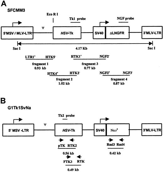
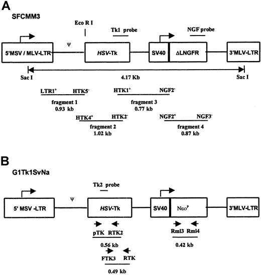
The GP+envAM12-derived producer cell line, SFCMM3 clone 16, was kindly provided by Dr Claudio Bordignon (Milan, Italy). The PA317-derived producer cell line containing the G1Tk1SvNa retroviral vector was provided by Genetic Therapy (Novartis, Gaithersburg, MD) (Figure 1). The vectors and their producer cell lines have been described previously.11,14 17
Schematic representation.
Proviral forms of SFCMM3 (A) and G1Tk1SvNa (B) vectors used for the transduction of CEM cells and primary T lymphocytes. LTR indicates long terminal repeats derived from Moloney murine leukemia virus (MLV) and Moloney sarcoma virus (MSV); HSV-Tk, herpes simplex virus gene sequence; SV40, simian virus 40 early promoter; ΔLNGFR, low-affinity nerve growth factor receptor cDNA truncated in the cytoplasmic domain; NeoR, neomycin phosphotransferase cDNA. Restriction enzyme sites for EcoRI and SacI used for the digestion of genomic DNAs derived from the CEM-Tk clones are indicated. The Tk1 and NGF probes were used to identify the provirus sequences in the genomic DNA. The Tk2 probe was also used to confirm the specificity of PCR products. The location of the PCR primers designed to amplify the SFCMM3 and G1Tk1SvNa provirus sequences are shown, together with the sizes of the PCR products.
Harvesting of virus supernatant
Retroviral supernatants were harvested in D-10 media (Dulbecco modified Eagle medium; GIBCO-BRL, Gaithersburg, MD) supplemented with 10% heat-inactivated fetal calf serum (FCS; Harlan Sera-Lab, Town, United Kingdom), 20 mM glutamine, 100 μg/mL streptomycin, and 100 U/mL penicillin after overnight incubation of the cells at 32°C. Virus-containing supernatants were filtered, snap-frozen with liquid nitrogen, and stored at −80°C until further use.
Isolation and culture of human T-cell lines and primary T lymphocytes
CEM cells (American Tissue Culture Collection, Manassas, VA) were cultured in RPMI (Gibco BRL; Life Technologies, Scotland) supplemented with 10% heat inactivated FCS, 20 mM glutamine, 100 μg/mL streptomycin, and 100 U/mL penicillin (RF-10).
Peripheral blood mononuclear cells were collected in heparinized (ICSM) or EDTA (EFSBFC) tubes from healthy donors or patients included in the phase I/II clinical trial11 38 with informed consent. Low-density mononuclear cells (less than 1.007 g/mL) were isolated by centrifugation (1500g, 30 minutes, 20°C) on Lymphoprep (Nycomed, Oslo, Norway). Peripheral blood mononuclear cells were cultured at a density of 2 × 10⁶ cells/mL in T-RF10 medium composed of RPMI containing 10% heat inactivated FCS, 5 μM β-mercaptoethanol, 25 μM HEPES (both from Sigma, St Louis, MO), glutamine, 100 μg/mL streptomycin, and 100 U/mL penicillin supplemented with 100 U/mL recombinant human interleukin 2 (rhIL-2) (Research & Development System Europe, Abingdon, United Kingdom; Prepotech EC, London, United Kingdom; or Chiron, Amsterdam, The Netherlands) at ICSM or 1000 U/mL day 0 to 3, then 500 U/mL rhIL-2 at EFSBFC.
Transduction of CEM and primary T lymphocytes
Peripheral blood mononuclear cells were stimulated with 1 μg/mL phytohemagglutinin-M (PHA) (Boehringer Mannheim, GmbH, Mannheim, Germany) and 100 U/mL rhIL-2 for 48 hours before transduction with SFCCM3. Nonadherent cells were collected by centrifugation and resuspended in T-RF10 at 2 × 10⁶cells/mL. Twenty-four hours before infection, CEM cells were fed with RF10 and were resuspended at a concentration of 5 × 10⁵cells/mL. Cell-free virus supernatants containing 4 μg/mL polybrene (Sigma) were added to the cell cultures and incubated for 16 hours at 37°C in a CO2 incubator. The infection process was repeated on 2 consecutive days. Cells were washed with fresh medium 24 hours after the last infection and further cultured for 2 to 3 days before FACS analysis.
Transduction of donor T cells with the G1Tk1SvNa retroviral vector was as previously described.18 Peripheral blood mononuclear cells were cultured with OKT3 (10 ng; Jasen-Cilag, Levalleis Perret, France) and rhIL-2 (500 U/mL, Chiron, UK Ltd) for 3 days. Infection with retroviral supernatant for 24 hours was followed by G418 (800 μg/mL; Sigma) selection of transduced cells for 7 days. Dead cells were removed, and the viable transduced product was cryopreserved for future use. The transduction of CEM cells with the G1Tk1SvNa vector involved identical infection conditions to those described for the SFCMM3 vector, followed by selection in G418 (800 μg/mL) for 7 to 14 days.
FACS analysis for the determination of ΔLNGFR expression
Transduced cells were incubated with the murine antihuman NGFR monoclonal antibody (HB8737, clone 20.4; ATCC) for 30 minutes at room temperature. Cells were washed twice with 1% bovine serum albumin (BSA)–phosphate-buffered saline (PBS) and stained with goat anti-immunoglobulin mouse fluorescein isothiocyanate-coupled antibody (Becton Dickinson, Mountain View, CA) for 20 minutes at 4°C. For dual-color analysis, cells were stained with phycoerythrin-coupled anti-CD3 monoclonal antibody (Becton Dickinson) for 20 minutes at 4°C and washed twice with 1% BSA-PBS. Finally, the cells were fixed with 1% paraformaldehyde–PBS solution. FACS analysis was performed using a flow cytometer (FACS Scan; Becton Dickinson). Within the CD3+ population, ΔLNGFR expression was measured using the CellQuest software (Becton Dickinson). For each reading at least 20 000 events were counted.
Selection of transduced cells
For the SFCMM3 vector, transduced cells were selected based on the expression of the ΔLNGFR on the cell surface using the MiniMACS system (Miltenyi Biotec, Germany) according to the manufacturer's instructions. Briefly, cells were incubated with the murine antihuman NGFR mAb for 30 minutes at room temperature, washed with MACS buffer (PBS supplement with 0.5% BSA and 2 mM EDTA), and incubated with a goat antimouse IgG microbeads (MACS; Miltenyi Biotec) for 15 minutes at 4°C. After washing, the ΔLNGFR-expressing cells were selected over MiniMACS MS+ separation columns (MACS; Miltenyi Biotec). Cells transduced with G1Tk1SvNa were selected by exposure to G418 (800 μg/mL) for 7 to 14 days. The transduction efficiency was evaluated by a competitive polymerase chain reaction (PCR) assay for theNeoR gene.19
Cloning of the transduced CEM cells
Cloning of the transduced and ΔLNGFR-selected CEM cells was performed by plating 400 cells in 1 mL methyl cellulose (MethoCult H4330; Stem Cell Technologies, Vancouver, Canada) in 35-mm Petri dishes. Semisolid cultures were incubated for 12 days at 37°C, 5% CO2. On day 13, individual colonies were picked, seeded in 100 μL RF10 in a 96-well plate, and subsequently expanded. These clones will be referred to as CEM-Tk clones.
Ganciclovir cytotoxic assay
Then 2 × 10⁴ cells/well were seeded into a 96-well plate in 100 μL RF-10 medium. Cells were cultured for 4 days with increasing concentrations of GCV ranging from 0.05 to 12.5 μg/mL (Cymevene; Hoffman-La Roche AG, Germany) in triplicate. On day 4, 1 μCi tritiated thymidine (methyl 3H-thymidine, TRA.120, 1.0 MBq/mL; Amersham International, United Kingdom) was added to each well 18 hours before harvesting the cells. Incorporation of3H-thymidine into the DNA was measured in a β-scintillation counter (Wallac 1410, Gaithersburg, MD). For each GCV concentration, the GCV-induced inhibition was calculated using the ratio (cpm culture with GCV/cpm culture without GCV) × 100.
Southern blot analysis
Genomic DNA was extracted using a QIAamp blood kit (Qiagen, Germany) according to the manufacturer's recommendations. After overnight digestion of 10 μg genomic DNA with the restriction enzymesSacI or EcoRI (New England Biolabs, United Kingdom), samples were size-fractionated by electrophoresis through a 0.8% agarose gel. Genomic DNA was transferred onto a Hybond N nylon filter (Amersham, Les Ullis, France), hybridized with an α-32P-dCTP random prime-labeled 1.1 kbMluI-XhoI fragment of the HSV-Tk gene sequence (Tk1 probe) or a 0.9 kb RsrII fragment of theΔLNGFR gene (NGF probe), and exposed to radiographic film (Biomax; Eastman Kodak, Rochester, NY) for at least 16 hours at −80°C.
Polymerase chain reaction and sequence analysis
The presence of the SFCMM3 proviral sequences was examined in genomic DNA extracted from transduced cells. Different regions of the provirus were amplified by PCR using primers (Figure 1A): fragment 1, LTR1+ 5′ GGTCTCCTCTGAGTGATTGACTA 3′ and HTK5-5′ AACGAATTCCGGCGCCTAGAGAA 3′; fragment 2, HTK4+ 5′ TTCTCTAGGCGCCGGAATTCGTT 3′ and HTK2-5′ ATCCAGGATAAAGACGTGCATGG3′; fragment 3, HTK1+ 5′ CCATGCACGTCTTTATCCTGGAT 3′ and NGF2-5′ TTGCAGCACTCACCGCTGTGTGT 3′; fragment 4, NGF2+ 5′ ACACACAGCGGTGAGTGCTGCAA 3′ and NGF3-5′ ATAGAAGGCGATGCGCTGCGAAT 3′.
PCRs were performed in a 20-μL reaction mixture containing 50 ng genomic DNA, 1× Taq DNA polymerase buffer with 15 mM MgCl2 (Boehringer Mannheim, Lewes, United Kingdom), 250 μM each dATP, dCTP, dGTP, dTTP, 0.25 μM each sense and antisense primer, and 0.025 U/μL Taq polymerase (Boehringer Mannheim). The thermocycling conditions used to amplify fragments 1 and 2 were 35 cycles of denaturation at 96°C for 30 seconds, annealing at 60°C for 50 seconds, and extension at 72°C for 1 minute, followed by a final 10-minute extension at 72°C. For fragments 3 and 4, annealing was performed at 64°C with a total of 31 cycles. The PCR products (10 μL) were electrophoresed on a 1% agarose gel stained with ethidium bromide (EtBr). PCR amplification of an 880-bp genomic fragment of the human ABL gene was performed as described elsewhere on negative clones to confirm the presence of amplifiable genomic DNA.20 The presence of the HSV-Tk gene in G1Tk1SvNa-transduced cells was examined by PCR using primers (Figure1B): pTK 5′ TAGACGGTCCTCACGGGATGGGGA 3′ and RTK2 5′ GCCAGCATAGCCAGGTCAAG 3′.
PCR assays were performed in a 50-μL reaction mixture containing 500 ng DNA, 1× Taq polymerase buffer with 15 mM MgCl2 (Eurogentec, Seraing, Belgium), 200 μM each dNTP, 0.25 μM each primer, and 0.5 U Taq DNA polymerase (Eurogentec). The thermocycling conditions used were 35 cycles at 94°C for 45 seconds, 60°C for 1 minute, 72°C for 1 minute, and a final extension of 5 minutes at 72°C. PCR products (18 μL) were electrophoresed on 2% EtBr-stained agarose gels. Genomic DNA was transferred to a nylon membrane, hybridized with an α-32P-dCTP end tail-labeled oligoprobe (Tk2 probe, 5′ATCGTCTACGTACCCGAGCCGATGA 3′, and washed at 60°C in 0.1 × SCC–0.1% sodium dodecyl sulphate before autoradiography. The sensitivity of this assay, determined by amplification of diluted DNA extracted from the packaging cell line, allowed the detection of one transduced cell in 105 unmodified cells, assuming that there was one Tk gene copy per genome.21
PCR products were cloned in duplicate into the pCR2.1 TA cloning vector (Invitrogen, Groningen, The Netherlands) or the pGEMT Easy vector from Promega (Charbonnières, France). Automated fluorescent DNA sequence analysis was carried out by the Advanced Biotechnology Centre (London, United Kingdom) and PE Biosystems (Courtaboeuf, France).
Frequency of the truncation event
Three hundred CEM cells that had been transduced by either G1Tk1SVNa or SFCMM3 vectors and selected by G418 or immunomagnetic procedures were mixed with 150 μL RF10 to 1 mL methyl cellulose (MethoCult H4320; Stem Cell Technologies, Vancouver, Canada). Cells were then plated in triplicate in 35-mm Petri dishes and grown for 15 days at 37°C in 5% CO2 and scored on day 15. Individual colonies were picked and expanded in 24-well plates for 3 days. Cells were then harvested for analysis by PCR for the presence of full-length or truncated HSV-Tk gene, as described above.
Clinical study
Escalating amounts of CD3+ gene-modified cells were infused together with T-cell–depleted bone marrow. Twelve patients with hematologic malignancies received 2 × 10⁵(n = 5), 6 × 10⁵ (n = 5), or 20 × 105(n = 2) transduced donor CD3+ cells per kilogram with bone marrow from an HLA-identical sibling. Blood samples were collected from all patients at 2-week intervals for the detection of gene-modified cells. In patients in whom GVHD developed and who were treated with GCV, peripheral blood samples were collected more frequently during and shortly after GCV administration.
Results
Transduction efficiency of CEM and primary cells
The average transduction efficiency of T lymphocytes and CEM cells based on the ΔLNGFR expression was 20% ± 5%. After 2 rounds of immunomagnetic selection, 95% to 98% of the transduced cells expressed ΔLNGFR.
The transduction efficiency of primary T cells with G1Tk1SvNa before G418 selection, as measured by quantitative PCR forNeoR, was 8.3% ± 1.4% (n = 12). The GCV sensitivity of these cells after G418 selection was 87% ± 1.2% (n = 12).38
ΔLNGFR expression in CEM-Tk clones
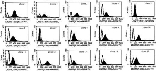
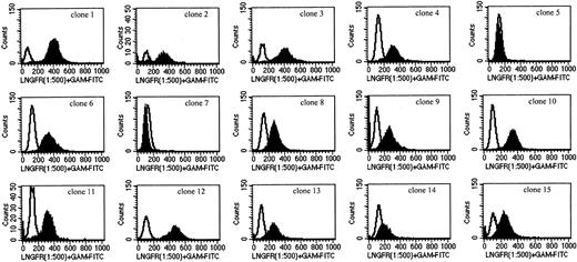
The expression of ΔLNGFR in individual CEM-Tk clones was determined by FACS analysis (Figure 2). Most (13 of 15) clones were positive for ΔLNGFR. Two clones (clones 5 and 7) showed no expression of ΔLNGFR, as demonstrated by the overlapping profiles with respect to the isotopic controls (Figure 2). In addition, different levels of expression of ΔLNGFR were observed within the positive clones.
FACs analysis for the expression of the ΔLNGFR transgene in the CEM-Tk–derived clones.
Cells were incubated with an unconjugated mouse antihuman NGFR mAb and subsequently stained with a fluorescein isothiocyanate–labeled goat antimouse mAb.
FACs analysis for the expression of the ΔLNGFR transgene in the CEM-Tk–derived clones.
Cells were incubated with an unconjugated mouse antihuman NGFR mAb and subsequently stained with a fluorescein isothiocyanate–labeled goat antimouse mAb.
HSV-Tk gene expression in CEM-Tk clones
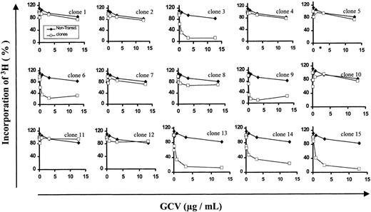
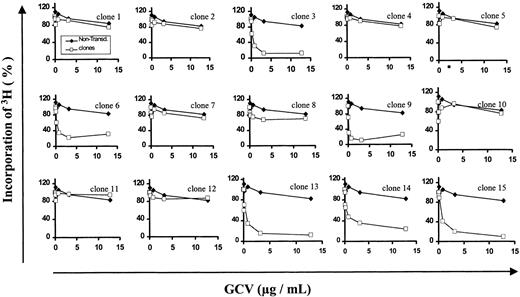
The expression of the HSV-Tk transgene in the CEM-Tk clones was determined by inhibition of cell proliferation when the cells were cultured at increasing concentrations of GCV (Figure3). The IC50 for inhibition of cell proliferation assessed by 3H-thymidine incorporation was reached at a concentration of GCV of 0.1 μg/mL for clones 3, 6, 9, 13, 14, and 15. In contrast, cell proliferation in clones 1, 2, 4, 5, 7, 8, 10, 11, and 12 was similar to that obtained in the nontransduced cells (negative control), indicating resistance to GCV.
GCV-induced cytotoxic assay for the determination of the
HSV-Tk transgene expression in the CEM-Tk derived clones. Cells were incubated for 4 days in the presence of increasing concentration of GCV (range, 0.05 to 12.5 μg/mL). Cell proliferation was measured by the incorporation of3H-thymidine into the cellular DNA. Results are expressed as percentage of incorporated 3H-thymidine at each GCV concentration with respect to the incorporation obtained in the absence of GCV. ♦, nontransduced CEM cells; ■, transduced CEM-Tk clones.
Integrity of the inserted SFCMM3 provirus by Southern blot and PCR analyses
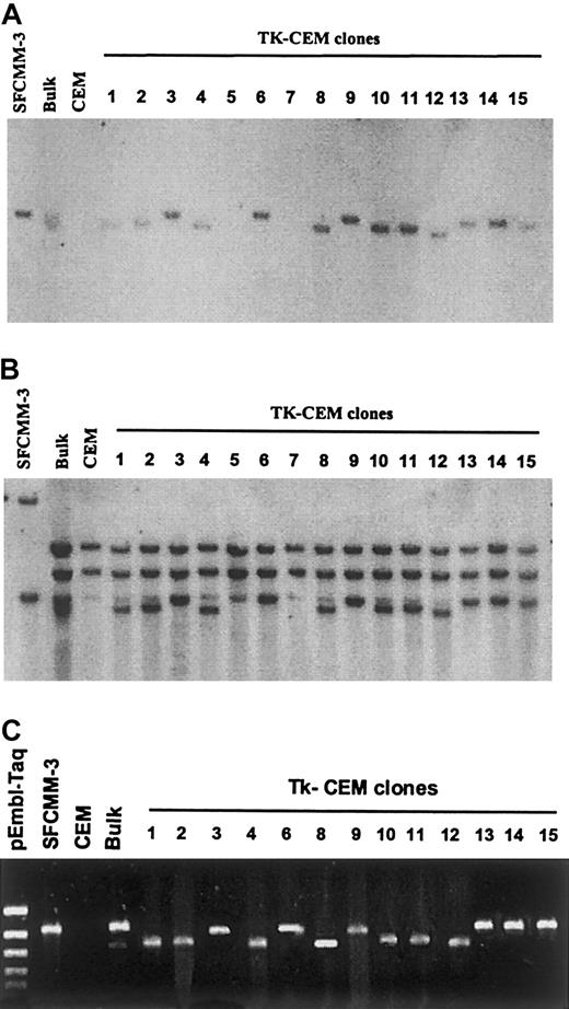
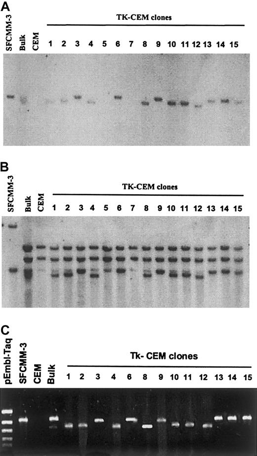
SacI-digested genomic DNA from the CEM-Tk clones was examined for the integrity of the inserted SFCMM3 provirus by Southern blot analysis. The SacI restriction enzyme cuts only in the 5′ and 3′ LTRs. Thirteen of the fifteen CEM-Tk clones contained the integrated provirus on hybridization with the Tk1 probe (Figure 4A). However, the expected 4.1-kb fragment was found in only 6 clones (3, 6, 9, 13-15). In the remaining clones (1, 2, 4, 8, 10-12), a smaller band was observed. When a probe specific for the ΔLNGFR gene was used (Figure 4B), multiple bands were observed, probably corresponding to the endogenous nerve growth factor receptor (NGFR) gene, and they appeared in all instances including the nontransduced parental CEM cells. The 4.1-kb fragment was seen in all clones except clones 5 and 7. The absence of the SFCMM3 vector after hybridization with both the Tk1 and the NGFR probes in CEM-Tk clones 5 and 7 confirmed our previous observations by FACS analysis and GCV cytotoxic assays.
Southern blot and PCR analyses of the CEM-Tk clones.
Genomic DNAs were digested with the SacI restriction enzyme. Southern blots were hybridized with Tk1 (A) and NGFR (B) specific probes. PCR amplification of the HSV-Tk gene sequence of SFCMM3 provirus (fragment 2, HTK4+ and HTK2−; 1020 bp) is shown (C).
To determine the number of insertions of the vector in each clone, genomic DNA was digested with the restriction enzyme EcoRI, which cuts once within the provirus sequence (Figure 1A). Southern blot analysis showed single independent integration events in each clone (results not shown).
PCR primers were designed along the SFCMM3 vector to amplify the entire provirus sequence (Figure 1A). PCR analysis of genomic DNA extracted from the CEM-Tk clones amplified bands of expected sizes when specific primers were used to amplify fragment 1 (LTR1+/ HTK5−, 934 bp), fragment 3 (HTK1+/ NGF2−, 775 bp), and fragment 4 (NGF2+/ NGF3−, 875 bp) (results not shown). As shown in Figure 4C, the bands corresponding to the HSV-Tk gene sequence (HTK4+/ HTK2−, 1020 bp) were as expected for CEM-Tk clones 3, 6, 9, 13, 14, and 15. Smaller bands of identical size (approximately 800 bp) were amplified in clones 1, 2, 4, 8, 10, 11, and 12. In the uncloned transduced CEM bulk populations, 2 bands of the same sizes as those seen in the clones were amplified. Proviral sequences were absent in clones 5 and 7 (results not shown). These results agree with those previously observed by Southern blot and FACS analyses.
DNA sequence analysis of the integrated provirus in CEM-Tk clones
To further analyze the provirus sequences, the HSV-Tkgene (fragment 2) was amplified from genomic DNA derived from the CEM-Tk clones and cloned. Sequence analysis showed that the small bands amplified by PCR (CEM-Tk clones 1, 2, 4, 8, 10, 11, and 12; Figure 4C) resulted from a deletion of 227 bp within the HSV-Tk gene sequence. The junctional region arose from the joining of a cryptic splice donor site (CAGG/GTGA, at position 1994 of the SFCMM3 vector) and a cryptic splice acceptor site (CCAG/GCCG, at position 2221 of the SFCMM3 vector).
PCR and sequence analysis of transduced primary T lymphocytes
Genomic DNA extracted from human primary T lymphocytes transduced with the SFCMM3 vector was analyzed by PCR for the HSV-Tkgene. One single band corresponding to the full-lengthHSV-Tk gene was amplified by PCR in the transduced primary T cells (results not shown). These results might indicate that the deletion observed in the CEM-Tk clones is a feature found only in the human T-cell lines. An alternative explanation is that the frequency of the deletion of the HSV-Tk gene in transduced primary T cells is below the level of detection of this PCR assay.
To address the above question, a primer was designed at the junction of the deletion in the HSV-Tk gene to specifically amplify the truncated gene found in some of the CEM-Tk clones. The CEM-Tk clones that amplified a short band for the HSV-Tk gene (fragment 2, Figure 4C) also amplified a band of the expected size (446 bp) for the truncated HSV-Tk gene (results not shown).
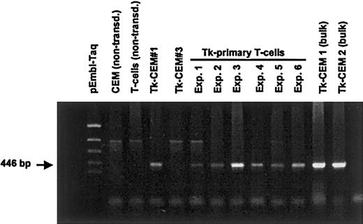
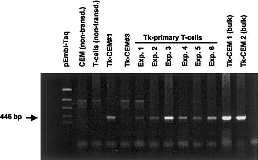
Using the primers specifically designed to amplify the truncatedHSV-Tk gene, the PCR assay was repeated on genomic DNA extracted from primary T lymphocytes transduced with the SFCMM3 vector. A band of the expected size for the truncated HSV-Tkgene was amplified from lymphocytes derived from 6 different persons (Figure 5). These results indicate that the truncated HSV-Tk gene is also present in the transduced primary T-lymphocyte populations. The frequency of this event in primary cells is probably lower than in the human T-cell lines because it was observed only when the specific primer to amplify the deletion junction of the truncated HSV-Tk gene was used. A fainter band of 970 bp was also amplified in all cases, including the nontransduced cells. Sequencing of this anomalous band revealed that it did not correspond to any sequence of the SFCMM3 retroviral vector or to any other sequence represented in the GenBank or EMBL databases. It may represent some cellular sequences as yet uncharacterized.
PCR amplification of the truncated
HSV-Tk gene from genomic DNA obtained from transduced and selected primary T lymphocytes. The PCRs were set up using primers (HTK8+ and HTK1−, 446 bp) that specifically amplified the spliced form of the HSV-Tk gene in the GCV-resistant CEM-Tk clones.
PCR amplification of the truncated
HSV-Tk gene from genomic DNA obtained from transduced and selected primary T lymphocytes. The PCRs were set up using primers (HTK8+ and HTK1−, 446 bp) that specifically amplified the spliced form of the HSV-Tk gene in the GCV-resistant CEM-Tk clones.
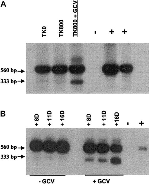
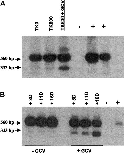
Genomic DNA extracted from human primary T lymphocytes transduced with the G1Tk1SvNa vector was also analyzed by PCR for the HSV-Tkgene. In this study 2 bands were identified in selected (TK800) and unselected (TK0) transduced cells (Figure6A). The 560-bp fragment corresponds to the full-length HSV-Tk gene and gave the strongest signal, compared with the smaller band of 333 bp, corresponding to the deleted form of the HSV-Tk gene. This may be due to competition in the PCR assay between the full-length and truncated HSV-Tkforms, when the target present in the larger amount is preferentially amplified. However, when transduced cells were first selected in G418 and then cultured in the presence of GCV (TK800 + GCV), the 333-bp band became more intense. This indicates that GCV treatment resulted in the enrichment of a population of cells expressing the truncatedHSV-Tk gene (Figure 6B). Interestingly, sequencing of the 333-bp PCR product showed the exact same deletion of 227 bp within theHSV-Tk sequence (donor and acceptor sites at position 1871 and 2098, respectively, in the G1Tk1SvNa vector) as in the CEM and primary T cells transduced with the SCFMM3 vector.
Southern blot of pTK/RTK2 PCR (35 cycles) products, from transduced primary T cells in culture with or without GCV.
(A) PCR amplification of the HSV-Tk sequence in transduced/unselected T cells (TK0), transduced/G418-selected T cells (TK800) cultured in the absence of GCV and with 1 μg/mL GCV for 7 days (TK800 + GCV). (B) Transduced primary T cells after 8, 11, and 16 days of culture in the presence (1 μg/mL) or absence of GCV. Positive (DNA from G1Tk1SvNa vector producer cells) and negative controls (nontransduced primary T cells) were used.
Frequency of truncation event
With the G1Tk1SvNa vector, PCR analysis of 153 CEM transduced colonies showed that 14 contained the truncated form of theHSV-Tk gene, whereas 139 colonies had the full-lengthHSV-Tk gene, giving a truncation frequency of 9.15%. Forty-five colonies transduced with the SFCMM3 vector were analyzed by PCR, and 7 were found to contain the truncated gene (frequency, 15.5%).
Identification of the truncated HSV-Tk gene in clinical samples
Twelve patients received transduced CD3+ cells at the time of stem cell transplantation. Acute toxicity was not observed. Quantitative PCR for NeoR revealed an early increase of the circulating transduced T cells, followed by a progressive decrease over time with persistent, detectable, long-term (more than 800 days) circulation of gene-modified cells. Acute GVHD grade II developed in 3 patients, whereas chronic GVHD (skin and salivary glands) developed in 1 patient. Treatment with GCV alone was associated with complete remission (CR) in 2 patients with acute GVHD, but the addition of steroids was necessary to achieve CR in one patient. Long-lasting CR was associated with GCV treatment in the patient with chronic GVHD. Ganciclovir treatment resulted in a significant rapid decrease in circulating transduced T cells.38
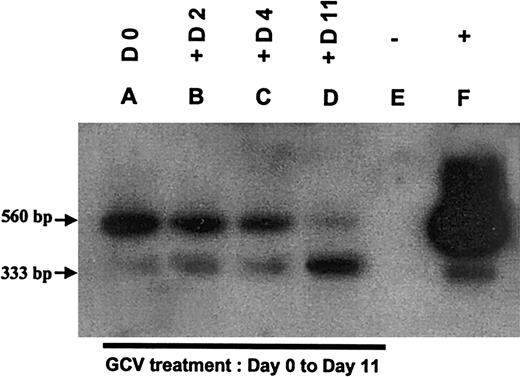
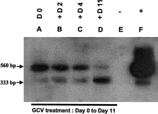
In vivo circulating transduced T cells containing the truncated form of the HSV-Tk gene were observed in all 12 patients as early as 30 minutes after infusion up to 800 days after transplantation. Although quantification of both forms of the HSV-Tk gene were not performed, PCR followed by Southern blot analysis suggested that the proportion of in vivo transduced T cells expressing the truncated HSV-Tk gene was present in a proportion similar to or less than that observed in vitro (less than 10%) (Figure7A). As illustrated in a representative sample from one patient (Figure 7), GCV administered as treatment for GVHD resulted in a decrease of the signal generated by the full-lengthHSV-Tk gene, whereas the signal associated with the truncated form of the HSV-Tk gene progressively increased. Importantly, GCV treatment always significantly reduced the percentage (85%-98%) and absolute number (76%-99.5%) of circulating transduced T cells as determined by quantitative PCR of theNeoR gene.19 Thus, an increasing proportion of transduced T cells containing the truncatedHSV-Tk gene was present in a reduced number of circulating transduced cells.
Southern blot analysis of TK PCR products of peripheral blood mononuclear cells from a representative patient of the clinical trial at time of GVHD (day 30 after allograft) during GCV treatment.
Lanes A-D: DNA samples extracted from peripheral blood mononuclear cells of the patient at time of GVHD (A), 2 days (B), 4 days (C), 11 days (D) after the beginning of the GCV treatment. Lane E: negative control (nontransduced primary T cells). Lane F: positive control (DNA from G1Tk1SvNa vector producer cells).
Discussion
The HSV-Tk gene encodes an enzyme able to convert nontoxic prodrugs such as GCV and acyclovir to cytotoxic metabolites. Gene transfer strategies using this and other suicide gene/prodrug systems have been proposed as novel therapeutic modalities for the treatment of cancer.22-24 More recently, the use of suicide gene therapy has been extended for allogeneic stem cell transplantation. Donor T lymphocytes transduced with theHSV-Tk gene can be selectively removed from circulation by the administration of GCV. This would allow modulation of GVHD while preserving a significant graft-versus-leukemia (GvL) effect and immune reconstitution.10,11,13,38 The efficacy of theHSV-Tk/GCV system has been demonstrated in a number of in vitro and in vivo models. The use of this system for the treatment of cancer may offer additional advantages derived from the so-called bystander effect.25,26 In this situation nontransduced tumor cells can be killed by phosphorylated GCV released from theHSV-Tk–transduced tumor cells through gap junctions. This is essential for complete regression of the tumor.27 We have demonstrated that in transduced primary T cells, GCV-induced growth inhibition is not mediated through a bystander effect.28 In this study, we examined the effect of GCV in a mixture of transduced and nontransduced T cells (50/50) as well as in HUT-78 cells (CD4+ T-cell lymphoma cell line). In both instances, no evidence for killing mediated by a bystander effect was found. In the context of allo-SCT the absence of a bystander-effect inHSV-Tk–transduced T lymphocytes has important implications for the successful application of suicide gene strategies because it specifically allows the depletion from circulation of those cells responsible for the GVHD.
The use of the HSV-Tk gene/GCV system was first proposed by Moolten and Wells.8 Later they observed in vivo the loss of HSV-Tk expression in transduced sarcoma and lymphoma tumor cells after GCV treatment.22,29 Similar observations have been confirmed in tumor cells derived from different tissues and animal models. Barba et al30 showed that genetically modified rat glioma cells expressing the HSV-Tk gene could be killed in culture after 14 days of GCV treatment. Eventually, some of the modified tumor cells became resistant to GCV. However, because of the bystander effect, inactivation of the HSV-Tk gene did not interfere with the outcome in vivo. In contrast, tumor regression after GCV treatment was not observed in nude mice bearingHSV-Tk–transduced adenocarcinoma cells.31 In this in vivo model, the bystander effect did not compensate for the faulty expression of the HSV-Tk gene in a small subset of transduced tumor cells.
The mechanisms underlying the lack of functional expression of theHSV-Tk gene in transduced tumor cells are poorly understood. Several explanations such as changes in cell susceptibility to GCV, transient exit of tumor cells from the cell cycle, inactivation or loss of the HSV-Tk gene, structural changes in theHSV-Tk mRNA, or transient methylation events have been proposed. Di Iani et al32 reported the lack ofHSV-Tk transgene expression in clones derived from U937 cells (a human hemopoietic malignant cell line) genetically engineered to express the HSV-Tk gene and the bacterial β-galactosidase gene (LacZ gene) in a bicistronic vector. The subclones showing resistance to GCV also failed to show expression of β-galactosidase. Culture of the resistant clones with 5-azacitidine (a demethylating agent) could not restore the expression of either of the 2 transgenes. The authors also speculated that rearrangements or mutations within the LTR of the bicistronic vector might also adversely affect expression.
GCV resistance in human primary T lymphocytes transduced with a retroviral vector carrying the HSV-Tk gene has been documented by different groups. We have previously described between 80% to 90% growth inhibition after GCV treatment of transduced rhIL-2–responding and NeoR T cells.28 These results suggest that at least 10% of the transduced populations are resistant to killing by GCV. In the first clinical trial using transduced donor T cells, Bonini et al12 observed that chronic GVHD developed in one patient. After the administration of GCV, the proportion of transduced donor cells fell from 11.9% to 2.8%. Complete depletion of transduced donor-derived T cells from circulation could not be achieved after further intensive treatment with GCV. Verzeletti et al14 further characterized this GCV resistance and suggested that it resulted from cell-cycle dependence of the HSV-Tk/GCV system rather than a molecular event occurring in the HSV-Tk gene sequence. GCV inhibition ofHSV-Tk–expressing T cells requires cell division and should leave noncycling cells unaffected by the action of the drug.33 In addition, recent studies have shown that viral LTR-driven expression of a transgene (HSV-Tk in the present case) is dependent on the state of activation of target T cells.34 Any of these mechanisms could account for in vivo resistance of the gene-modified T cells to GCV.
Using the SFCMM 3 retroviral vector, we found that some of the clones derived from transduced and selected CEM-Tk populations were not sensitive to GCV. The Southern blots revealed a shortened integrated provirus in the cell genome (Figure 5). PCR analysis of these clones indicated that the molecular events resulting in GCV-resistance occurred in the HSV-Tk gene sequence of the provirus (fragment 2; Figure 7). Comparison of the wild-type HSV-Tkgene with the deleted gene revealed a 227-bp deletion in the DNA sequence of the HSV-Tk gene. Mapping of the junctional region showed recombination at the cryptic splice donor and acceptor sites at positions 1994 and 2221 of the SFCMM3 or positions 1871 and 2098 for the G1Tk1SvNa vector, respectively. These findings suggest that in the producer cells, part of the mRNA derived from the provirus is recognized by the splicing machinery of the GP+envAm12 or PA317 cells, and this results in spliced, vector-derived mRNA. This results in the production of virus particles containing the full-lengthHSV-Tk gene, together with a small proportion carrying the aberrant HSV-Tk gene. This mechanism explains the transfer of the truncated HSV-Tk gene to the target cells. Northern blot analysis performed on the CEM-Tk clones has revealed the formation of aberrant HSV-Tk transcripts in those clones carrying the truncated gene (results not shown). Because these clones are resistant to killing by GCV a nonfunctional protein may be encoded from the aberrant HSV-Tk gene sequence.
Yang et al35 have found resistant colonies inHSV-Tk transduced gastrointestinal tumor cells. Characterization of the resistant colonies demonstrated that theHSV-Tk gene was either partially (a 220-bp deletion) or completely deleted from the resistant HSV-Tk transduced cells. Interestingly, to rule out the possibility of an unknown cellular mechanism participating in the GCV resistance, they infected the retrovirally transduced-GCV resistant clones with an adenoviral vector containing the HSV-Tk gene. The GCV sensitivity was restored, suggesting the continuing capability of these cells to express a functional HSV-Tk protein. The demonstration that GCV treatment increases the proportion of truncatedHSV-Tk–expressing donor T cells in vivo confirms that truncation of the HSV-Tk gene results in GCV resistance in vivo and so contributes to the persistence of gene-modified T cells after GCV treatment.
Sorrentino et al36 and Abonour et al37have reported similar observations when using retroviral vectors containing the MDR-1 cDNA for the transduction of hemopoietic stem cells. Expression of truncated and full-length MDR-1 mRNAs were found in bone marrow and spleen colony-forming cells in mice and also in the peripheral blood of patients undergoing autologous transplantation with transduced cells. They also demonstrated the formation of spliced and full-length transcripts in the producer cells, leading to the presence of truncated and full-length versions of the MDR-1 gene in the transduced products.
The in vitro and the clinical data suggest that the use of theHSV-Tk gene/GCV system for cancer gene therapy and allo-SCT represents a realistic improvement for the clinic.38In particular, GCV-sensitive acute and chronic GVHD were observed both in the Bonini study12 and in our study, suggesting that the proportion of GCV-sensitive donor T cells present in vivo are sufficient for therapeutic efficacy. However, GCV-induced selection of truncated HSV-Tk expressing donor T cells might result in subsequent GCV-resistance and deleterious alloreactivity. These results strongly support the idea that new suicide genes or optimized versions of the ones in use should be developed to achieve optimal killing efficiency of the genetically engineered cells. We are in the process of modifying the HSV-Tk gene sequence to remove one or both of the cryptic splice sites. This strategy will allow the development of an improved HSV-Tk gene for future clinical use.
Supported by the European Community (Biomed grant BMH4-CT97-2760); the Kay Kendall Leukaemia Fund; the Programme Hospitalier de Recherche Clinique; l'Association pour la Recherche sur le Cancer; la Ligue National contre Cancer; l'Association Française contre la Myopathie; le Minsistère de l'Enseignement Supérieur et de la Recherche; and Genetic Therapy/Novartis.
The publication costs of this article were defrayed in part by page charge payment. Therefore, and solely to indicate this fact, this article is hereby marked “advertisement” in accordance with 18 U.S.C. section 1734.
References
Author notes
Jane F. Apperley, Department of Haematology, Imperial College School of Medicine, Hammersmith Hospital, Ducane Road, London W12 0NN, United Kingdom; e-mail: j.apperley@ic.ac.uk.

