Abstract
Shear-induced platelet aggregation (SIPA) involves the sequential interaction of von Willebrand factor (VWF) with both glycoprotein Ib (GPIb) and αIIbβ3 receptors. Type 2B recombinant VWF (2B-rVWF), characterized by an increased affinity for GPIb, induces strong SIPA at a high shear rate (4000 s–1). Despite the increased affinity of 2B-rVWF for GPIb, patients with type 2B von Willebrand disease have a paradoxical bleeding disorder, which is not well understood. The purpose of this study was to determine if SIPA induced by 2B-rVWF was associated with αIIbβ3-dependent platelet activation. To this end, we have addressed the influence of 2B-rVWF (Val553Met substitution) on SIPA-dependent variations of tyrosine protein phosphorylation (P-Tyr) and the effect of αIIbβ3 blockers. At a high shear rate, 2B-rVWF induced a strong SIPA, as shown by a 92.7% ± 0.4% disappearance of single platelets (DSP) after 4.5 minutes. In these conditions, increased P-Tyr of proteins migrating at positions 64 kd, 72 kd, and 125 kd were observed. The band at 125 kd was identified as pp125FAK using anti–phospho-FAK antibody. This effect, which required a high level of SIPA (> 70% DSP), was observed at 4000 s–1 but not at 200 s–1. Monoclonal antibodies (MoAbs) 6D1 (anti-GPIb) and 328 (anti-VWF A1 domain), completely abolished SIPA and p125FAK phosphorylation mediated by 2B-rVWF. In contrast, neither RGDS peptide nor MoAb 7E3, both known to block αIIbβ3 engagement, had any effect on SIPA and pp125FAK. The size of aggregates formed at a high shear rate in the presence of 2B-rVWF was decreased by genistein, demonstrating the biologic relevance of pp125FAK. These findings provide a unique mechanism whereby the enhanced interaction of 2B-rVWF with GPIb, without engagement of αIIbβ3, is sufficient to induce SIPA but does not lead to stable thrombus formation.
Introduction
Platelets play a critical role in the arrest of bleeding by adhering to vascular matrix proteins and to other activated platelets at sites of vessel wall injury. High shear rates generate forces between platelets and the damaged vessel wall that induce specific ligand-receptor interactions required for platelet adhesion, activation, and aggregation. A key adhesive protein initiating platelet-vessel wall interactions is the large multimeric protein, von Willebrand factor (VWF). When immobilized at the site of injury, VWF exhibits its A1 domain, allowing the capture of platelets from rapidly flowing blood by an interaction with the glycoprotein Ib/V/IX (GPIb/V/IX) complex. This binding generates signals leading to αIIbβ3 integrin activation which becomes competent to interact with VWF and to promote irreversible platelet adhesion.1, 2, 3, 4 In vitro, the change in VWF can be mimicked by the binding of VWF to its modulators ristocetin or botrocetin.5,6
High shear rates are important not only in the initial contact of platelets with the vessel wall, but also in the process of platelet aggregation, where fluid-phase VWF is required to stabilize platelet-platelet interactions and thrombus growth.7 The mechanism of shear-induced platelet aggregation (SIPA) differs from conventional platelet aggregation observed under stirring conditions, essentially because the latter involves the binding of fibrinogen to the activated αIIbβ3 receptor.8 Based on studies performed with viscometers in which uniform shear fields can be applied in the absence of exogenous modulators, the following working model has been proposed: (1) in high shear rate conditions, fluid-phase VWF becomes able to bind to GPIb; (2) αIIbβ3 becomes activated through intracellular signals generated by VWF-GPIb complex formation; (3) activated αIIbβ3 binds to the Arg-Gly-Asp (RGD) sequence of VWF, thereby stabilizing platelet aggregation.9,10 Using antibodies against VWF, GPIb, or αIIbβ3 as well as platelet-rich plasma (PRP) from patients with von Willebrand disease, Glanzmann thrombasthenia, or Bernard-Soulier syndrome, an interaction of VWF with both its receptors has been confirmed in SIPA.10, 11, 12
Under high shear conditions, evidence has been provided that the VWF-GPIb interaction leads to activation of αIIbβ3 through the initiation of signaling pathways that involve transmembrane calcium influx or protein kinase C activation.13,14 In addition, protein tyrosine phosphorylations have been demonstrated resulting from the interaction between VWF and both GPIb and αIIbβ3 integrin.15,16 Other studies have described tyrosine-phosphorylated proteins of platelets in the presence of VWF and nonphysiologic agonists such as ristocetin, botrocetin, or alboaggregin which induce binding to GPIb.17, 18, 19, 20, 21 More recently, signaling events resulting from the interaction between GPIb and VWF were distinguished from those initiated by αIIbβ3 engagement with VWF, the latter requiring phosphoinositide 3-kinase (PI 3-kinase) activation.22,23 Thus, it remains to be determined whether under high shear conditions engagement of αIIbβ3 is an obligatory consequence of the signaling pathway triggered by the VWF-GPIb interaction.
We have previously established that recombinant VWF reproducing type 2B von Willebrand disease mutations (2B-rVWF), characterized by an increased affinity for GPIb, induced complete SIPA at high shear rates by a mechanism that was primarily mediated by GPIb.24 In the present study, we addressed the issue of a signal transduction pathway that sustains the platelet response induced by the VWF-GPIb axis. Our results showed that SIPA induced by 2B-rVWF was associated with tyrosine phosphorylations of platelet proteins, and in particular of p125FAK. A major finding was that in the presence of 2B-rVWF, activation of p125FAK was correlated with a high level of platelet aggregation, which could not be abrogated by the synthetic peptide Arg-Gly-Asp-Ser (RGDS), suggesting that activation of p125FAK occurred independently of αIIbβ3 engagement.
Materials and methods
Antibodies and reagents
Synthetic RGDS peptide, leupeptin, aprotinin, wortmannin, genistein, sodium orthovanadate (Na3VO4), β-glycerophosphate, sodium fluoride (NaF), apyrase, adenosine 5′ phosphate (ADP), and adenosine 2′,5′-diphosphate (A2P5P) were purchased from Sigma (St Louis, MO). Electrophoresis reagents and nitrocellulose sheets were obtained from Bio-Rad (Ivry-sur-Seine, France) and chemical products were from Merck Eurolab (Strasbourg, France). Sheep anti–monoclonal antibody (MoAb) horseradish peroxidase–labeled antibody was from Amersham (Little Chalfont, Bucks, United Kingdom). Goat antirabbit horseradish peroxidase–labeled antibody was from Bio-Rad. Antiphosphotyrosine MoAbs PY20 and 4G10 were from Chemicon International (Tecula, CA) and Upstate Biotechnology (Lake Placid, NY), respectively. Rabbit polyclonal antiserum anti–phospho-FAK, raised against peptide 392-399 of human phospho-FAK, which recognizes p125FAK phosphorylation at Tyr397, was purchased from Upstate Biotechnology. The absence of cross-reactivity with Pyk2 at Tyr402 has been established (unpublished information provided by Upstate Biotechnology, March, 2002). For FAK detection we used a polyclonal or a monoclonal anti-FAK antibody, both from Upstate Biotechnology. Human thrombin was from Diagnostica-Stago (Asnières, France). MoAb anti-GPIb 6D125 and MoAb anti-αIIbβ3 7E326 were a generous gift of Dr B. S. Coller (The Rockefeller University, New York, NY). MoAb 328, which blocks VWF binding to GPIb in the presence of ristocetin but not botrocetin, was produced in our laboratory.27 PAC-1 fluorescein isothiocyanate (FITC) recognizing an epitope on the αIIbβ3 complex of activated platelets was purchased from Becton Dickinson (San Jose, CA). Kistrin was purified from the venom of the viper Agkistrodon rhodostoma as described.28
Plasmid constructs, cell culture, and transfection
Plasmids with full-length complementary DNA (cDNA) coding for wild-type (WT) rVWF (WT-pNUT) or mutated 2B-rVWF with substitution of methionine in position 553 for valine (Val553Met) were constructed as previously described.29,30 The mutant was confirmed by restriction enzyme digests.
For stable transfection of WT- or 2B-rVWF Val553Met, a baby hamster kidney (BHK) cell line overexpressing furin was established using the lipofectamine method as previously described.29 The culture supernatant of these cell lines was harvested every 3 to 4 days, and the amount of rVWF was estimated by enzyme-linked immunosorbent assay (ELISA) using a pool of 12 MoAbs to VWF.24 The concentration of rVWF was estimated between 2 μg/mL and 8 μg/mL in the cell expression medium. In some experiments we used 2B-rVWF Met540MetMet (duplication of methionine at position 540) expressed by COS cells.24
Purification of WT- and 2B-rVWFs
WT-rVWF and 2B-rVWF were purified by immunoaffinity chromatography on CNBr-activated sepharose 4B (Amersham) coupled to a MoAb 453, directed against the C-terminal part of the VWF subunit and produced in our laboratory. The MoAb 453 sepharose column was equilibrated with 50 mM Tris/100 mM NaCl (TBS), pH 7.4. Expression medium was applied to the column and bound rVWF was eluted in 1 mL fractions using 50 mM triethylamine (TEA), pH 11-12, which was immediately neutralized with 2 M glycine, pH 3. rVWF-containing fractions were stored at –80°C.
Characterization of WT- and 2B-rVWFs
The multimeric composition of rVWFs, analyzed by 0.1% sodium dodecyl sulfate (SDS) and 1% agarose gel electrophoresis, was similar in 2B-rVWF and WT-rVWF.31 Subunit analysis was performed by electrophoresis on 0.1% SDS–5% polyacrylamide gel under reducing conditions followed by immunotransfer to a nitrocellulose membrane (Bio-Rad),32 and showed that both WT- and 2B-rVWF were secreted as full-length mature VWF. Binding of mutated and WT-rVWFs to formalin-fixed platelets was performed using 125I-MoAb 505 (anti-VWF) as a tracer in the presence of ristocetin or botrocetin as described.24 In the presence of 1 mg/mL ristocetin, WT-rVWF as well as 2B-rVWF exhibited binding to GPIb, reaching 69.2% and 80.1%, respectively. At low concentration of ristocetin, 2B-rVWF showed increased binding to GPIb (65.4%), whereas WT-rVWF was unable to bind to platelets (5.9%).
Preparation of washed platelets
Blood was obtained from healthy individuals who had not ingested any medication for 2 weeks before donation, with acid-citrate-dextrose (ACD) 15% as an anticoagulant. Washed platelets were prepared as previously described.24 Briefly, platelets were isolated from platelet-rich-plasma by centrifugation at 220g for 15 minutes at 37°C and washed twice with HEPES buffer pH 6.7 (10 mM HEPES; N-[2-hydroxyethyl]piperazine-N′-[ethanesulfonic acid], 136 mM NaCl, 2.7 mM KCl, 2 mM MgCl2) containing 0.35% bovine serum albumin (BSA) in the presence of apyrase (2 U/mL) and ACD (2.5%). Platelets were resuspended in HEPES buffer pH 7.5 containing 0.025% BSA, and the concentration was counted with an electronic particle counter (Model 1; Coulter Electronics, Margency, France).
Shear-induced platelet aggregation
The rotating device is a Couette type viscometer used as previously described with minor modifications.12 Washed platelets (2.8 × 10⁸/mL) were exposed for 4.5 minutes at 37°C to a continuous shear rate of 200 s–1 or 4000 s–1 in the presence of rVWF and 1 mM CaCl2. Recombinant proteins were adjusted to a final concentration of 0.7 μg/mL (2B-rVWF) or 4 μg/mL (WT-rVWF) in cell expression medium. In some experiments, purified rVWF was used at final concentrations varying from 0.7 μg/mL to 5 μg/mL for 2B-rVWF or 10 μg/mL to 34 μg/mL for WT-rVWF. Following exposure to shear, an aliquot (40 μL) was fixed with 1.25% paraformaldehyde for 15 minutes and diluted 50-fold in fluorescence activated cell sorting (FACS)–flow buffer (Becton Dickinson, Le Pont-de Claix, France) for flow cytometry analysis in a FACScan flow cytometer as reported.12 Data acquisition was performed by counting the particle number during a constant time (30 seconds) to measure identical volumes in different samples. Platelets were analyzed by forward light scatter and side light scatter, and the single platelet region (n) was determined using as reference (no) a platelet sample incubated in the rotating viscometer without exposure to shear, and in the absence of rVWF. SIPA was calculated as [(no – n)/no] × 100, which represents the percentage of disappearance of single platelets (DSP). For inhibition studies, platelets were incubated for 5 minutes with MoAb 6D1 or 7E3 (20 μg/mL), RGDS (1 mM), or EDTA (ethylenediaminetetraacetic acid; 2 mM). Alternatively, platelets were preincubated for 30 minutes at room temperature with genistein (100 μM-1000 μM), or for 15 minutes at 37°C with wortmannin (100 nM) before being submitted to shear. Means ± SEM were calculated from 3 experiments. In some experiments, after exposure to shear an aliquot of the fixed platelet suspension was deposited in 96-multiwell plastic plates (Nunc, Roskilde, Denmark). Following sedimentation in the wells, aggregates were observed at a 10-fold magnification using an Axiovert 135 inverted microscope (Zeiss, Oberkochen, Baden-Wuerttemberg, Germany). Images were observed with a charged-coupled-device (CCD) camera (Sony, Tokyo, Japan) and vizualized using a standard display VGA monitor (Microvision Instruments, Evry, France).
ADP and thrombin-induced platelet aggregation
Washed platelets (2.8 × 10⁸/mL) were supplemented with 1 mM CaCl2 and preincubated with either purified WT-rVWF or 2B-rVWF (8 μg/mL) for 2 minutes at 37°C. Platelet aggregation was initiated by adding thrombin (0.01 U/mL-0.1 U/mL), monitored under constant stirring in a lumi-aggregometer (Chrono-Log, Coultronics, France) for 2 minutes at 37°C, and quantitated by measuring maximal intensity. For positive control of specific inhibitors, platelets were preincubated with each blocker as described in the previous paragraph and platelet aggregation was initiated by adding thrombin (0.5 U/mL) alone or ADP (5 μM) with fibrinogen (0.7 mg/mL) (Merck Eurolab).
Preparation of cell lysates and Western blot analysis
Following exposure to shear, platelet suspensions (160 μL) were solubilized in the cylinder in the absence of shear with 40 μL of denaturing buffer (10% SDS, 100 mM NaCl, 50 mM Tris, 50 mM NaF, 5 mM EDTA, 40 mM β-glycerophosphate, 3 mM Na3VO4, 5 μg/mL leupeptin, 10 μg/mL aprotinin, pH 7.4), and then transferred to microcentrifuge Eppendorf tubes. Samples were left on ice at 4°C for 30 minutes, followed by addition of Laemmli buffer (25 mM Tris pH 6.8, 10% [vol/vol] glycerol, 2% SDS, 10 μg/mL bromophenol blue) containing 10 mM dithiothreitol. Samples were heated at 100°C for 15 minutes and proteins were separated by SDS–polyacrylamide gel electrophoresis (SDS-PAGE), in an 8% polyacrylamide gel. Separated proteins were transferred electrophoretically to a nitrocellulose membrane. The membrane was incubated overnight at 4°C with a blocking buffer containing 5% skimmed low-fat milk, 2% Tween 20, 100 mM NaCl, and 20 mM Tris pH 7.4 (T-TBS). Membranes were then incubated for 2 hours at room temperature with the combination of 2 antiphosphotyrosine antibodies 4G10 and PY20 (1:10 000), washed extensively with T-TBS, and incubated with horseradish peroxidase–conjugated secondary antibody (1:10 000) for one hour at room temperature. Immunoreactive bands were detected by chemiluminescence system (Amersham). In some experiments, the membranes were stripped, and bound antibody was removed by incubation in buffer containing 2% SDS, 62.5 mM Tris, pH 6.8, and 100 mM 2-mercaptoethanol for 45 minutes at 60°C. After extensive washing, the membrane was reprobed with anti–phospho-FAK antibody (1:10 000) as described above. Quantitation of p125FAK phosphorylation was performed by scanning autoradiographs using a Hyrys 2 densitometer (Sebia, Issy les Moulineaux, France) and expressed as arbitrary units.
Statistical analysis
Means values and their standard errors were calculated from at least 3 experiments. Statistical analysis using the Student t test for paired samples was performed on 14 experiments and a P value less than .05 was considered significant (Statview Software; Abacus Concepts, Berkeley, CA).
Results
2B-rVWF–dependent shear-induced platelet aggregation
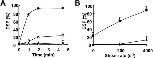
In order to assess the ability of 2B-rVWF bearing the Val553Met substitution to induce SIPA, we determined the percent of DSP in various time and shear conditions. At a high shear rate of 4000 s–1, 2B-rVWF–dependent SIPA increased with time, reaching almost 80% DSP at 1 minute and increasing between 1 and 2 minutes. Finally, 2B-rVWF induced a strong SIPA leading to a DSP of 92.7% ± 0.4% at 4.5 minutes (Figure 1A). In agreement with previous findings, this mutant induced a spontaneous aggregation, since in the absence of shear, 2B-rVWF was able to reach a DSP of 22.7% ± 8.8% at 4.5 minutes.24 In contrast, in the absence of 2B-rVWF, no detectable SIPA was observed at 4000 s–1 (Figure 1A). In addition, 2B-rVWF induced a significant SIPA at a lower shear, as shown by 60.1% ± 8.8% DSP at 200 s–1 (Figure 1B). When intermediate shear rates of 500 s–1 and 1000 s–1 were applied to the platelets, we found that DSP increased as a function of shear rate, as shown by intermediate values of 82.4% and 84.3% DSP, respectively. We have previously shown that conditioned medium from cells transfected with plasmid pcDNA3-without full-length cDNA of VWF did not induce platelet aggregation.24
Kinetics of SIPA and effect of increasing shear rates on platelet aggregation. (A) Washed platelets (2.8 × 10⁸/mL) were submitted to 0 s–1 (open symbols) or 4000 s–1 (closed symbols) for various times as indicated, in the presence of buffer (▵, ▴) or 2B-rVWF (□, ▪). (B) Platelets were submitted to a shear rate of 200 s–1 or 4000 s–1 for 4.5 minutes at 37°C in the absence (▴) or presence (▪) of 0.7 μg/mL 2B-rVWF. Following exposure to shear rates, an aliquot was fixed with 1.25% paraformaldehyde for 15 minutes. The results were expressed as the percentage of disappearance of single platelets (% DSP). Results are means ± SEM of 3 experiments.
Kinetics of SIPA and effect of increasing shear rates on platelet aggregation. (A) Washed platelets (2.8 × 10⁸/mL) were submitted to 0 s–1 (open symbols) or 4000 s–1 (closed symbols) for various times as indicated, in the presence of buffer (▵, ▴) or 2B-rVWF (□, ▪). (B) Platelets were submitted to a shear rate of 200 s–1 or 4000 s–1 for 4.5 minutes at 37°C in the absence (▴) or presence (▪) of 0.7 μg/mL 2B-rVWF. Following exposure to shear rates, an aliquot was fixed with 1.25% paraformaldehyde for 15 minutes. The results were expressed as the percentage of disappearance of single platelets (% DSP). Results are means ± SEM of 3 experiments.
SIPA is associated with tyrosine phosphorylations in the presence of 2B-rVWF
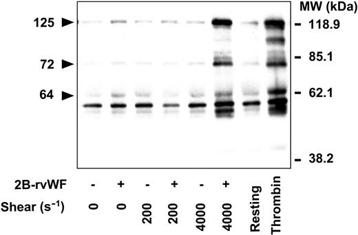
We examined tyrosine phosphorylations resulting from the interaction between platelets and 2B-rVWF in the presence or absence of shear. Whole-platelet lysates were run on SDS-PAGE and immunoblotted with MoAbs directed against tyrosine-phosphorylated proteins. Influence of increasing shear rates was compared with thrombin-induced platelet tyrosine phosphorylations (Figure 2). Interestingly, in the presence of 2B-rVWF at 4000 s–1 we showed a significant increase in protein tyrosine phosphorylations. In particular, we found major modifications of band intensities corresponding to proteins with molecular masses of 125 kd, 72 kd, and 64 kd. In contrast, a faint increase in tyrosine phosphorylation was observed in platelet lysates containing 2B-rVWF in the presence of a lower shear rate (200 s–1) or in the absence of shear. Furthermore, in the absence of 2B-rVWF, no detectable modification in tyrosine phosphorylation was obtained at 200 s–1 and 4000 s–1 (Figure 2). As described in “2B-rVWF–dependent shear-induced platelet aggregation” for SIPA, when the conditioned medium of mock-transfected cells was tested instead of buffer, no increase in tyrosine phosphorylation was seen (data not shown). To verify that increased tyrosine phosphorylations obtained in the presence of expression medium of cells transfected with the cDNA of 2B-rVWF (Val553Met) was not due to a contaminant, we have also tested purified mutant. Interestingly, similar bands of tyrosine-phosphorylated proteins were obtained using purified 2B-rVWF (5 μg/mL-10 μg/mL). Identical results were also obtained using another 2B-rVWF mutant (Met540MetMet), which was previously described to induce high SIPA24 (data not shown). As a control of tyrosine-phosphorylated proteins following agonist-induced activation, we prepared platelet samples stimulated with thrombin (0.5 U/mL). In particular, compared with resting (nonstimulated) platelets, we observed the appearance of a 97-kd protein, which was previously reported to be associated specifically with the presence of large aggregates following activation with strong agonists.33 This 97-kd protein was not seen in the samples exposed to a high shear rate in the presence of 2B-rVWF, despite the almost complete SIPA (>90% DSP) (Figure 2).
Effect of increasing shear rates on platelet tyrosine phosphorylations. Platelets (2.8 × 10⁸/mL) in the absence (–) or presence (+) of 2B-rVWF (0.7 μg/mL), were submitted to increasing shear rates (0 s–1, 200 s–1, and 4000 s–1) for 4.5 minutes at 37°C. Lysates were prepared as described in “Materials and methods.” On the right side of the figure, platelets were either unstimulated (Resting) or stimulated by 0.5 U/mL thrombin for 2 minutes in a lumi-aggregometer. Positions of the molecular mass markers are indicated on the right side and the positions of 3 bands that are strongly phosphorylated are indicated on the left side by arrowheads. MW indicates molecular weight. Data are representative of 3 experiments.
Effect of increasing shear rates on platelet tyrosine phosphorylations. Platelets (2.8 × 10⁸/mL) in the absence (–) or presence (+) of 2B-rVWF (0.7 μg/mL), were submitted to increasing shear rates (0 s–1, 200 s–1, and 4000 s–1) for 4.5 minutes at 37°C. Lysates were prepared as described in “Materials and methods.” On the right side of the figure, platelets were either unstimulated (Resting) or stimulated by 0.5 U/mL thrombin for 2 minutes in a lumi-aggregometer. Positions of the molecular mass markers are indicated on the right side and the positions of 3 bands that are strongly phosphorylated are indicated on the left side by arrowheads. MW indicates molecular weight. Data are representative of 3 experiments.
2B-rVWF–dependent p125FAK phosphorylation is specific to high shear rates
In the presence of 2B-rVWF, at 4000 s–1 tyrosine phosphorylation of 64-kd, 72-kd, and 125-kd proteins increased in a time-dependent manner, from 1 minute to 4.5 minutes (Figure 3A). By stripping and reprobing with a polyclonal antibody that recognizes pp125FAK, the tyrosine-phosphorylated form of p125FAK, we found a strong staining for a protein with a molecular mass of 125 kd (Figure 3B, top panel). In contrast, a faint phosphorylation of p125FAK was observed in samples containing 2B-rVWF in the absence of shear rates, and no increase was seen in samples devoid of 2B-rVWF, whether in the presence or absence of shear. As a positive control, we showed an increased phosphotyrosine intensity in the sample stimulated with 0.5 U/mL thrombin, as previously reported for p125FAK phosphorylation in thrombin-stimulated platelets.34 We probed the lysates for their p125FAK protein content using an antibody against the nonphosphorylated form, and found that differences in phosphorylation were not related to variations in protein content (Figure 3B, bottom panel). (In subsequent figures, no differences in FAK protein content were observed among lysates and are not shown.)
Time course of protein tyrosine phosphorylation induced by 2B-rVWF. Platelets (2.8 × 10⁸/mL) were either mixed with buffer (–) or with 2B-rVWF at 0.7 μg/mL (+) in the absence (0 s–1) or in the presence (4000 s–1) of high shear rate. Proteins of the lysates were separated on a 8% polyacrylamide gel, transferred to a nitrocellulose membrane, and blotted with antiphosphotyrosine (A) or anti-pFAK/FAK antibody (B), using either anti–phospho-FAK or anti-FAK. On the right side, resting or thrombin-stimulated platelets are shown. Data are representative of 3 experiments.
Time course of protein tyrosine phosphorylation induced by 2B-rVWF. Platelets (2.8 × 10⁸/mL) were either mixed with buffer (–) or with 2B-rVWF at 0.7 μg/mL (+) in the absence (0 s–1) or in the presence (4000 s–1) of high shear rate. Proteins of the lysates were separated on a 8% polyacrylamide gel, transferred to a nitrocellulose membrane, and blotted with antiphosphotyrosine (A) or anti-pFAK/FAK antibody (B), using either anti–phospho-FAK or anti-FAK. On the right side, resting or thrombin-stimulated platelets are shown. Data are representative of 3 experiments.
Comparison of the effect of 2B-rVWF and WT-rVWF on SIPA and p125FAK tyrosine phosphorylation
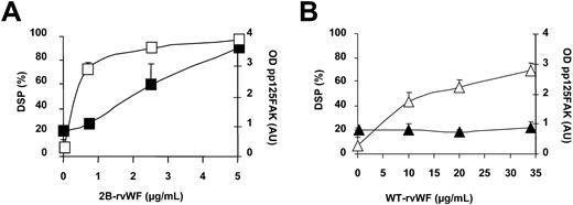
We next attempted to understand the relationship, if any, between the extent of SIPA and that of pp125FAK, and to determine if 2B-rVWF was able to modify p125FAK phosphorylation. In the presence of 2B-rVWF, at 4000 s–1 SIPA reached 97.1% ± 0.4% (n = 14), a value significantly higher than that (32.6% ± 1.9%) in the absence of shear (P < .0001). These results were compared with variations of pp125FAK intensities, quantitated in the corresponding samples. We found that at 0 s–1 optical density (OD) of pp125FAK corresponded to 0.9 ± 0.1 arbitrary units (AUs), while it increased to 2.9 AU ± 0.2 AU at a high shear rate (P < .0001). Thus, in order to establish whether the increased p125FAK phosphorylation at high shear was specific to 2B-rVWF, we compared the ability of increasing concentrations of purified 2B-rVWF and WT-rVWF, to induce an increase in p125FAK phosphorylation (Figure 4).
Effect of various concentrations of 2B-rVWF or WT-rVWF on SIPA and p125FAK phosphorylation. Washed platelets (2.8 × 10⁸/mL) were exposed to 4000 s–1 for 4.5 minutes at 37°C in the presence of various concentrations of 2B-rVWF (A; □, ▪) or WT-rVWF (B; ▵, ▴). An aliquot was taken for quantitation of SIPA expressed as % DSP (left vertical axis; □, ▵). Corresponding lysates were analyzed by Western blotting with anti–phospho-FAK antibody, and autoradiographs were scanned with a laser densitometer and expressed as OD arbitrary units (AU) (right vertical axis; ▪, ▴). Results are representative of 2 experiments.
Effect of various concentrations of 2B-rVWF or WT-rVWF on SIPA and p125FAK phosphorylation. Washed platelets (2.8 × 10⁸/mL) were exposed to 4000 s–1 for 4.5 minutes at 37°C in the presence of various concentrations of 2B-rVWF (A; □, ▪) or WT-rVWF (B; ▵, ▴). An aliquot was taken for quantitation of SIPA expressed as % DSP (left vertical axis; □, ▵). Corresponding lysates were analyzed by Western blotting with anti–phospho-FAK antibody, and autoradiographs were scanned with a laser densitometer and expressed as OD arbitrary units (AU) (right vertical axis; ▪, ▴). Results are representative of 2 experiments.
Between 0.7 μg/mL and 5 μg/mL 2B-rVWF, SIPA increased from 70% to 98%; in this range of 2B-rVWF, the extent of p125FAK phosphorylation increased almost linearly from 0.9 AU to 3.8 AU, and did not reach a plateau (Figure 4A). In the presence of WT-rVWF, SIPA increased in a dose-dependent manner between 0 μg/mL and 34 μg/mL, up to a DSP of 67.9%. It should be noted that despite the 7-fold-higher concentration of WT- than of 2B-rVWF, SIPA remained under the plateau observed at 2B-rVWF concentrations between 0.7 μg/mL and 5 μg/mL. The OD of pp125FAK was 0.9 AU ± 0.1 AU, and did not increase as a function of WT-rVWF concentration (Figure 4B). Thus, in this range of WT-rVWF concentrations, SIPA increased whereas pp125FAK did not.
Effect of GPIb-VWF blockers on tyrosine phosphorylations induced by 2B-rVWF
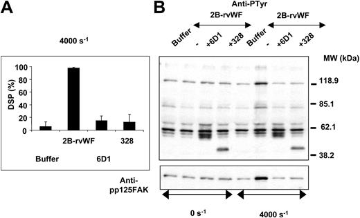
We have previously established that SIPA in the presence of 2B-rVWF predominantly involves the interaction between GPIb and 2B-rVWF.24 We confirmed the inhibitory effect of either anti-GPIb MoAb 6D1 or anti-VWF MoAb 328 (known to block VWF-GPIb interaction) on SIPA in the presence of 2B-rVWF at 4000 s–1 (Figure 5A). The effect of MoAb 6D1 or MoAb 328 was further evaluated on shear-induced tyrosine phosphorylation. At 4000 s–1 2B-rVWF–dependent tyrosine phosphorylations were totally blocked by MoAb 6D1 as well as by MoAb 328 (Figure 5B). An additional band was seen in the presence of MoAb 328, suggesting that its heavy chain could react with the secondary antibody used in the Western blot. Interestingly, p125FAK tyrosine phosphorylation was also strongly inhibited by MoAbs 6D1 and 328 (Figure 5B). These results suggested that tyrosine phosphorylations and p125FAK phosphorylation were primarily dependent on the interaction between GPIb and 2B-rVWF at high shear rates.
Effect of VWF-GPIb interaction blockers on shear-induced platelet aggregation and tyrosine phosphorylations in the presence of 2B-rVWF. Washed platelets (2.8 × 10⁸/mL) were submitted to high shear rates in the presence of 2B-rVWF (0.7 μg/mL) at 37°C without (–) or with (+) MoAb 6D1 (20 μg/mL) or MoAb 328 (20 μg/mL). Buffer indicates samples without 2B-rVWF. SIPA was measured in aliquots fixed by 1.25% paraformaldehyde and means ± SEM of 3 experiments are shown (A). Proteins were immunoblotted with antiphosphotyrosine MoAbs 4G10 and PY20 (B, top) or anti–phospho-FAK (B, bottom). Results are representative of 3 experiments.
Effect of VWF-GPIb interaction blockers on shear-induced platelet aggregation and tyrosine phosphorylations in the presence of 2B-rVWF. Washed platelets (2.8 × 10⁸/mL) were submitted to high shear rates in the presence of 2B-rVWF (0.7 μg/mL) at 37°C without (–) or with (+) MoAb 6D1 (20 μg/mL) or MoAb 328 (20 μg/mL). Buffer indicates samples without 2B-rVWF. SIPA was measured in aliquots fixed by 1.25% paraformaldehyde and means ± SEM of 3 experiments are shown (A). Proteins were immunoblotted with antiphosphotyrosine MoAbs 4G10 and PY20 (B, top) or anti–phospho-FAK (B, bottom). Results are representative of 3 experiments.
Effect of tyrosine kinase inhibitors and ADP receptor blockers
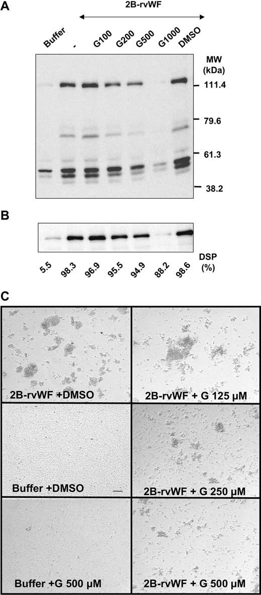
In order to further understand the relationship between SIPA and tyrosine phosphorylations, we investigated the effect of the tyrosine kinase inhibitor genistein on SIPA in the presence of 2B-rVWF. Genistein inhibited in a dose-dependent manner protein tyrosine phosphorylations (Figure 6A) and tyrosine phosphorylation of p125FAK (Figure 6B).
Effect of genistein on protein tyrosine phosphorylation and FAK phosphorylation. Washed platelets (2.8 × 10⁸/mL) were preincubated without (–) or with genistein (G; 100 μM-1000 μM) or vehicle (DMSO) for 30 minutes at 37°C, prior to exposure to 2B-rVWF (0.7 μg/mL) for 4.5 minutes at 37°C under high shear rates. Buffer indicates samples without 2B-rVWF. Total cell lysates were analyzed by immunoblotting with antiphosphotyrosine antibodies (4G10 and PY20) upon protein separation on a 8% polyacrylamide gel (A). Membranes were then stripped and reblotted with anti–phospho-FAK polyclonal antibody (B). DSP are indicated on the bottom part in the corresponding samples. In separate experiments, platelets were preincubated with genistein at the indicated concentration, exposed to shear, and fixed with PFA. An aliquot was deposited in microtiter plates for microscopic observation of aggregates. Images were recorded following sedimentation in the wells. Bar represents 50 μm (C). Results are representative of 3 experiments.
Effect of genistein on protein tyrosine phosphorylation and FAK phosphorylation. Washed platelets (2.8 × 10⁸/mL) were preincubated without (–) or with genistein (G; 100 μM-1000 μM) or vehicle (DMSO) for 30 minutes at 37°C, prior to exposure to 2B-rVWF (0.7 μg/mL) for 4.5 minutes at 37°C under high shear rates. Buffer indicates samples without 2B-rVWF. Total cell lysates were analyzed by immunoblotting with antiphosphotyrosine antibodies (4G10 and PY20) upon protein separation on a 8% polyacrylamide gel (A). Membranes were then stripped and reblotted with anti–phospho-FAK polyclonal antibody (B). DSP are indicated on the bottom part in the corresponding samples. In separate experiments, platelets were preincubated with genistein at the indicated concentration, exposed to shear, and fixed with PFA. An aliquot was deposited in microtiter plates for microscopic observation of aggregates. Images were recorded following sedimentation in the wells. Bar represents 50 μm (C). Results are representative of 3 experiments.
Preincubation of platelets with genistein did not result in a significant variation of the percent DSP from 98% in the absence of genistein, to 88% in the presence of 1000 μM genistein (Figure 6B). However, microscopic observation of platelet aggregates showed that the size of aggregates was reduced by genistein in a dose-dependent manner (Figure 6C). It is interesting to note that 2B-rVWF–dependent aggregate formation at a high shear rate was reversible when the exposure to shear was discontinued. Indeed, the fixation step by paraformaldehyde (PFA) was required to prevent loss of aggregates (data not shown).
These results suggest that in conditions where genistein efficiently blocked activation of tyrosine kinases involved in p125FAK phosphorylation, it was able to decrease the size of aggregates resulting from 2B-rVWF–dependent SIPA at a high shear rate.
In separate experiments, we determined the effect of inhibitors of ADP on SIPA and p125FAK phosphorylation in samples exposed to high shear and 2B-rVWF. We found that apyrase or A2P5P was without effect on SIPA and p125FAK phosphorylation (data not shown). These results suggest that 2B-rVWF–dependent SIPA and tyrosine phosphorylations were unlikely to involve ADP secretion from dense granules, but as it was obtained using washed platelets, may differ from studies in whole blood or PRP showing a role for ADP receptors in SIPA.35
Effect of αIIbβ3-VWF blockers on SIPA and tyrosine phosphorylations induced by 2B-rVWF at high shear rates
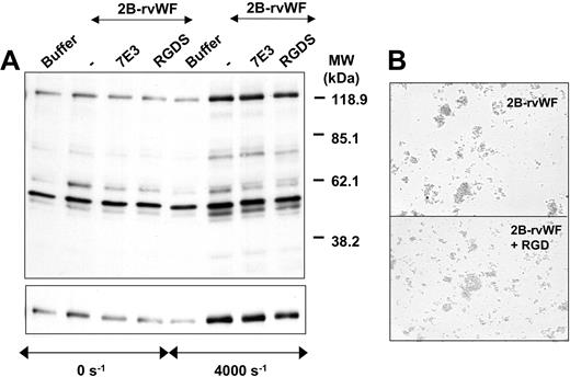
We and others have previously established that SIPA at high shear rates involves partially an interaction between plasma-derived VWF and αIIbβ3.12, 13, 14 In the present study, using 2B-rVWF at 4000 s–1 we found a different property of established inhibitors of VWF-αIIbβ3 interaction. Indeed, DSP, which started at 97.3% ± 1.2% in the absence of inhibitor, remained unchanged at values of 95.8% ± 1.6% in the presence of 7E3 (20 μg/mL), and of 96.1% ± 1.8% in the presence of RGDS (1 mM). Accordingly, we found no inhibitory effect of MoAb 7E3 or RGDS on tyrosine phosphorylations and p125FAK phosphorylation (Figure 7A). We also showed that 7E3 and RGDS peptide did not influence tyrosine phosphorylation in the presence of 2B-rVWF and in the absence of shear (Figure 7A). However, we verified that the latter blockers were still able to block thrombin-induced tyrosine phosphorylations, as reported.33,36 In separate experiments, kistrin, an RGD-containing cystein-rich peptide and a potent antagonist of αIIbβ3,28 was studied for its effect on 2B-rVWF–dependent SIPA and on tyrosine phosphorylation. In agreement with the absence of effect by RGDS, no inhibition was found on percent of DSP and tyrosine phosphorylation (not shown). Microscopic observation of platelet aggregates which were formed in the presence of 2B-rVWF at a high shear rate did not indicate any difference in the size of aggregates in the presence of 50 nM kistrin (Figure 7B). Accordingly, when we measured PAC-1 binding to platelets exposed to high shear in the presence of 2B-rVWF, we found less than 3% platelets bound to PAC-1, while 35% of platelets were positive for PAC-1 after thrombin-stimulation (data not shown).
Inhibitory effect of αIIbβ3 inhibitors on SIPA-associated protein tyrosine phosphorylation and p125FAK phosphorylation. (A) Platelets (2.8 × 10⁸/mL) were preincubated with MoAb 7E3 (20 μg/mL) or RGDS peptide (1 mM) for 5 minutes at room temperature, prior to exposure to 2B-rVWF (0.7 μg/mL) for 4.5 minutes at 37°C under high shear rates. Buffer indicates samples without 2B-rVWF. Lysates were prepared as described in “Materials and methods,” separated by an 8% polyacrylamide gel, transferred to nitrocellulose membrane, and blotted with antiphosphotyrosine MoAbs 4G10 and PY20 (top). Membranes were then stripped and reprobed with anti–phospho-FAK polyclonal antibody (bottom). Data are representative of 3 experiments. (B) In separate experiments, platelets and 2B-rVWF, in the absence (top) or the presence (bottom) of the RGD-containing disintegrin kistrin at 50 nM, were exposed to shear and fixed with PFA. An aliquot was deposited in microtiter plates for microscopic observation of aggregates, as described in the legend to Figure 6C.
Inhibitory effect of αIIbβ3 inhibitors on SIPA-associated protein tyrosine phosphorylation and p125FAK phosphorylation. (A) Platelets (2.8 × 10⁸/mL) were preincubated with MoAb 7E3 (20 μg/mL) or RGDS peptide (1 mM) for 5 minutes at room temperature, prior to exposure to 2B-rVWF (0.7 μg/mL) for 4.5 minutes at 37°C under high shear rates. Buffer indicates samples without 2B-rVWF. Lysates were prepared as described in “Materials and methods,” separated by an 8% polyacrylamide gel, transferred to nitrocellulose membrane, and blotted with antiphosphotyrosine MoAbs 4G10 and PY20 (top). Membranes were then stripped and reprobed with anti–phospho-FAK polyclonal antibody (bottom). Data are representative of 3 experiments. (B) In separate experiments, platelets and 2B-rVWF, in the absence (top) or the presence (bottom) of the RGD-containing disintegrin kistrin at 50 nM, were exposed to shear and fixed with PFA. An aliquot was deposited in microtiter plates for microscopic observation of aggregates, as described in the legend to Figure 6C.
Finally, platelets stimulated with ADP (1 μM) were able to aggregate in the presence of purified 2B-rVWF; this aggregation was inhibited by RGDS or MoAb 7E3, suggesting that 2B-rVWF exhibited a functional binding site toward activated αIIbβ3. Indeed, this low ADP concentration was not sufficient to induce a release from alpha granules, ruling out that aggregation involved released fibrinogen or VWF (data not shown). Altogether, these results suggest that αIIbβ3 engagement was not required in 2B-rVWF–induced tyrosine phosphorylations and p125FAK phosphorylation at a high shear rate.
2B-rVWF–dependent tyrosine phosphorylations are independent of PI 3-kinase
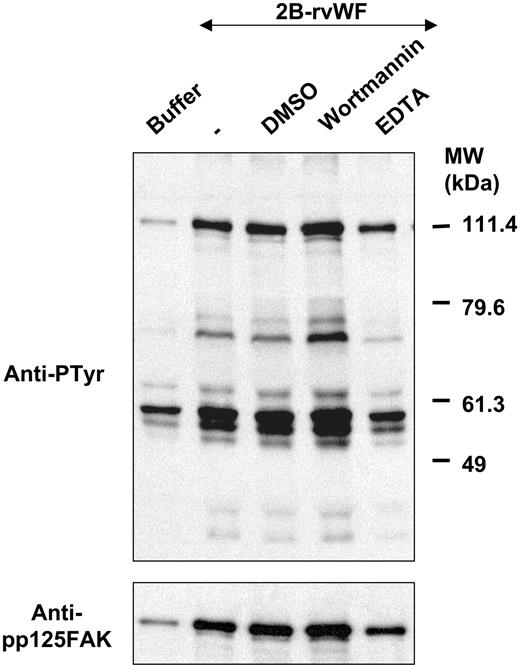
Previous studies have demonstrated the requirement of PI 3-kinase activation and calcium flux in mechanisms resulting from the interaction between GPIb and VWF and leading to αIIbβ3 activation.22 In order to elucidate a potential role of PI 3-kinase, we have tested the effect of wortmannin, a potent inhibitor of PI 3-kinase.
At 4000 s–1, shear-induced tyrosine phosphorylation (Figure 8, top panel) as well as p125FAK phosphorylation (Figure 8, bottom panel) were unaffected by wortmannin. In contrast, EDTA was able to decrease both tyrosine phosphorylation and p125FAK phosphorylation, indicating a calcium-dependent effect. Wortmannin as well as EDTA were without effect on SIPA (data not shown). As positive control, these blockers, as well as 1 mM RGDS peptide, inhibited thrombin-induced platelet tyrosine phosphorylation and p125FAK phosphorylation (data not shown). Thus, these results suggest that 2B-rVWF–dependent tyrosine phosphorylations and p125FAK phosphorylation under high shear rates did not involve PI 3-kinase activation.
Influence of PI 3-kinase activation and calcium chelator on shear-induced tyrosine phosphorylation. Washed platelets (2.8 × 10⁸/mL) were preincubated with either wortmannin (100 nM) or vehicle (DMSO) for 15 minutes at 37°C, or EDTA (2 mM) for 5 minutes at room temperature. Platelets were submitted to high shear rates for 4.5 minutes at 37°C in the presence of 2B-rVWF (0.7 μg/mL), lysed, and separated on an 8% polyacrylamide gel. Immunoblotting was performed using antiphosphotyrosine MoAbs 4G10 and PY20 (top panel), stripped, and reprobed with anti–phospho-FAK polyclonal antibody (bottom panel). Results are representative of 3 experiments.
Influence of PI 3-kinase activation and calcium chelator on shear-induced tyrosine phosphorylation. Washed platelets (2.8 × 10⁸/mL) were preincubated with either wortmannin (100 nM) or vehicle (DMSO) for 15 minutes at 37°C, or EDTA (2 mM) for 5 minutes at room temperature. Platelets were submitted to high shear rates for 4.5 minutes at 37°C in the presence of 2B-rVWF (0.7 μg/mL), lysed, and separated on an 8% polyacrylamide gel. Immunoblotting was performed using antiphosphotyrosine MoAbs 4G10 and PY20 (top panel), stripped, and reprobed with anti–phospho-FAK polyclonal antibody (bottom panel). Results are representative of 3 experiments.
Discussion
The interaction between GPIb and VWF plays a critical role in platelet adhesion and aggregation under high shear rates. Signals resulting from this interaction are required to maintain stable platelet interaction with VWF.11,14,22,37 In the present study we describe tyrosine phosphorylations in a SIPA model allowing to show predominantly the contribution of mutated 2B-rVWF binding to GPIb. There are 2 main findings described in this study: (1) the interaction between GPIb and 2B-rVWF at a high shear rate induced p125FAK phosphorylation and (2) the involvement of αIIbβ3 integrin was not required for 2B-rVWF–dependent SIPA and p125FAK phosphorylation.
Type 2B von Willebrand disease (VWD) is a bleeding disorder characterized by the ability of mutated VWF to bind platelet GPIb at lower ristocetin concentrations than normal VWF does.38 Mutations are localized in the A1 domain of VWF, which contains the binding site for GPIb and extends from amino acids 497 to 716. Recent structural studies of the GPIbα amino-terminal domain and its complex with the VWF A1 domain have shed a new light on the mechanism of these gain-of-function mutations related to bleeding disorders. These mutations are located on an amino-terminus extension, which in the wild-type A1 normally covers an important contact site of GPIb, and should be dislodged for maximal binding of GPIb. Mutations would favor the bound conformation at this GPIb contact site, and presumably shear could increase the exposure of the second binding site of GPIb.39,40 We show that 2B-rVWF induced SIPA in a shear-dependent manner from 200 s–1 to 4000 s–1, an effect that was abrogated in the presence of MoAbs blocking VWF-GPIb binding. We have previously established that 2B-rVWF–dependent SIPA is closely correlated with 2B-rVWF ability to bind GPIb at low ristocetin concentrations.24 In the present study, we extend our previous data concerning the important role of GPIb in promoting SIPA by demonstrating a dose-dependent response of SIPA with increasing concentrations of purified 2B-rVWF.
The bleeding disorder of type 2B VWD is paradoxical, since individuals with increased interaction between VWF and GPIb could be expected to display increased thrombus formation. Although little is known about the mechanism of thrombus formation in type 2B VWD, a heterogenous defect in thrombus growth has been recently demonstrated in perfusion studies on collagen. Indeed, 2 of 4 patients' blood leads to an impaired thrombus generation measured by epifluorescence microscopy, whereas confocal microscopy, which required fixation of the blood sample, allowed us to identify in all patients' blood a homogenous defect in thrombus spatial growth.41 It is our hypothesis that this requirement for fixation is in favor of reversible aggregate formation in type 2B VWD. Indeed, we have observed the formation of unstable aggregates induced by type 2B VWD at a high shear rate, since in the absence of PFA, aggregates were no longer seen over time after cessation of the shear. Interestingly, we could extend this observation to Glanzmann thrombasthenia type 1 platelets. Indeed, aggregates were formed in the presence of 2B-rVWF at 4000 s–1, and these aggregates were unstable in the absence of fixation by PFA (data not shown).
A key question raised by the present study on the pathophysiologic mechanism is the possible lack of involvement of αIIbβ3 activation in SIPA. We establish that under high shear rates, 2B-rVWF–dependent SIPA was unaffected by RGDS peptide or MoAb 7E3, which block αIIbβ3 integrin function. In favor of our hypothesis that binding of 2B-rVWF to GPIb does not lead to αIIbβ3 activation, we found that PAC-1 binding to platelets exposed to 2B-rVWF at a high shear rate did not exceed 3%. To rule out a defect of the mutant, we have verified that 2B-rVWF exposes normally its functional binding site to αIIbβ3, since it was able to promote platelet aggregation at low ADP concentrations, unable to induce the release of VWF or fibrinogen. Our results showing the lack of inhibition of 2B-rVWF–dependent SIPA by αIIbβ3 blockers are in agreement with previous studies showing that using porcine VWF, which has a high affinity for human GPIb, thus mimicking the 2B VWD phenotype in humans, platelet aggregation was hardly affected by anti-αIIbβ3 MoAbs.42 Furthermore, a rVWF mutant that does not interact with αIIbβ3, RGGS-rVWF, induced higher thrombus formation than WT-rVWF at a high shear rate, but not at a low shear rate.43 Thus our results indicate that platelet aggregates induced by type 2B VWD at a high shear rate are formed without a need for αIIbβ3 activation, and are reversible in vitro when exposure to shear is discontinued. Therefore we propose that the absence of thrombotic complications in these patients and their paradoxical bleeding disorders are supported by our findings of a reversible aggregation induced by type 2B VWD at a high shear rate.
Having established a model that enables us to study signal transduction dependent on the interaction between GPIb and 2B-rVWF, we show an increase in tyrosine phosphorylations and in particular at positions 125 kd, 72 kd, and 64 kd at a high shear rate of 4000 s–1. No increased phosphorylation of platelet proteins (relative to the absence of shear) was observed at the intermediate shear rate of 200 s–1 despite a significant extent of SIPA (60% DSP) and the presence of small aggregates. Therefore, SIPA induced signaling pathways that resulted in different activated proteins from the 3 phosphorylated proteins of 97 kd, 95 kd, and 84 kd observed using strong agonists, which were dependent on stirring and aggregation.33,36 Indeed, these authors reported that these phosphorylated proteins were not seen in platelets in the presence of VWF and ristocetin.33 Therefore, we propose a model where SIPA induced by 2B-rVWF in high shear rate conditions results in protein tyrosine phosphorylation profiles distinct from those previously found specifically in the presence of large aggregates, and presumably linked to engagement of activated αIIbβ3.36 Interestingly, when SIPA was obtained at a high shear rate in the presence of 2B-rVWF, we observed tyrosine phosphorylations which were dependent on GPIb, since they were abolished in the presence of GPIb blockers. Other groups have reported an increase in protein tyrosine phosphorylations at positions 130 kd, 100 kd, 85 kd, 74 kd, 70 kd, 64 kd, 58 kd, and 40 kd in the presence of plasma-derived VWF and high shear rates.15,16 In the absence of shear, VWF-dependent platelet aggregation in the presence of nonphysiologic agonists (ristocetin or botrocetin) was also associated with increased protein tyrosine phosphorylations at position 64 kd or 72 kd.19,20,44 More precisely, p125FAK, p72Syk, PLCγ2, and SHIP were phosphorylated in the presence of ristocetin.19 However, phosphorylation of the latter 3 proteins was also strongly inhibited by MoAb IV.3 which inhibits FcγRII activation.19 Indeed, GPIb cross-linking with the FcγRII receptor could contribute to increased tyrosine phosphorylations, since a physical proximity of both receptors has been reported,45 and cross-linking of FcγRII with secondary antibodies triggers the tyrosine phosphorylation of multiple cellular proteins.46,47 We have studied the effect of MoAb IV.3 on SIPA induced by 2B-rVWF using blood samples from 6 different donors, but we were unable to find an inhibition of SIPA (data not shown). This result is in agreement with a previous finding that p125FAK phosphorylation was unaffected by MoAb IV.3,19 but differs from another one obtained with PRP exposed to shear of 10 000 s–1 during 60 seconds.48 Thus it remains to be clarified if tyrosine phophorylation of FAK is independent of FcγRII engagement, as shown in flow conditions in a model of platelet adhesion mediated by α2β1.49
We demonstrated that at 4000 s–1 increased SIPA and p125FAK phosphorylation were strongly dependent on 2B-rVWF interaction with GPIb, since MoAbs 6D1 and 328 completely blocked both events. Furthermore, whereas thrombin-induced p125FAK phosphorylation was completely inhibited by 1 mM RGDS and 7E3 in an aggregometer, these 2 inhibitors were unable to block p125FAK phosphorylations at 4000 s–1 in the presence of 2B-rVWF. Our results suggest that αIIbβ3 engagement was not required for both SIPA and p125FAK phosphorylation in the presence of 2B-rVWF under high shear rates. Our results are in agreement with previous data showing that RGDS did not inhibit ristocetin-induced platelet aggregation.50 However, they are at variance with those showing that SIPA-associated tyrosine phosphorylations were inhibited by RGDS, although there was no evidence of p125FAK phosphorylation.15,16 It has been shown that p125FAK phosphorylation is dependent on postoccupancy events in thrombin-stimulated platelets,51 or in platelets costimulated by epinephrine and by an anti-β3 LIBS antibody.34 However, there is also evidence that phosphorylation may occur independently of αIIbβ3 engagement, such as the p125FAK tyrosine phosphorylation in collagen-stimulated platelets involving GPVI.52 Furthermore, it has been shown that a peptide fused to the cytoplasmic tail of αIIb, which allows conditional clustering of αIIbβ3, could also promote FAK phosphorylation without fibrinogen binding to αIIbβ3.53 Platelets from patients with Glanzmann thrombasthenia showed similar low density lipoprotein–induced FAK phosphorylation as control platelets.54 Integrin-independent phosphorylation of p125FAK has also been reported in platelets activated using immobilized human immunoglobulin G (IgG), an event that depends on FcγRII.55
An intriguing finding was that p125FAK phosphorylation was detected in 2B-rVWF–dependent SIPA but not in WT-rVWF–dependent SIPA. This finding was observed whether using Val553Met or Met540MetMet 2B-rVWF, thus it was not related to a particular mutation responsible for the type 2B VWD phenotype. Several hypotheses could explain this discrepancy. First, p125FAK phosphorylation may correspond to a mechanism similar to a postaggregation event. Indeed, p125FAK phosphorylation may require the very high level of SIPA obtained at high shear rates and in the presence of 2B-rVWF, but not that obtained in the presence of WT-rVWF, which remained always lower. In that manner, the mechanism of p125FAK phosphorylation would be close to the relationship previously established between tyrosine phosphorylations and the intensity of platelet aggregation.36 Second, an activation of p125FAK kinase activity may be triggered by high shear rates and in the presence of 2B-rVWF, whereas a phosphatase activity may be down-regulated by 2B-rVWF and normally activated by WT-rVWF. A third hypothesis is that p125FAK phosphorylation involves different mechanisms in either WT-rVWF– or 2B-rVWF–dependent SIPA. We found that RGDS or 7E3 was without effect on 2B-rVWF–dependent p125FAK phosphorylation as well as SIPA. In contrast, we have previously reported that plasma-derived VWF-dependent SIPA was reduced 2-fold by inhibitors of αIIbβ3-VWF interaction, MoAb 9, or MoAb 10E5.12 Finally, in keeping with situations where SIPA reached 60% to 70% DSP without inducing p125FAK phosphorylation, we may also suggest that the p125FAK phosphorylation requires both a threshold DSP (eg, > 80%) and an additional signaling mechanism. In favor of this mechanism, we found that at a shear rate of 500 s–1 and 1000 s–1, 2B-rVWF–associated p125FAK phosphorylation was also significantly increased (data not shown).
Finally, we found that genistein inhibited p125FAK phosphorylation in a dose-dependent manner and decreased the size of aggregates, indicating that modifications of tyrosine phosphorylations were relevant for shear-induced aggregate formation induced by 2B-rVWF. In addition, we found that the shedding of microparticles and the procoagulant activity of platelets initiated by 2B-rVWF were increased in the presence of genistein (C.B.-L. and D.B., unpublished observation, October 2002). The contribution of the changes in tyrosine phosphorylations induced by 2B-rVWF at a high shear rate on the regulation of platelet procoagulant activity is presently unknown. To better understand how p125FAK phosphorylation obtained upon platelet activation by 2B-rVWF at a high shear rate was relevant in terms of platelet functional state, we found that genistein was able to lower the size of aggregates. This finding raises the question how p125FAK phosphorylation could induce larger aggregates independently of αIIbβ3. Although somewhat speculative, we like to propose an explanation for aggregate formation and downstream events associated to one or several signaling pathways directly involving GPIb, based on new structural findings. Indeed, crystallization of the VWF-A1 domain complex with the amino-terminal part of GPIb required the use of gain-of-function mutants GPIbMet239Val and A1-Arg543Gln, associated with platelet-type and type 2B von Willebrand disease. There were 2 contact sites involved, one depending on conformational changes in a beta-switch of GPIb and the other site requiring the dislodging of the termini of the A1 domain to uncover the site of interaction.39 Therefore, shear-induced platelet activation could involve conformational changes on the A1 domain, propagating to GPIb and allowing amplification of the A1-GPIb interaction, but this model will require further elucidation.
By comparing 2B-rVWF–dependent and thrombin-dependent signals, we suggest that increasing the affinity of VWF for GPIb could substitute for mechanisms of signal transduction depending on αIIbβ3 integrin engagement. This hypothesis is also supported by the finding that 2B-rVWF-dependent signal transduction did not involve PI 3-kinase activation, whereas it has been shown that platelet adhesion to VWF under a high shear rate leads to αIIbβ3 integrin activation through PI 3-kinase activation.22,56 We found that the interaction between 2B-rVWF and GPIb at high shear was able to induce FAK phosphorylation without requiring αIIbβ3 integrin engagement. This suggests that 2B-rVWF–dependent SIPA did not require tyrosine phosphorylations to maintain stable aggregation but induced signal transduction independently of αIIbβ3 integrin engagement. Similar results have been previously described using ristocetin as agonist.19
Altogether, our findings indicate that 2B-rVWF is able to induce p125FAK phosphorylation without affecting the 2B-rVWF–αIIbβ3 interaction. This effect involves different elements, the increased affinity of 2B-rVWF for GPIb, the high level of aggregate formation, the clustering of platelet GPIb, as well as pp125FAK as shown by the effect of genistein on the size of aggregates. This study provides additional evidence for the paradoxical bleeding disorder and instability of thrombi in patients with type 2B VWD, and are in favor of a unique mechanism whereby circulating aggregates are independent of 2B-rVWF interaction with αIIbβ3 integrin, and thus, do not lead to thrombus formation.
Prepublished online as Blood First Edition Paper, January 23, 2003; DOI 10.1182/blood-2002-06-1879.
Supported by EC Biomed No. CT98-3517 (D.B.). M.M. was supported by the French Society of Atherosclerosis.
The publication costs of this article were defrayed in part by page charge payment. Therefore, and solely to indicate this fact, this article is hereby marked “advertisement” in accordance with 18 U.S.C. section 1734.
We are grateful to Dr B. Coller (The Rockefeller University, New York, NY) for the gift of monoclonal antibodies 6D1 and 7E3, and Drs L. Hilbert and C. Mazurier (LFB, Lille, France) for the kind gift of plasmid coding for Val553Met. The help of G. Rastegar-lari in the production of stable cell lines expressing WT- and 2B-rVWF is gratefully acknowledged.


This feature is available to Subscribers Only
Sign In or Create an Account Close Modal