Vascular endothelial growth factor (VEGF) is a major growth factor for developing endothelial cells (ECs). Embryonic lethality due to haploinsufficiency of VEGF in the mouse highlighted the strict dose dependency of VEGF on embryonic vascular development. Here we investigated the dose-dependent effects of VEGF on the differentiation of ES cell–derived fetal liver kinase 1 (Flk-1)/VEGF receptor 2+ (VEGFR2+) mesodermal cells into ECs on type IV collagen under a chemically defined serum-free condition. These cells could grow even in the absence of VEGF, but differentiated mostly into mural cells positive for α-smooth muscle actin. VEGF supported in a dose-dependent manner the differentiation into ECs defined by the expression of VE-cadherin, platelet-endothelial cell adhesion molecule 1 (PECAM-1)/ CD31, CD34, and TIE2/TEK. VEGF requirement was greater at late than at early phase of culture during EC development, whereas response of VEGFR2+ cells to VEGF-E, which is a virus-derived ligand for VEGFR2 but not for Flt-1/VEGFR1, was not dose sensitive even at late phase of culture. Delayed expression of VEGFR1 correlated with increased dose dependency of VEGF. These results suggested that greater requirement of VEGF in the maintenance than induction of ECs was due to the activity of VEGFR1 sequestering VEGF from VEGFR2 signal. The chemically defined serum-free culture system described here provides a new tool for assessing different factors for the proliferation and differentiation of VEGFR2+ mesodermal cells.
Introduction
Vascular endothelial growth factor (VEGF), also known as vascular permeability factor,1-3 has been implicated as the major growth factor for developing endothelial cells (ECs). Targeted disruption of the VEGF gene in mice did not only show the important role for VEGF in embryonic vascular development, but also presented embryonic lethality due to haploinsufficiency of VEGF.4,5 Carmeliet et al4 indeed emphasized that VEGF deficiency constituted the most severe haploid-insufficient phenotype reported to date. Moreover, deletion of a single VEGF allele in collagen2a1-Cre-expressing cells was reported to result in embryonic lethality around E10.5.6 These studies highlighted the strict dose-dependent effects of VEGF on embryonic vascular development.
Fetal liver kinase 1 (Flk-1)/VEGF receptor 27 (VEGFR2) is one of the receptors of VEGF.8 The role for VEGFR2 in the development of ECs and hematopoietic cells (HCs) has been intensively studied as VEGFR2 is a receptor tyrosine kinase expressed by a subset of mesodermal cells9,10 and gene-targeting studies showed that VEGFR2 was essential for the development of ECs and HCs during embryogenesis.11,12 Consistent with the in vivo studies, we and another group previously reported that VEGFR2+mesodermal cells derived from mouse embryonic stem (ES) cells or isolated from the early chicken gastrula could differentiate into ECs and HCs in vitro.13,14 The blast colony-forming cells expressing VEGFR2 derived from ES cells were reported to give rise to both lineages.15 In addition, we demonstrated that VEGFR2+ cells derived from ES cells could differentiate into mural cells such as pericytes and vascular smooth muscle cells.16
Fms-like tyrosine kinase (Flt-1)/VEGFR117 is another receptor of VEGF.18 The expression of VEGFR1 is first turned on in the extraembryonic mesoderm and is eventually confined to ECs.19,20 Targeted disruption of VEGFR1 gene in mice resulted in embryonic lethality with overpopulation of ECs, and chimeric analysis using VEGFR1 null mutant ES cells showed that the EC development was affected in a cell nonautonomous manner.21,22 Moreover, VEGFR1 lacking the tyrosine kinase domain is sufficient for normal vascular development in mice.23 These studies indicated that a major function of VEGFR1 during embryogenesis is to regulate VEGFR2 signal negatively by sequestering VEGF rather than to transduce VEGFR1 signal itself. This was also suggested by the biochemical analysis showing that the affinity of VEGFR1 to VEGF is at least 10-fold higher than that of VEGFR2, but its tyrosine kinase activity is 10-fold weaker than that of VEGFR2.24
To investigate the dose-dependent effects of VEGF on EC development, the in vitro cellular events of VEGFR2+ mesodermal cells from ES cells or from chicken embryos were analyzed in the presence of various doses of VEGF.14,25 By using a feeder cell–dependent culture system, we have demonstrated the dose-dependent effects of VEGF on cellular events of VEGFR2+ mesodermal cells. However, because VEGF did not support the growth of VEGFR2+ mesodermal cells on a certain feeder cell layer, the cellular events on the feeder cell layer were induced by VEGF in combination with undefined molecules derived from animal sera and feeder cells.25 Establishment of a culture system in a defined culture medium without feeder cells has been awaited to further address the issue on the response of VEGFR2+ mesodermal cell to VEGF.
Here we report that ES cell–derived VEGFR2+mesodermal cells could grow and differentiate on type IV collagen–coated dishes under a chemically defined serum-free condition. In the serum-free culture, sorted VEGFR2+ cells could differentiate in a VEGF dose-dependent manner into ECs defined by the expression of vascular endothelial–cadherin (VE-cadherin),26 platelet–endothelial cell adhesion molecule 1 (PECAM-1)/CD31,27 CD34,28 and tyrosine kinase with immunoglobulin and endothelial growth factor (EGF) homology domains 2/tunica interna endothelial cell kinase (TIE2/TEK).29 VEGF requirement in culture of VEGFR2+ mesodermal cells was greater in the maintenance than induction of ECs, which correlated with the delayed expression of VEGFR1. The chemically defined serum-free culture system described here provides a new tool for assessing different factors for the proliferation and differentiation of VEGFR2+ mesodermal cells.
Materials and methods
Monoclonal antibodies
Monoclonal antibodies (MoAbs) for murine E-cadherin (ECCD2), murine VEGFR2 (AVAS12), and murine VE-cadherin (VECD1) were described previously.10,30,31 These MoAbs were prepared and labeled in our laboratory. Biotinylated MoAb for murine TIE2 (TEK4)32 was kindly provided by Dr Toshio Suda (Kumamoto University School of Medicine, Japan). MoAb for murine VEGFR1 (5B12; characterization will be reported elsewhere) was kindly provided by Drs Dan Hicklem and Peter Bolen (Imclone Systems, New York, NY) and was labeled with biotin in our laboratory. Fluorescein isothiocyanate (FITC)–conjugated MoAb for murine PECAM-1 (Mec13.3) and FITC-conjugated MoAb for murine CD34 (RAM34) were purchased from Pharmingen (San Diego, CA). MoAb for murine α-smooth muscle actin 1A4 was purchased from Sigma (St Louis, MO).
Cell culture
An embryonic stem (ES) cell line, CCE,33was maintained as described previously.13VEGFR2+ E-cadherin–negative cells were generated from ES cells cultured for 4 days on type IV collagen–coated dishes (Becton Dickinson Labware, Bedford, MA) in α minimal essential medium (MEM; Gibco, Grand Island, NY) supplemented with 10% fetal calf serum (FCS; Gibco) and 5 × 10⁻⁵ M 2-mercaptoethanol (2-ME; Merck, Darmstadt, Germany) in the absence of leukemia inhibitory factor.
To induce the differentiation of VEGFR2+ mesodermal cells under a chemically defined condition, VEGFR2+ cells were sorted and recultured on type IV collagen–coated dishes in RPMI 1640/Dulbecco modified Eagle medium (DMEM)/F12-based defined medium SFO3 (Sanko Junyaku, Chiba, Japan)34 containing 0.1% bovine serum albumin (BSA; Sigma) and 5 × 10⁻⁵ M 2-ME, which we term SFO3 medium in this study. To assess the effects of growth factors, sorted VEGFR2+ cells were cultured on type IV collagen–coated dishes in SFO3 medium in the presence of human VEGF (R & D Systems, Minneapolis, MN) or VEGF-E.35
Flow cytometry and cell sorting
After 4 days for ES cell differentiation, cultured cells were harvested by incubating with cell dissociation buffer (Gibco). The harvested cells were incubated in mouse serum for 20 minutes on ice to block the nonspecific antibody binding, incubated with allophycocyanin (APC)-conjugated AVAS12 and FITC-conjugated ECCD2 for 15 minutes on ice. Cells were washed with Hanks balanced salt solution (Gibco) containing 1% BSA (Seikagaku Kogyo, Tokyo, Japan) and 0.05% sodium azide (Wako Chemical, Osaka, Japan). Living VEGFR2+E-cadherin-negative cells excluding propidium iodide (Sigma) were sorted by FACS Vantage (Becton Dickinson).
After 4 days for ES cell differentiation or 2 to 4 days for VEGFR2+ cell differentiation, cultured cells were harvested and stained with a mixture of MoAbs containing APC-conjugated AVAS12, FITC-conjugated Mec13.3, FITC-conjugated RAM34, biotinylated VECD1, biotinylated TEK4, or biotinylated 5B12 and developed by streptavidin-conjugated R-phycoerythrin (Gibco). Cells were analyzed by FACS Vantage while being gated to exclude small debris, dying cells, and other sources of background interference.
Double immunostaining for smooth muscle cell and endothelial cell markers
The cultured VEGFR2+ cells were fixed in situ by 5% dimethyl sulfoxide (Nacalai Tesque, Kyoto, Japan) in methanol for 3 minutes at 4°C. After washing with phosphate-buffered saline (PBS), 2% skim milk in PBS was incubated as a blocking solution for 1 hour at room temperature. The fixed dishes were incubated with 1A4 and VECD1 MoAbs overnight at 4°C, followed by incubating with horseradish peroxidase (HRP)–conjugated antimouse immunoglobulin G (IgG; Zymed Laboratories, San Francisco, CA) and alkaline phosphatase (ALP)-conjugated antirat IgG (Jackson Immunoresearch Laboratories, West Grove, PA) for 1 hour at room temperature. After each step, the cultured cells were washed 3 times with PBS containing 0.05% Tween 20 (Wako Chemical). Cells were visualized by using first 3,3′-diaminobenzidine (DAB) substrate (Dojindo, Kumamoto, Japan) for HRP staining and next nitroblue tetrazolium/5-bromo-4-chloro-3-inodolyl phosphate (NBT/BCIP) substrate (Boehringer Mannheim, Germany) for ALP staining. Endogenous HRP or ALP activity was blocked by hydroperoxide (Wako Chemical) or 2 mM levamisole (Sigma), respectively.
Quantification of VEGFR1 mRNA
Cells were directly sorted into tubes containing Trizol (Sigma) and mRNA was extracted according to the manufacturer's protocol. First-strand cDNA was synthesized from 2 μg total RNA using Superscript First-Strand Synthesis System for reverse transcriptase–polymerase chain reaction (RT-PCR; Gibco BRL). Quantification of VEGFR1 and VEGFR2 mRNA was performed by the LightCycler (Roche Diagnostics, Basel, Switzerland) using FITC- and LcRed-labeled hybriprobes. We measured mRNA of VEGFR1, VEGFR2, and GAPDH. Sense/antisense primer sets used for VEGFR1, VEGFR2, and GAPDH were 5′-ATTACATCCCCCTCAATGCC-3′/5′-GCTCAGATTCATCGTCCTGC-3′, 5′-AGGCTCCAACCAGACCAGT-3′/5′-CTAAGCAGCACCTCTCTCGT-3′, and 5′-TGAACGGGAAGCTCACTGG-3′/5′-TCCACCACCCTGTTGCTGTA-3′, respectively. Hybriprobes used for VEGFR1, VEGFR2, and GAPDH were 5′-CCCCTTGCTGAAGCGGTTCAC-3′-FITC/LcRed640-5′-TGGACTGAGACCAAGCCCAAGG-3′-P, 5′-TCCAGCGACGAGGCAGGACTTTTA-3′-FITC/LcRed640-5′-AGATGGTGGATGCTGCAGTTCACG-3′-P, and 5′-GCATCTTGGGCTACACTGAGGACC-3′-FITC/LcRed640-5′-GGTTGTCTCCTGCGACTTCAACAG-3′-P, respectively. A standard curve was prepared with 10 to 105copies of purified plasmids containing VEGFR1, VEGFR2, and GAPDH. The amount of mRNA was normalized against the GAPDH mRNA content.
Results
Serum-free culture of ES cell–derived VEGFR2+mesodermal cells
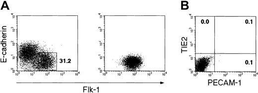
VEGFR2+ E-cadherin–negative mesodermal cells were generated from ES cells on type IV collagen–coated dishes by incubating for 4 days in the culture supplemented with FCS and were purified by fluorescence-activated cell sorting (Figure1A). At this stage, VEGFR2+cells did not express EC or HC markers such as PECAM-1, TIE2, VE-cadherin, CD34, or CD45 (Figure 1B and data not shown). Our previous studies demonstrated that the differentiation of ES cells through VEGFR2+ mesodermal cells into EC and HC lineages occurred in the medium supplemented with FCS without addition of exogenous VEGF.13,25 On the other hand, gene knock-out studies demonstrated that VEGF was essential for EC differentiation.4,5 It is thus likely that animal sera contain VEGF or its equivalents that supported the growth of VEGFR2+ cells in the culture described previously.10 This highlighted the importance of using defined culture conditions for assessing activities of soluble factors, particularly such a molecule as VEGF that exerts its effect in a dose-dependent manner.
Flow cytometric analysis of the in vitro differentiation of ES cells into VEGFR2+ mesodermal cells.
CCE ES cells were cultured on type IV collagen-coated dishes in the absence of leukemia inhibitory factor. (A, left panel) VEGFR2+ E-cadherin–negative cells were generated after 4 days for ES cell differentiation. In this experiment, the proportion of this population reached 31.2%. (A, right panel) The VEGFR2+ E-cadherin-negative cells were sorted and reanalyzed. (B) The expression of PECAM-1 and TIE2 in the purified VEGFR2+ cells were analyzed. The numbers indicate the percentage of cells that appeared in each quadrant.
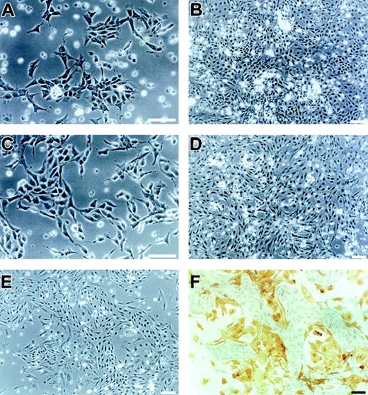
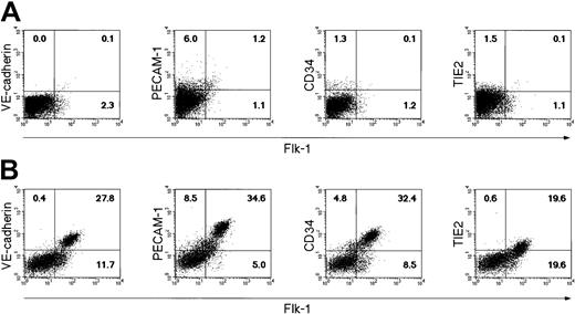
To start searching for the defined medium for EC differentiation, we selected first RPMI 1640/DMEM/F12-based defined medium SFO3, which we have developed for culturing hematopoietic stem cells.34 To test whether or not SFO3 could support differentiation of VEGFR2+ cells into ECs, sorted VEGFR2+ cells were cultured on type IV collagen–coated dishes in SFO3 medium. As observed under phase contrast microscopy, SFO3 supported growth and differentiation of these cells to some extent in the absence of VEGF (Figure 2A-B). However, the expressions of VEGFR2 as well as of other EC markers were barely observed in the cultured cells for 3 days (Figure3A). These results are consistent with previous in vivo studies showing absolute requirement of VEGF for the differentiation of ECs from the mesoderm.11 12
Differentiated cells in a chemically defined culture of ES cell–derived VEGFR2+ cells.
Sorted VEGFR2+ cells were cultured on type IV collagen–coated dishes under a chemically defined condition without (A-B) or with (C-F) 50 ng/mL VEGF. After 1 day (A,C), 3 days (B,D), or 2 days (E) of culture, differentiated cells were observed by phase-contrast microscopy. (F) Cells shown in panel E were doubly stained with MoAbs for VE-cadherin (purple) and α-smooth muscle actin (brown). Note that spindle-shaped or dendritic-shaped cells were positive for VE-cadherin or α-smooth muscle actin, respectively. Scale bars = 100 μm.
Flow cytometric analysis of the in vitro differentiation of sorted VEGFR2+ cells into EC lineage under a chemically defined condition.
Sorted VEGFR2+ cells were cultured on type IV collagen–coated dishes under a chemically defined condition without additional factors (A), or with 50 ng/mL VEGF (B). The expression of VE-cadherin, PECAM-1, TIE2, and CD34 was analyzed after 3 days of culture. The numbers indicate the percentage of cells that appeared in each quadrant.
Effect of VEGF on the differentiation of VEGFR2+cells under serum-free conditions
We next investigated whether or not the addition of VEGF to the culture of VEGFR2+ mesodermal cells could support their differentiation to ECs. Sorted VEGFR2+ cells were cultured on type IV collagen-coated dishes in SFO3 medium supplemented with 50 ng/mL VEGF. As observed under phase contrast microscopy, these cells could grow and form a population mostly with spindle-shaped cells (Figure 2C-D). In contrast to culture without VEGF, nearly 40% of cells after 3 days of culture with VEGF were positive for VEGFR2 and coexpressed VE-cadherin, PECAM-1, CD34, and TIE2 (Figure 3B), although the expression of EC markers in VEGFR2+ cell population was variable among markers. As we demonstrated previously,16mural cells positive for α-smooth muscle actin were other lineage cells derived from VEGFR2+ mesodermal cells. Almost all the cells negative for EC markers after 3 days of serum-free culture in the absence of VEGF were actually positive for α-smooth muscle actin (data not shown). Double staining with anti–VE-cadherin and anti–α-smooth muscle actin MoAbs showed that ECs with spindle shape and mural cells with dendritic shape were the major populations generated after 2 days of this serum-free culture in the presence of VEGF (Figure 2E-F). These results indicates that VEGF alone is sufficient to induce differentiation of VEGFR2+mesoderm cells into ECs, although involvement of other cellular factors generated from the cultured cells themselves should be taken into account.
Effects of VEGF concentration on EC differentiation
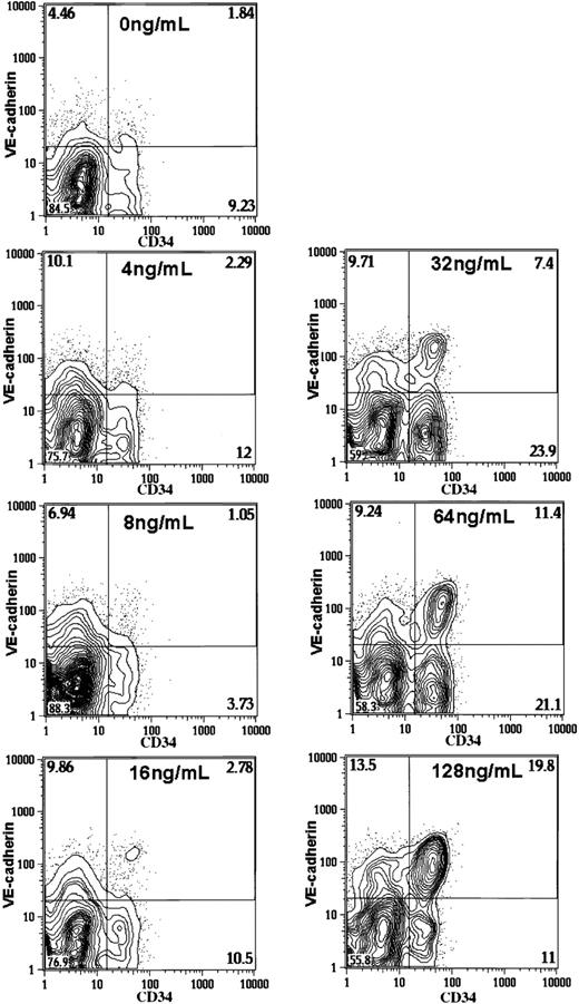
We next investigated whether or not VEGF-induced EC differentiation from VEGFR2+ mesodermal cells occurs in a VEGF dose-dependent manner. VEGFR2+ mesodermal cells were cultured in the presence of serially diluted VEGF, and the generation of VE-cadherin–positive CD34+ ECs was measured after 4 days of culture. Cell recoveries at day 4 of culture were comparable over those VEGF concentrations (1.3, 1.6, 1.4, 1.0, 1.1, 1.2, or 1.1 × 10⁶ at 0, 4, 8, 16, 32, 64, or 128 ng/mL VEGF, respectively). However, the proportion of VE-cadherin–positive CD34+ cells increased in a dose-dependent manner over the range of VEGF concentrations tested here (Figure4). Such a stoichiometric VEGF dependency in EC generation may be relevant to the previous observation that a 50% reduction of VEGF in the VEGF+/− embryos resulted in impaired vascular development.
Flow cytometric analysis of the in vitro differentiation of sorted VEGFR2+ cells into EC lineage under a chemically defined condition.
Sorted VEGFR2+ cells were cultured on type IV collagen–coated dishes under a chemically defined condition in the presence of serially diluted VEGF. The expression of VE-cadherin and CD34 was analyzed after 4 days of culture. The numbers indicate the percentage of cells that appeared in each quadrant.
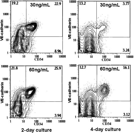
Interestingly, when the assay was performed after 2 days of culture, the stoichiometric VEGF dependency was less evident and the comparable number of VE-cadherin–positive CD34+ cells was generated both at 30 and 60 ng/mL VEGF (Figure5; 2-day culture; cell recoveries at 30 or 60 ng/mL VEGF was 8.4 or 9.0 × 10⁵/culture, respectively). On the other hand, VE-cadherin–positive C34+ cells could not be maintained for the next 2 days of culture with lower concentration of VEGF (Figure 5, 4 day culture; cell recoveries at 30 or 60 ng/mL VEGF was 8.0 or 7.6 × 10⁶/culture, respectively). These results suggest that stoichiometric VEGF dependency is more evident in the maintenance rather than induction of ECs.
Flow cytometric analysis of the in vitro differentiation of sorted VEGFR2+ cells into EC lineage under a chemically defined condition.
Sorted VEGFR2+ cells were cultured on type IV collagen–coated dishes under a chemically defined condition in the presence of 30 or 60 ng/mL VEGF. The expression of VE-cadherin and CD34 was analyzed after 2 or 4 days of culture. The numbers indicate the percentage of cells that appeared in each quadrant.
Role for VEGFR1 in VEGF-dependent EC differentiation
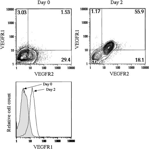
A line of evidence indicated that a major function of VEGFR1 during embryogenesis was to regulate VEGFR2 signal negatively by sequestering VEGF rather than to transduce the VEGFR1 signal itself. Because the in vivo expression of VEGFR1 was suggested to be later than that of VEGFR2 during EC differentiation,19 it is plausible that the failure of EC maintenance in the late phase of culture was due to the antagonistic activity of VEGFR1 that is newly synthesized in developing ECs. To quantify the change of mRNA level of VEGFR1 compared with that of VEGFR2 during EC maturation, VEGFR2+ mesodermal cells were cultured in the presence of 30 ng/mL VEGF and VEGFR2+ cells were purified from the culture after 1 and 2 days. The amount of VEGFR2 mRNA was comparable during EC maturation (17.19 ± 0.44, 17.55 ± 0.55, and 17.21 ± 0.24 copies/cell, for the initiation, day 1, and day 2 of culture, respectively). On the other hand, the amount of VEGFR1 mRNA expressed in the VEGFR2+ cells rapidly increased during first 2 days of culture (0.902 ± 0.02, 6.03 ± 0.09, and 11.9 ± 1.4 copies/cell, for the initiation, day 1 and day 2 of culture, respectively). This result is consistent with a previous report that VEGF up-regulates VEGFR-1 expression at the transcriptional level in ECs.36 To confirm that this increase in mRNA of VEGFR1 is represented by the surface expression at the protein level, we analyzed VEGFR1 expression in the VEGFR2+ population before and after the culture in the serum-free medium containing 30 ng/mL VEGF. As shown in Figure6, VEGFR1 expression increased during the culture (4.5-fold in this particular experiment). This increase was observed only in the VEGFR2+ population. The quantification of mRNA and protein levels showed that expression of VEGFR1 was later than that of VEGFR2 and the amount of VEGFR1 became close to that of VEGFR2 during in vitro EC maturation.
Flow cytometric analysis of the in vitro differentiation of sorted VEGFR2+ cells into EC lineage under a chemically defined condition.
Sorted VEGFR2+ cells were cultured on type IV collagen–coated dishes under a chemically defined condition in the presence of 30 ng/mL VEGF. The expression of VEGFR1 and VEGFR2 was analyzed before cell sorting (day 0) and after 2 days of culture (day 2). The numbers indicate the percentage of cells that appeared in each quadrant. The change of VEGFR1 expression in VEGFR2+population was shown in a histogram (lower panel).
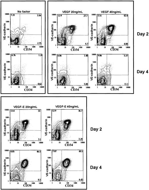
To investigate whether or not delayed expression of VEGFR1 was responsible for the stoichiometric relationship between dose of VEGF and maintenance of VE-cadherin–positive CD34+ cells, we took advantage of VEGF-E that is a virus-derived ligand for VEGFR2 but not for VEGFR1. VEGFR2+ cells were cultured in the presence or absence of VEGF or VEGF-E (Figure 7). Twenty or 40 ng/mL VEGF or VEGF-E induced comparable levels of VE-cadherin–positive CD34+ cells after 2 days of culture. When the assay was performed after 4 days of culture, VEGF at these doses failed to maintain VE-cadherin–positive CD34+ cells in vitro, whereas no marked reduction of this population was observed in the culture with VEGF-E. On the contrary, its proportion increased slightly during another 48 hours from day 2 of culture with VEGF-E. These results suggested that greater requirement of VEGF at late phase of EC differentiation was due to the delayed expression of VEGFR1 in VEGFR2+ cells.
Flow cytometric analysis of the in vitro differentiation of sorted VEGFR2+ cells into EC lineage under a chemically defined condition.
Sorted VEGFR2+ cells were cultured on type IV collagen–coated dishes under a chemically defined condition in the presence or absence of VEGF or VEGF-E. The expression of VE-cadherin and CD34 was analyzed after 2 or 4 days of culture. The numbers indicate the percentage of cells that appeared in each quadrant.
Discussion
The main purpose of this study is to establish an experimental system that allows assessment of the effects of soluble factors such as VEGF on the ECs and their progenitors. Fortunately, the serum-free culture medium SFO3, which we have developed for culturing hematopoietic stem cells, could also support the proliferation and differentiation of ECs from VEGFR2+ mesoderm cells.
VEGF has been implicated as the major growth factor for developing ECs. The number of VE-cadherin–positive CD34+ cells harvested after 3 to 4 days of incubation correlated well with the dose of VEGF under the defined culture condition. Because embryonic lethality of VEGF+/− mice suggested that vascular development is highly vulnerable to the decrease of VEGF level, the stoichiometric relationship between EC recovery and VEGF concentration observed in our defined culture condition may be relevant to this in vivo phenomenon. However, Carmeliet et al4 reported in the analysis of VEGF+/− embryos that the EC density and proliferation rate were normal during vasculogenesis but subsequent remodeling process was defective. This suggested that EC generation at the early phase of vascular development was supported equally over a wide range of VEGF dose. Indeed, when we measured the number of EC generated in the 2-day culture of VEGFR2+ mesodermal cells, EC recoveries were nearly the same from 8 to 64 ng/mL VEGF concentration. Thus, EC generation is not severely affected by the change of VEGF dose at the early phase of development in vitro as well as in vivo. In contrast, relatively strict dose dependency appeared between EC recovery and VEGF concentration in the late phase of culture. This observation in the late phase of our culture system may correlate with the report of Carmeliet et al4demonstrating that the process affected by the decreased dose ofVEGF gene is the remodeling of the vascular system, a process occurring at a later phase of vascular development.
What is the molecular basis underlying the delayed appearance of the stoichiometric pattern of VEGF dose responsiveness? One possibility is that the property of the signal transduction pathway in the downstream of VEGFR2 changes along with EC differentiation. This possibility, however, cannot explain our observation that the in vitro response of VEGFR2+ cells to VEGF-E, another ligand for VEGFR2, differed from that of VEGF in terms of dose dependency. It is of note that the level of VEGFR1 expression increases rapidly during first 2 days of culture in VEGFR2+ cells. Because VEGFR1 has been shown to play a role in antagonizing VEGFR2 during early embryogenesis, another possibility is that the increase of VEGFR1 expression level during EC differentiation is the reason for the shift of VEGF dose responsiveness. This possibility is strongly supported by the observation that response of VEGFR2+ mesodermal cells to VEGF-E that is not antagonized by VEGFR1 was not dose sensitive even at late phase of culture. It was reported that the expression of VEGFR1 was first turned on at the early primitive streak stage and that the expression level reached a maximum at about midsomite stage when the extraembryonic vascular plexus has already developed.20This kinetics of VEGFR1 expression is consistent with our results on ES cells cultured in vitro. A previous in vitro experiment also demonstrated that VEGF up-regulates VEGFR1 expression in ECs.36 Taken together, it is most likely that VEGF acts on VEGFR2+ mesodermal cells to induce the differentiation into ECs as well as to up-regulate the expression of VEGFR1 that consequently sequesters VEGF from VEGFR2 signal. To our knowledge, the present result would be the first case demonstrating the correlation between differential level of VEGFR1 expression and differential dose of VEGF requirement. It should be emphasized that this in vitro finding was enabled only by the establishment of the defined culture condition that supported EC differentiation.
Our experiment using a serum-free culture system clearly demonstrated that VEGF alone is sufficient for supporting the generation of ECs from VEGFR2+ mesoderm cells, whereas factors produced by cultured cells themselves may be also involved. We observed that ECs and mural cells were the major cell types in this culture. Because smooth muscle cells are known to produce VEGF in vitro,37we have to take the effects of VEGF family ligands produced in culture on VEGFR2+ cells into account. In the absence of VEGF, VEGFR2+ cells gave rise mostly to mural cells (data not shown). If the effects of VEGFs produced by these cells were significant, the growth factors could induce the differentiation of VEGFR2+ cells into ECs in a paracrine fashion. However, it was not the case. The stoichiometric VEGF dependency in EC generation shown here also suggested that VEGF added exogenously was a more significant growth factor than VEGFs produced endogenously in this culture.
We are well aware that the scheme drawn in the in vitro system is far simpler than what happens in the actual embryo. For example, VEGF-C38 binds to VEGFR2 and Flt4/VEGFR339but not to VEGFR1. It is likely that in the local region where VEGF-C is expressed together with VEGF, the VEGFR2 signal may remain less affected by the increase of VEGFR1 expression. It is also the case with VEGF-D40 in the human, whereas it may not be the case with mouse VEGF-D that does not bind to VEGFR2.41 On the other hand, in the situation where only VEGF is available, ECs are highly sensitive to a change of VEGF dose. Reconstitution of those signal systems in the response of ECs in culture should be essential for investigating the role of all these signals in EC behavior, and the defined culture system described here will provide a useful system for this purpose.
We thank Dr M. J. Evans for providing the CCE ES cell line, Dr A. Nagafuchi for ECCD2 MoAb, Dr N. Matsuyoshi for VECD1 MoAb, Dr T. Suda for TEK4 MoAb, and Drs D. Hicklem and P. Bolen for 5B12 MoAb. We also thank Dr M. Osawa for technical assistance and all the members in our laboratory for helpful discussions.
Prepublished online as Blood First Edition Paper, October 24, 2002; DOI 10.1182/blood-2002-01-0003.
Supported by grants from the Ministry of Education, Culture, Sports, Science, and Technology (12219209) and the Organization of Pharmaceutical Safety and Research, and the Ministry of Health, Labor, and Welfare of Japan. M.H. was a recipient of a postdoctoral fellowship from Tsumura and Company.
The publication costs of this article were defrayed in part by page charge payment. Therefore, and solely to indicate this fact, this article is hereby marked “advertisement” in accordance with 18 U.S.C. section 1734.
References
Author notes
Shin-Ichi Nishikawa, Department of Molecular Genetics, Graduate School of Medicine, Kyoto University, Shogoin-Kawaharacho 53, Sakyo-Ku, Kyoto, Japan; e-mail:snishika@virus.kyoto-u.ac.jp.

This feature is available to Subscribers Only
Sign In or Create an Account Close Modal