Abstract
The Kit (White) gene encodes the transmembrane receptor of stem cell factor/Kit ligand (KL) and is essential for the normal development/maintenance of pluripotent primordial germ cells (PGCs), hematopoietic stem cells (HSCs), melanoblasts, and some of their descendants. The molecular basis for the transcriptional regulation of Kit during development of these important cell types is unknown. We investigated Kit regulation in hematopoietic cells and PGCs. We identified 6 DNase I hypersensitive sites (HS1-HS6) within the promoter and first intron of the mouse Kit gene and developed mouse lines expressing transgenic green fluorescent protein (GFP) under the control of these regulatory elements. A construct driven by the Kit promoter and including all 6 HS sites is highly expressed during mouse development in Kit+ cells including PGCs and hematopoietic progenitors (erythroid blast-forming units and mixed colony-forming units). In contrast, the Kit promoter alone (comprising HS1) is sufficient to drive low-level GFP expression in PGCs, but unable to function in hematopoietic cells. Hematopoietic expression further requires the addition of the intronproximal HS2 fragment; HS2 also greatly potentiates the activity in PGCs. Thus, HS2 acts as an enhancer integrating transcriptional signals common to 2 developmentally unrelated stem cell/progenitor lineages. Optimal hematopoietic expression further requires HS3-HS6.
Introduction
Stem cells are traditionally considered to be either multipotent (eg, embryonic stem [ES] cells) or restricted in their differentiation potential (tissue stem cells). Reports on “transdifferentiation” and the discovery of multipotent adult progenitor cells in bone marrow, brain, and muscle have recently challenged this view.1-5 One central issue underlying the debate on developmental options of stem cells concerns the molecular mechanisms responsible for establishing and maintaining their transcriptional programs and decisions to differentiate. So far, few of the key regulatory genes active in stem cells and/or multipotent progenitors have been studied at the level of transcriptional regulation.6-17 Identification and comparison of several such genes will provide important insights into the transcriptional programs of stem and multipotent progenitor cells.
The Kit (White spotting locus) gene, encoding the transmembrane receptor of the cytokine stem cell factor/Kit ligand (KL), is an important regulator of proliferation/survival and/or migration of several stem cell types such as the primordial germ cells (PGCs),18-22 the multipotent hematopoietic stem cells (HSCs),20,23 the neural crest, and the intestinal Cajal cells.20 Null mutations in the Kit or the KL (Steel) gene result in severe hematopoietic and germ cell defects and in utero or perinatal death, whereas mutations that diminish Kit tyrosine kinase activity or KL production affect mainly hematopoiesis and the development of germ cells, melanocytes, and the intestinal Cajal cells.20 During mouse development Kit is expressed at a low level in pluripotent inner cell mass cells (and in epiblast-derived ES cells in culture)24,25 and, at relatively higher levels, in PGCs, early hematopoietic progenitors, and other cells.25 In adults, Kit is expressed in a variety of cell types including HSCs, immature hematopoietic progenitors and mast cells, oocytes, a subpopulation of male germ cells, and melanocytes.18-23,25
In the mouse embryo, hematopoietic progenitors are detected both extraembryonically in the yolk sac, after embryonic day 7 (E7), and intraembryonically, first in the para-aortic splanchnopleura and aorta-gonad-mesonephros (AGM) regions, then in fetal liver and vitelline vessels,26-28 and finally in bone marrow. The earlier hematopoietic cells (primitive hematopoietic cells) are morphologically and biologically distinct from the later cells (definitive hematopoietic cells). Definitive hematopoiesis is seeded by HSCs, which arise intraembryonically in the AGM region and in the vitelline vessels around E11.28-30 Immature precursors to definitive HSCs, however, have also been detected both intraembryonically31,32 and in the yolk sac32 at earlier stages of development. Murine PGCs first become visible around E7 in the extraembryonic mesoderm, then migrate through the allantois (E8) to the hindgut, from where they move to reach the gonadal ridges (E9.5-E11.5).22,33,34
An important characteristic of Kit is that its expression is common to, and critical for, various unrelated multipotent stem/progenitor cells, belonging to different lineages. To identify the Kit DNA regulatory elements responsible for this common expression, we studied the transcriptional regulation of Kit in early hematopoietic and germ cells. Fusing the promoter and enhancer elements from the first intron of the mouse Kit gene to a green fluorescent protein (GFP)-reporter gene, we obtained a construct that correctly recapitulates Kit expression throughout the development of both these lineages. Our results highlight the essential role of a DNA region, centered on a single DNase I hypersensitive (HS) site, for gene expression in 2 stem/multipotent lineages, PGCs and hematopoietic cells.
Materials and methods
DNase I hypersensitivity studies
Nuclei were prepared from exponentially growing cells according to Forrester et al.35 Aliquots were incubated for 5 minutes at 37°C either without or with DNase I (Boehringer-Mannheim, Mannheim, Germany) over a range of 0.2 to 10 μg/mL. gDNA was isolated and digested with appropriate enzymes and analyzed by Southern blotting. Kit+ cells were KL-dependent SV40T-immortalized hematopoietic cells from bone marrow and yolk sac,36,37 mel-c melanocytes,38 and CCE ES cells.
Kit-GFP constructs
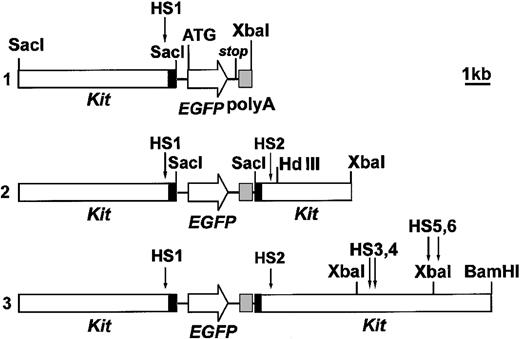
Mouse Kit genomic fragments were cloned from a λ Fix II genomic library (Stratagene, La Jolla, CA). An enhanced GFP (EGFP) gene carrying at its 5′ an HA epitope followed by a nuclear localization signal and, at its 3′, stop codons, and a polyA addition site, was used as a reporter. The coding sequence of the EGFP gene was fused, at its initial ATG, to the first ATG of the first exon (and 6.7-kb 5′ flanking sequence) of the Kit gene. Fusion was obtained through a linker inserted at the SacI site of the Kit first exon and including 26 nucleotides from the SacI site itself to the first ATG of the first Kit exon, appropriately modified to generate a NcoI site. The EGFP gene, cut by NcoI at its initial ATG, was ligated into the linker NcoI site, thus generating an in-frame Kit-EGFP fusion. This is construct 1. Construct 2 was obtained by adding, downstream to the EGFP polyA-addition site, the 3′ part of the first Kit exon downstream to SacI, and approximately 3.5 kb of the first intron. Construct 3 further included a 4.5-kb XbaI-BamHI fragment of the first intron. All constructs were flanked by SalI sites.
Transgenic mice
Stable transgenic lines of BDF1 mice were obtained by injecting plasmidfree fragments generated by SalI digestion, according to standard techniques.39 The number of integrated constructs varied between 2 and 20, as evaluated by Southern blot comparison of the intensities of hybridization to GFP reporter gene, between transgenic DNA and appropriate dilutions of the constructs.
Cell isolation
Cells were obtained from E9.5 AGM and E8.5 to 10.5 yolk sac by mechanical disaggregation. The morning when the vaginal plug was detected was taken as E0.5. PGCs were purified from AGM of E10.5 embryos using the mini magnetic-activated cell sorting (MACS) method,40 and from E11.5 gonadal ridges by the EDTA (ethylenediaminetetraacetic acid)-stab method.41
Immunohistochemistry
Cells were fixed using freshly prepared 4% paraformaldehyde in phosphate-buffered saline (PBS) for 10 minutes at room temperature. Prior to fixation, cells from gonadal ridges were attached onto a poly-l-lysine-coated coverslip. Alkaline phosphatase (AP) staining was performed as described.41 Immunostaining was performed by incubating the cells with solutions containing the primary antibody in PBS with 1% bovine serum albumin (BSA) for 1 hour at room temperature. Primary antibodies included mouse IgM TG-1 (kindly provided by Dr P. Donovan, Kimmel Cancer Center, Thomas Jefferson University, Philadelphia, PA), biotinconjugated rat anti-Kit antibody (Becton Dickinson, Heidelberg, Germany), and anti-HA epitope mouse monoclonal IgG 2a (Santa Cruz Biotechnology, Santa Cruz, CA). Secondary antibodies were antimouse-IgM-TRITC (Sigma, St Louis, MO) for TG-1, streptavidin-phycoerythrin (PE; Sigma) for anti-Kit, and antimouse IgG fluorescein isothiocyanate (FITC; Sigma) for HA. Incubation was 30 minutes at room temperature. For cryostat sections, E9.5 embryos were fixed in 4% paraformaldehyde for 15 minutes at 4°C, washed in PBS, immersed in OCT embedding matrix (Sakura, Zoeterwoude, The Netherlands), and frozen in liquid nitrogen vapor. Serial 7-μm sections were stained for AP activity and with anti-HA and TG-1 antibodies.
FACS analysis
Fetal liver cells from transgenic mice were obtained by disaggregation in PBS, stained with anti-Kit (45 minutes, 4°C), washed, incubated with streptavidin-PE (45 minutes, 4°C), and analyzed with a Becton Dickinson fluorescence-activated cell sorting (FACS) scan for both GFP and Kit fluorescence.
Sorting of GFP+ cells and colony assays
Fetal liver cells were sorted into a GFP+ (> 5 × 101 fluorescence units) and a GFP- population (< 2 × 101 fluorescence units), using FACS Vantage; adult bone marrow cells were sorted into a GFP- population, and 2 GFP+ populations of intermediate and high fluorescence, respectively. Cells were cultured in Iscove modified Dulbecco medium (IMDM) containing α-thioglycerol, methylcellulose, BSA, 5% fetal calf serum (FCS), iron-saturated transferrin, lecithin, oleic acid, cholesterol, 2 U/mL human recombinant erythropoietin, 10 ng/mL murine KL, and 10 ng/mL murine interleukin 3 (IL-3). Small erythroid colonies from erythroid colony-forming units (CFU-Es) were counted after 2 days of culture, and larger erythroid (BFU-E), mixed (CFU-mix), and granulocyte-macrophage (CFUGM) colonies were scored on days 7 to 14.42
Results
DNase I hypersensitive sites in Kit chromatin
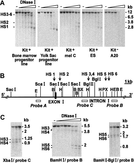
The mouse Kit gene is over 80-kb long and includes at least 21 exons; the first 2 exons are separated by a large (about 20 kb) intron.43 Inherited Kit expression defects due to distant upstream deletions suggested the existence of long-range-acting regulatory sequences.44,45 To additionally identify proximal regulatory elements we cloned about 10 kb of 5′ flanking sequences and the whole first exon and intron, and explored DNase I sensitivity of this DNA region in chromatin from Kit-expressing hematopoietic, melanocytic, and ES cells as well as nonexpressing hematopoietic cells. Digestion with EcoRI and hybridization with probe A (Figure 1A-B) causes the appearance of 2 bands (HS1, HS2) with DNA from Kit+ cells, ES cells, and melanocytes, and an additional band (HS3-HS6) with DNA from Kit+ hematopoietic cells. No bands are seen in Kit- cells (lymphoid A20, Figure 1A and other lines, not shown). Further experiments with other enzymes and probes B and C (Figure 1C) show that HS1 maps in the promoter within approximately 300 nucleotides upstream to the cap site, HS2 just downstream (500-700 nucleotides) to the first exon, whereas the hematopoietic-specific HS3-6 band splits into 4 different downstream sites (HS3-HS6; Figure 1C).
DNase I hypersensitivity of Kit 5′-flanking, exon I, and first intron region in chromatin from Kit-expressing and nonexpressing cells. (A) DNA from nuclei treated with DNase I was digested with EcoRI and hybridized to probe A (see panel B). HS sites are indicated. (B) Restriction map of the region analyzed. (C) Fine mapping of HS3 to HS6. Probes and restriction enzymes used are indicated. Molecular weight markers correspond to bands obtained by double digestion with EcoRI/XbaI, EcoRI/HindIII, and EcoRI/SacI of plasmids containing the cloned EcoRI fragment (A), run and hybridized in parallel to the gDNA. Similarly, double digestions (C) were XbaI/Bgl I, XbaI/Bgl II, and XbaI/HindIII for plasmids containing the cloned XbaI fragment; BamHI/BglII, BamHI/PstI (BamHI fragment); and BamHI/PstI, BamHI/XbaI BamHI/HindIII (BamHI/Bgl I fragment). The nucleotide sequence of the first exon/first intron of the Kit gene is reported in GenBank (NT_039306). Nucleotide 1 of the first intron corresponds to nucleotide 559 of this sequence; the positions of the HindIII (near HS2), Bgl II (near HS3-4) and XbaI (near HS5-6) sites correspond to nucleotides 1071, 4941, and 7047 of the first intron, respectively.
DNase I hypersensitivity of Kit 5′-flanking, exon I, and first intron region in chromatin from Kit-expressing and nonexpressing cells. (A) DNA from nuclei treated with DNase I was digested with EcoRI and hybridized to probe A (see panel B). HS sites are indicated. (B) Restriction map of the region analyzed. (C) Fine mapping of HS3 to HS6. Probes and restriction enzymes used are indicated. Molecular weight markers correspond to bands obtained by double digestion with EcoRI/XbaI, EcoRI/HindIII, and EcoRI/SacI of plasmids containing the cloned EcoRI fragment (A), run and hybridized in parallel to the gDNA. Similarly, double digestions (C) were XbaI/Bgl I, XbaI/Bgl II, and XbaI/HindIII for plasmids containing the cloned XbaI fragment; BamHI/BglII, BamHI/PstI (BamHI fragment); and BamHI/PstI, BamHI/XbaI BamHI/HindIII (BamHI/Bgl I fragment). The nucleotide sequence of the first exon/first intron of the Kit gene is reported in GenBank (NT_039306). Nucleotide 1 of the first intron corresponds to nucleotide 559 of this sequence; the positions of the HindIII (near HS2), Bgl II (near HS3-4) and XbaI (near HS5-6) sites correspond to nucleotides 1071, 4941, and 7047 of the first intron, respectively.
Expression of transgenic constructs in the hematopoietic system and in the germ cell line
To evaluate the role of these sites in Kit regulation we cloned the promoter in front of a nuclear EGFP gene, carrying an HA epitope (construct 1) and further added a fragment comprising HS2 (construct 2) or HS2 to HS6 (construct 3; Figure 2). All 3 constructs are similarly active in transfected undifferentiated ES cells (not shown). To investigate expression during development, we generated several transgenic mouse lines (8 with construct 1, 5 with construct 2, and 6 with construct 3; Table 1). The numbers of integrated constructs ranged between 2 and 20 for construct 1, 3 and 15 for construct 2, and 2 and 12 for construct 3.
Schematic representation of constructs 1, 2, and 3.EGFP not to scale. The indicated ATG corresponds to the first ATG of the Kit gene and is fused in frame (at NcoI site) to the open reading frame of the EGFP construct. Exon 1 (black box) is split by the Sac I cut.
Schematic representation of constructs 1, 2, and 3.EGFP not to scale. The indicated ATG corresponds to the first ATG of the Kit gene and is fused in frame (at NcoI site) to the open reading frame of the EGFP construct. Exon 1 (black box) is split by the Sac I cut.
Expression of Kit-GFP in transgenic mice: frequency of mouse lines expressing in various sites
. | Construct . | . | . | ||
|---|---|---|---|---|---|
| Expression sites* . | 1 . | 2 . | 3 . | ||
| Allantoid E8.0-8.5 | 1/4 | 2/2 | 3/3 | ||
| AGM E9.5 | 3/6 | 5/5 | 6/6 | ||
| Gonadal ridge/gonad E10.5-12 | 3/6 | 4/4 | 6/6 | ||
| Yolk sac E9.5-12 | 0/7 | 5/5 | 6/6 | ||
| Fetal liver E10.5-12 | 0/8 | 1/4 | 5/6 | ||
| Bone marrow | 0/6 | 1/5 | 6/6 | ||
| Ectopic | 5/8 | 1/5 | 1/5 | ||
Numbers show the proportion of mouse lines expressing the transgene in the indicated sites. The construct 3 line described in detail in this work was line 6 (2 integrated DNA copies). Construct 3 line 5 (8 construct copies) gave very similar results. All other lines had approximately 1% (line 4, 20 copies), 2% (line 3, 9 copies), and 10% to 15% (lines 1 and 2, 8-10 copies) positive cells in the bone marrow. Values in fetal liver ranged between 0% and 2% (lines 4 and 3, respectively), 5% to 15% (lines 1 and 2), and 50% to 65% (lines 5 and 6). Results in gonads, AGM, and yolk sac are qualitatively similar to those presented for line 6, although some lines (3 and 4) gave weaker fluorescence. For construct 2, line 5 (12 construct copies) is described. Other examined lines were negative in fetal liver and bone marrow, but all lines (25 construct copies) were significantly fluorescent in the other regions examined. The construct 1 lines gave clear, but weak, fluorescence in gonads and AGM (line 4, 5 copies [Figure 6], and line 2, 8 copies). Line 4 also gave expression in allantoid. Ectopic expression was mainly in the central nervous system, after E11.5; it was detected in the 5 expressing construct 1 lines, in construct 2, line 4, and in construct 3, line 4.
Not all lines were examined in all sites. Other sites, possibly representing physiologic expression, were peripheral nerve ganglia, epidermis (melanocytes?), and heart
As shown in Table 1, all 3 constructs are active in the developing germline (although construct 1 is weaker than constructs 2 and 3), whereas only constructs 2 and 3 are active in hematopoietic cells. Other types of expressing cells (melanocytes, neural crest, cranial ganglia, etc) were not studied in detail. Ectopic expression was rarely seen with constructs 2 and 3; with construct 1 it was seen more often, particularly as strong localized spots in the central nervous system. The level of expression was not related to the copy number of integrated construct.
We characterized in detail the expression pattern during development using one construct 3 mouse line (carrying 2 copies of the construct), one construct 2 line (12 copies), and 2 construct 1 lines (5 and 8 copies). The data obtained with constructs 2 and 3 are essentially superimposable, with only minor specific differences.
GFP-expressing cells in the AGM region
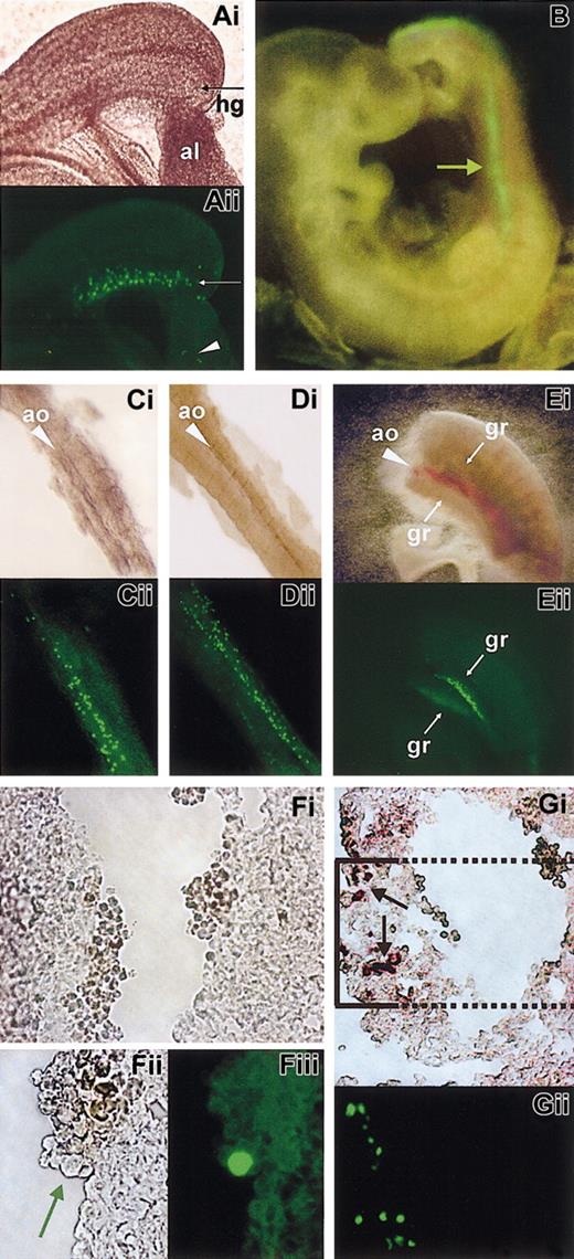
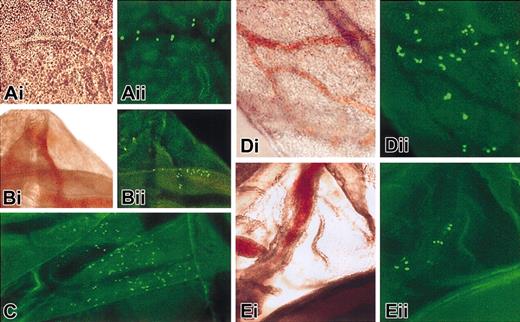
Using constructs 2 and 3, GFP+ cells apparently migrating from the allantois into the embryo are initially detected at E8 to 8.5 (Figure 3A). Later (E9.5-10), a fluorescent streak becomes visible in the dorsal posterior part of the embryo, corresponding to the gut or aorta (Figure 3B), where PGCs and hematopoietic progenitors, respectively, are located.
GFP expression of construct 3 in the allantoid, aorta, and gonadal ridges regions. Each panel shows the image in bright field (i) and fluorescence (ii). Panel B shows fluorescence only, and panels Fi and Fii are bright field images. (A) E8 allantoid. An arrow points to the hindgut region. An arrowhead in Aii points to a group of fluorescent cells in the allantoid. (B) E9.5 embryo. An arrow points to the fluorescent streak. (C-D) E9.5 dissected aortas with some adjacent mesenchyme. (E) lateral view of an embryo (E10.5) truncated below and above the AGM region. One of the fluorescent gonadal ridges is out of focus. (F) Cross-section of E9.5 aorta and magnification of a detail; note the large fluorescent cell in Fiii, shown by an arrow in Fii. (G) Cross-section of the aorta. (Gi) AP+ cells in the ventral part are indicated by arrows. The box indicates the area corresponding to the fluorescence image (enlarged) shown in panel Gii. (Gii) GFP (anti-HA) staining of the same region of the adjacent section. In the panels, ao indicates aorta; al, allantoid; gr, gonadal ridge; and hg, hindgut. Note that nontransgenic control embryos (and dissected organs) were completely nonfluorescent, even with exposure times of 90 seconds (versus 8-15 seconds as used here). Original magnifications are × 100 (A), × 20 (B), × 40 (C-E), × 100 (Fi), × 400 (Fii-iii), and × 100 (G).
GFP expression of construct 3 in the allantoid, aorta, and gonadal ridges regions. Each panel shows the image in bright field (i) and fluorescence (ii). Panel B shows fluorescence only, and panels Fi and Fii are bright field images. (A) E8 allantoid. An arrow points to the hindgut region. An arrowhead in Aii points to a group of fluorescent cells in the allantoid. (B) E9.5 embryo. An arrow points to the fluorescent streak. (C-D) E9.5 dissected aortas with some adjacent mesenchyme. (E) lateral view of an embryo (E10.5) truncated below and above the AGM region. One of the fluorescent gonadal ridges is out of focus. (F) Cross-section of E9.5 aorta and magnification of a detail; note the large fluorescent cell in Fiii, shown by an arrow in Fii. (G) Cross-section of the aorta. (Gi) AP+ cells in the ventral part are indicated by arrows. The box indicates the area corresponding to the fluorescence image (enlarged) shown in panel Gii. (Gii) GFP (anti-HA) staining of the same region of the adjacent section. In the panels, ao indicates aorta; al, allantoid; gr, gonadal ridge; and hg, hindgut. Note that nontransgenic control embryos (and dissected organs) were completely nonfluorescent, even with exposure times of 90 seconds (versus 8-15 seconds as used here). Original magnifications are × 100 (A), × 20 (B), × 40 (C-E), × 100 (Fi), × 400 (Fii-iii), and × 100 (G).
To better understand the location of the fluorescent cells, we dissected the dorsal aorta, with some adjacent tissue. At E9.5, most fluorescent cells are located in close proximity to the aorta and, occasionally, within it (Figure 3C-D). Later (E10.5), very few fluorescent cells reside in the aorta region, and most of the fluorescent cells form 2 streams ventrally and laterally to it (corresponding to the gonadal ridges, which are seen in a lateral view of a truncated embryo; Figure 3E).
To further investigate the nature of the fluorescent cells in the aorta region, we made transverse sections of the embryo. At E9.5, most of the fluorescent cells are seen as clusters located ventrally to the aorta (Figure 3G), which correspond to groups of cells positively stained for alkaline phosphatase activity (Figure 3G) or for TG1 antibody (not shown). Thus, these cells are mostly or exclusively PGCs. However, some rare fluorescent cells are also present inside the aorta (Figure 3F, and within vessels in general, not shown), occasionally adjacent to the endothelium, and may represent circulating hematopoietic progenitors (or endothelial cells or both).
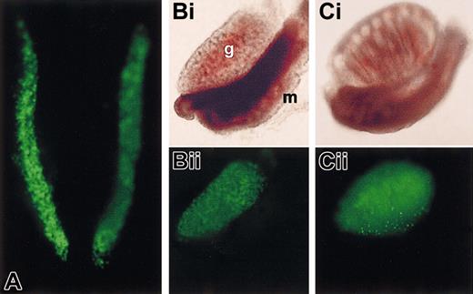
At further later stages, fluorescent PGCs progressively accumulate in the gonadal ridges (Figure 4A) and then in the developing gonads, with a clearly demarcated border with the mesonephros (Figure 4B-C). Fluorescence is most intense at E12.5 and persists up to E14 to 14.5.
GFP expression of construct 3 in the gonads. Bright field (i) and fluorescence (ii) images are shown. Panel A shows fluorescence image only. (A) E11.5 dissected AGM. Only the gonadal ridges are fluorescent. (B-C) E13.5 female and male gonads with the adjacent mesonephros. Note the absence of staining of the mesonephros. Note that qualitatively similar results are obtained with constructs 1 and 2; g indicates gonad, and m indicates mesonephros. Original magnifications are × 40 (A) and × 20 (B-C).
GFP expression of construct 3 in the gonads. Bright field (i) and fluorescence (ii) images are shown. Panel A shows fluorescence image only. (A) E11.5 dissected AGM. Only the gonadal ridges are fluorescent. (B-C) E13.5 female and male gonads with the adjacent mesonephros. Note the absence of staining of the mesonephros. Note that qualitatively similar results are obtained with constructs 1 and 2; g indicates gonad, and m indicates mesonephros. Original magnifications are × 40 (A) and × 20 (B-C).
Overall, with constructs 2 and 3 the pattern of GFP expression within the AGM region is largely superimposable to that expected for PGCs, a comparatively smaller number of putative hematopoietic cells can also be detected. Notably, in contrast to the results obtained with constructs 2 and 3, GFP expression with construct 1 is weak in the gonadal ridges (E10.5-11.5) and very low—hardly detectable—in the aorta region at E9.5 (not shown).
GFP expression in hematopoietic sites
With constructs 2 and 3, fluorescent cells are visible extraembryonically in the yolk sac starting at E8-8.5 through E11.5 to 12, and in vitelline vessels (Figure 5). In the yolk sac (Figure 5A-D), many positive cells are spread in the mesenchyme, but often tend to cluster within or along small vessels. In vitelline vessels (Figure 5E), cells are within the wall or in the bloodstream. In the fetal liver, fluorescence appears between E10 and 10.5 persisting up to E12.5 to 13 (see “Correlation of GFP fluorescence with Kit expression”). Postnatally, the bone marrow is strongly fluorescent (not shown). Importantly, no expression is detected with construct 1 in these sites (Table 1).
GFP expression of construct 3 in yolk sac and vitelline vessels (E11.5). Brightfield (i) and fluorescence (ii) images are shown. (A-D) Yolk sac. (E) Vitelline vessels. Note that qualitatively similar results are obtained with construct 2 (C), but no expression is seen with construct 1, and no nonspecific fluorescence is detected with nontransgenic embryos. Original magnification, × 40 for all panels.
GFP expression of construct 3 in yolk sac and vitelline vessels (E11.5). Brightfield (i) and fluorescence (ii) images are shown. (A-D) Yolk sac. (E) Vitelline vessels. Note that qualitatively similar results are obtained with construct 2 (C), but no expression is seen with construct 1, and no nonspecific fluorescence is detected with nontransgenic embryos. Original magnification, × 40 for all panels.
Correlation of GFP fluorescence with Kit expression
PGCs and early hematopoietic progenitors are mostly Kit+. To evaluate if the Kit transgene is expressed within the proper cells, we correlated Kit membrane expression (or AP and TG1 immunostaining, which identify PGCs) with GFP fluorescence.
PGCs purified from the AGM (E10.5) by immunomagnetic cell sorting using TG1 antibody40 (Figure 6A) or from the gonadal ridges (E11.5; Figure 6B) of transgenic mice (constructs 2 and 3) are mostly GFP+ (Figure 6 and Table 2; compare the AP or TG-1 staining [red] with GFP [green] fluorescence). Similarly, PGCs from mice transgenic for construct 1, although much less fluorescent than those obtained with constructs 3 (Figure 6C-E) and 2 (not shown), are mostly (> 80%) GFP+ as well. The TG-1- somatic cell fractions from all 3 transgenic lines are negative (not shown).
GFP+ PGCs in the gonadal ridge. (A) GFP (green) and AP (red) staining in TG1 immunopurified gonadal ridge PGCs (E10.5). (B) GFP (green; Bi) and TG1 (red, membrane staining; Bii) of PGCs from E11.5 gonads; in panel Biii the images are merged. (C-E) Comparison of GFP fluorescence of gonadal ridge from construct 3 (panel C, left image) and 1 (panel C, right image) lines and of dissociated cells (panels D [construct 3] and E [construct 1]) at E11.5. Panels Di and Ei show GFP fluorescence, and panels Dii and Eii show TG1 fluorescence. Results with construct 2 are essentially identical to those with construct 3. All the construct 1 lines show very weak fluorescence. No nonspecific fluorescence is detected with nontransgenic embryos. Original magnifications, × 400 (A-B, D-E) and × 40 (C).
GFP+ PGCs in the gonadal ridge. (A) GFP (green) and AP (red) staining in TG1 immunopurified gonadal ridge PGCs (E10.5). (B) GFP (green; Bi) and TG1 (red, membrane staining; Bii) of PGCs from E11.5 gonads; in panel Biii the images are merged. (C-E) Comparison of GFP fluorescence of gonadal ridge from construct 3 (panel C, left image) and 1 (panel C, right image) lines and of dissociated cells (panels D [construct 3] and E [construct 1]) at E11.5. Panels Di and Ei show GFP fluorescence, and panels Dii and Eii show TG1 fluorescence. Results with construct 2 are essentially identical to those with construct 3. All the construct 1 lines show very weak fluorescence. No nonspecific fluorescence is detected with nontransgenic embryos. Original magnifications, × 400 (A-B, D-E) and × 40 (C).
GFP+ cells among various progenitor cell types
. | Cell type . | GFP+ cells, % . |
|---|---|---|
| Yolk sac, E8.5 | Kit+ | 30-70 |
| AGM, E9.5 | Kit+ | 50-60 |
| PGC, E10.5 | Kit+ | 90-95 |
| PGC, E11.5 | Kit+ | 90-95 |
| Fetal liver, E11-12 | Kit+ | 25-85 |
| Bone marrow, adult | Kit+ | 65-80 |
| Fetal liver | CFU-mix | 57*; 16† |
| Fetal liver | BFU-E | 57*; 37† |
| Fetal liver | CFU-GM | 52*; 6† |
| Fetal liver | CFU-E | NA*; 64† |
Data shown for construct 3 transgenics. NA indicates not acquired.
E11
E12-12.5. Average of 2 experiments
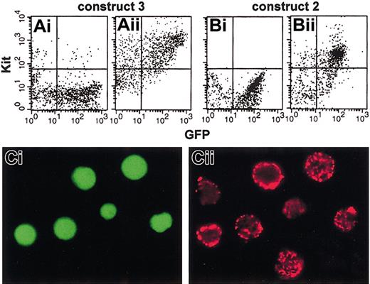
Yolk sac and AGM region from E8.5 and E9.5 embryos of transgenic construct 2-3 mice were dissected, disaggregated as single-cell suspensions, stained with anti-Kit antibodies, and analyzed by fluorescence microscopy. The results show that a large proportion of the Kit+ cells (from both regions) are also GFP+ (Figure 7C; Table 2).
GFP expression in Kit+ fetal liver and yolk sac cells. (A-B) FACS analysis of GFP and Kit fluorescence in fetal liver cells at E11.5. (A) Construct 3. (B) Construct 2. (Ai,Bi) Second antibody; (Aii,Bii) first and second antibodies. (C) GFP (green; Ci) and Kit (red; Cii) fluorescence in E9.5 yolk sac cells. No expression is seen with construct 1. Original magnification, × 400.
GFP expression in Kit+ fetal liver and yolk sac cells. (A-B) FACS analysis of GFP and Kit fluorescence in fetal liver cells at E11.5. (A) Construct 3. (B) Construct 2. (Ai,Bi) Second antibody; (Aii,Bii) first and second antibodies. (C) GFP (green; Ci) and Kit (red; Cii) fluorescence in E9.5 yolk sac cells. No expression is seen with construct 1. Original magnification, × 400.
In fetal liver cells, obtained from E11.5 embryos, about 80% of Kit+ cells are also highly GFP+, as shown by FACS analysis (Figure 7A, constructs 2 and 3); however, the intensity of GFP fluorescence with construct 2 is only about 35% of that seen with construct 3. In adult bone marrow (of construct 3 mice), 30% to 40% of the cells are GFP+ and up to 80% of the Kit+ population (which represents 12%-15% of total cells) express GFP (Table 2). Some Ter-119+ and Mac-1+ cells are also GFP+, but they are mostly located in a region of intermediate fluorescence (mean fluorescence intensity [MFI], 3.8 × 101; range, 2 × 101 to 2 × 10²), whereas Kithigh cells are mostly located in a region of high fluorescence (MFI, 3.2 × 10²; range, 2 × 10² to 3 × 10³; data not shown). The presence of some GFP in Ter-119+ and Mac-1+ cells, which are Kit low or negative, might be due to a low level of inappropriate expression of the construct; however, the data might also be compatible with a slow decay of the high level of GFP synthesized in early Kit+ progenitors. The fact that GFP expression is extinguished in differentiating erythroid and myeloid cells during in vitro culture (see “GFP expression in early hematopoietic progenitors”) supports, in our view, the latter possibility.
GFP expression in early hematopoietic progenitors
Early multilineage hematopoietic progenitors expressing Kit represent a very minor proportion of total hematopoietic cells. To identify different progenitors among GFP+ cells, we FACS purified GFP+ and GFP- cells from fetal livers (E11-12.5) of mice transgenic for constructs 2 and 3, and plated them for in vitro clonogenic assays. Early progenitors (CFU-mix, BFU-Es, etc) are significantly enriched (> 50%; Table 2) in the GFP+ fraction (representing about 25% of the total cells) in construct 3 line at E11; this proportion somewhat declined at E12.5, when only 4% to 5% of the total cell population was GFP+ (and Kit+ cells were only 10%-15% of the total population). This decline may represent decreased GFP expression in a more mature cell population with a threshold effect on fluorescence detection. A comparable enrichment was detected with construct 2 (not shown).
In a similar experiment, clonogenic assays were carried out using bone marrow cells from 2 adult construct 3 mice (line 6) sorted into 3 fractions: a negative, an intermediate, and a high fluorescence fraction. All the pluripotent progenitors (CFU-mix) were present in the fluorescent fractions, with the great majority in the GFP high population (Table 3).
GFP+ progenitors in bone marrow cells of adult transgenic mice
. | BFU-E, % . | CFU-GM, % . | CFU-mix, % . |
|---|---|---|---|
| GFP-high | 58 | 39 | 73 |
| GFP-intermediate | 36 | 50 | 27 |
| GFP-negative | 6 | 11 | 0 |
. | BFU-E, % . | CFU-GM, % . | CFU-mix, % . |
|---|---|---|---|
| GFP-high | 58 | 39 | 73 |
| GFP-intermediate | 36 | 50 | 27 |
| GFP-negative | 6 | 11 | 0 |
Average of 2 experiments. Percentages of erythroid, myelomonocytic, and pluripotent progenitors present in different fractions of FACS bone marrow cells from construct 3 (line 6) transgenic mice.
Similarly, early erythroid precursors (BFU-Es) were mostly highly fluorescent, whereas myelomonocytic progenitors (granulocyte-macrophage colony-forming units [CFU-GMs]), although highly enriched in the fluorescent population, peaked mainly in the GFP intermediate fraction.
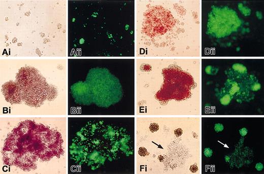
Examination of developing colonies from adult bone marrow and unfractionated fetal liver (construct 3 line) shows that almost all early cell clusters appearing at days 3 to 5 of the culture are intensely fluorescent (Figure 8A). At later stages of culture (day 9-12), most CFU-mix (97% ± 6%) and BFU-Es (78% ± 4%; Figure 8B-D) are positive, whereas few CFU-GMs (not shown) are fluorescent (11% ± 8%). Intensely hemoglobinized erythroid cells (Figure 8E) and mature myelomonocytic cells in developing mixed colonies (Figure 8F) become mostly GFP-, indicating that the gene is down-regulated according to cell type and stage of maturation. With construct 2, similar results are obtained for CFU-mix (84% ± 20% GFP positivity), but a lower proportion of BFU-Es (45% ± 19%) and a higher proportion of CFU-GMs (28% ± 15%) express the GFP gene.
GFP expression in hematopoietic colonies from mice transgenic for construct 3. Cells were observed in the visible (i) and fluorescent (ii) light at day 4 to 5 of culture (A) and day 9 to 12 (B, CFU-mix; C-E, BFU-E; F, CFU-mix). Original magnification, × 100. Similar results are obtained also with construct 2, both using fetal liver and marrow cells. No fluorescence is seen with cells from construct 1 mice, and from nontransgenic controls. Arrows in panel F point to a group of myelomonocytic cells expressing little or no GFP.
GFP expression in hematopoietic colonies from mice transgenic for construct 3. Cells were observed in the visible (i) and fluorescent (ii) light at day 4 to 5 of culture (A) and day 9 to 12 (B, CFU-mix; C-E, BFU-E; F, CFU-mix). Original magnification, × 100. Similar results are obtained also with construct 2, both using fetal liver and marrow cells. No fluorescence is seen with cells from construct 1 mice, and from nontransgenic controls. Arrows in panel F point to a group of myelomonocytic cells expressing little or no GFP.
As expected, no fluorescent colonies are obtained with hematopoietic progenitors from construct 1 lines (not shown).
Discussion
A molecular understanding of the development and maintenance of stem cells and early progenitors will require the elucidation of the transcriptional network controlling the cascade of regulatory factors, such as cytokine receptors and transcription factors themselves. The Kit receptor plays a unique role because its expression is critical for the development of several stem and early multipotent cell types belonging to unrelated lineages (such as HSCs and PGCs). The large size and complexity of the Kit gene has so far precluded transgenic studies and the delineation of functional elements involved in its differential regulation during development. Previous transgenic studies of other large hematopoietically expressed genes, such as CD34, GATA-2, and GATA-3, used long DNA fragments cloned in YAC vectors to reproduce, partly or fully, developmentally regulated expression.46-49 In this work, we used the alternative approach50 of first identifying cell type-specific DNase I hypersensitive sites of the Kit gene, encompassing potential regulatory regions, and then combining them into transgenic constructs driving GFP expression. We thus obtained a construct that is correctly expressed throughout the development of hematopoietic and germ cell lineages. Our findings highlight the role of a 3.5-kb fragment (HS2), which is absolutely necessary for activity in multipotent hematopoietic progenitors and is required for high-level expression in PGCs.
Requirement for correct Kit expression in hematopoiesis
The results obtained with construct 1 (Figures 2 and 6; Table 1) show that the Kit promoter alone is sufficient to direct GFP expression to PGCs but is unable to provide activity in hematopoietic sites. However the addition of a single element, the HS2 fragment, to generate construct 2 (Figures 2, 5, and 7; Table 1), is both sufficient and necessary to drive expression in early hematopoietic progenitors. Further, the HS2 fragment greatly enhances the activity of the promoter in PGCs (Table 1; Figure 6). Thus, the HS2 fragment is a key element for expression in both lineages; indeed, this site is “open” in the chromatin of all examined Kit-expressing cell types (Figure 1). HS2 may be required to enhance a weak cell type-specific activity of the promoter, as observed in PGCs. However, HS2 itself might also provide cell type-specific activity, for example, in hematopoietic cells (Table 1). The HS2 fragment contains sequences showing enhancer activity when introduced into multipotent ES cells as well as into hematopoietic progenitor cell lines (E.M., L.A.C., B.G., et al, manuscript in preparation); this regulatory element might thus be responsive to signals common to several multipotent cell types.
HS3 to HS6 are open only in hematopoietic cells (Figure 1); these sites are not strictly required for expression, but their addition results in a greater proportion of transgenic lines expressing GFP in hematopoietic cells (Table 1), a higher activity, and a greater percentage of GFP+ hematopoietic colonies (Table 2), relative to construct 2. Further, as expected, GFP expression is appropriately down-regulated in mature erythroid cells and in most differentiated myeloid colonies derived from construct 3 transgenics (Figure 8). In contrast, construct 2 shows a less tight regulatory ability because it is relatively more expressed in myeloid colonies. These data suggest that HS3 to HS6 might potentiate expression in hematopoietic cells and possibly modulate Kit activity during cell maturation.
Kit is a large gene, and we have so far explored the function of only about 20 kb surrounding the transcription initiation site. It is thus unlikely that we have identified all the elements required for correct Kit expression in hematopoietic and germ cells. Indeed, a number of deletions or rearrangements, mapping from 21 to over 150 kb upstream to the first exon, have been identified in mouse.44,45 These mutations severely affect Kit expression in melanocytes or in mast cells. In addition, some of these mutations decrease by 25% to 40% Kit expression in Kit+ early hematopoietic progenitors and cause the disappearance of several mast cell-specific chromatin DNase I hypersensitive sites.45 In view of the complexity of the Kit locus it is remarkable that a relatively short region of DNA as that represented in our constructs 2 and 3 is able to direct proper expression in hematopoietic and germ cells.
Recently, Bernex et al25 inserted by homologous recombination alacZ reporter (lacZ knock in) into the endogenous mouse Kit gene allowing evaluation of Kit regulation under the control of all Kit regulatory sequences. In addition to detecting reporter expression in yolk sac, AGM, and fetal liver (peaking at E11.5), as we do with our transgenic constructs, they also observed significant activity in endothelia, where our constructs are poorly, if at all, expressed. Endothelia and hematopoietic cells are thought to derive from a common progenitor, the hemangioblast. Our results therefore suggest that different elements are required for Kit expression after the divergence of the 2 lineages. Finally, it is interesting that only a very low lacZ activity was detected by Bernex et al25 between E10.5 and E13.5 in PGCs, a site of abundant expression of Kit and of our transgenes (constructs 2 and 3). They suggested that this unexpected lack of activity was due to the replacement of part of the first exon and 200 nucleotides of the first intron by the lacZ cassette, which might have deleted or disturbed a PGC regulatory region. Significantly, this is just the 5′ portion of the HS2 element and is immediately upstream to the HS2 site and to a highly conserved human-mouse homology sequence, spanning the first approximate 900 nucleotides of the intron.
Correlation between GFP expression and Kit positivity
The analysis of GFP+ cells during development identifies at least 2 highly expressing cell types: hematopoietic cells and PGCs. All PGCs are expected to express Kit, whereas only some hematopoietic progenitors do.20,51 Direct analysis of immunoselected PGCs at early (E10.5) and more advanced (E11.5) stages shows that almost all cells express GFP (Table 2; Figure 6), whereas no expression in somatic gonadal cells was detectable. This strong correlation indicates that the Kit promoter and (other regulatory sequences) included in our constructs are sufficient for faithful expression in PGCs at these early stages. The proportion of hematopoietic Kit+ cells expressing GFP is also high, though not as much as in PGCs (Table 2), peaking in fetal liver Kit+ cells (80%-85% at E11.5). The reasons for the discrepancy in the expression levels between PGCs and hematopoietic cells are not yet known. One possibility is that the construct is still subject, particularly in hematopoietic cells, to variegation of expression due to position of integration effects, a common observation in transgenics. An alternative, though not mutually exclusive, explanation is that hematopoietic Kit+ cells are more heterogeneous than PGCs, and thus their ability to express the transgene may depend on the developmental stage and degree of maturation levels.
The latter hypothesis is consistent with the progressive variation in GFP expression observed along development. In sites such as yolk sac and AGM, where hematopoietic cells coexist with other Kit-expressing cells, GFP expression is detected in the Kit+ fraction already at early stages (Table 2). Although in these early sites a large proportion of the Kit+ cells express GFP, not all do so (Table 2). In the AGM region, for example, few GFP+ cells are detected intravascularly at E9.5 (Figure 3) and most GFP+ cells are likely PGCs (Figure 3G). At slightly later stages (E10.5), a small proportion (5%) of fetal liver cells express the transgene; these cells include early colony-forming progenitors (not shown). On the other hand, in fetal liver at E11.5, most Kit+ cells express GFP (Figure 7). Our results in colony assays indicate that GFP is expressed in a high proportion of early progenitors, such as CFU-mix and BFU-Es from fetal liver and adult marrow (Tables 2 and 3), and in immature cells (Figure 8), which are known to be Kit+, whereas, as expected, maturation into late erythroblasts or myeloid cells leads to extinction of GFP expression. Taken together, our findings are consistent with the possibility that we have identified regulatory sequences sufficient for recapitulating several aspects of the normal regulation of Kit expression in development and maturation of hematopoietic cells. It remains possible, however, that additional regulatory elements may be required to ensure optimal levels of expression in all Kit+ cells. Experiments are now in progress to evaluate, by transplantation of FACS cells, if GFP+ cells found in fetal liver or AGM after E11 or in bone marrow mark not only multipotent erythroid-myelomonocytic progenitors (Tables 2 and 3), but also HSCs.
Comparison with other early hematopoietic and germ cell transgenes
There are few studies of the transcriptional regulation of genes in stem cells and early progenitors. Among the genes studied are those encoding the transcription factors Sox2 (ES and neural stem cells),13 Oct-4 (inner cell mass, PGC),6,9,14 SCL-Tal-1, GATA-1 and GATA-2 (HSC and hematopoietic progenitors),7,8,10-12,15,16,37 and the surface protein Sca-1.17,30
Among transgenes expressed in early hematopoietic sites, the best characterized are those dependent on regulatory sequences from the GATA-1 and SCL-Tal-1 genes.7,10-12,15,16 Although the precise identity of the transcription factors regulating these genes during development and in adult cells is not yet known,11,12,15 it is thought that expression in a subset of hematopoietic embryonicfetal progenitors requires a GATA factor (possibly GATA-2, expressed in several hematopoietic cells) in combination with other not yet identified factors.11,12,16 Could the expression of Kit/GFP transgenes be regulated similarly to GATA-1 and SCL/Tal-1 transgenes? Within the hematopoietic system, the expression of Kit constructs 2 and 3 is similar in several respects to that of the SCL-Tal-1 transgene: expression is detected in the yolk sac, within dorsal aorta and vitelline vessels, in fetal liver, and in a large proportion of early Kit+ progenitors (CFU-mix, BFU-Es, etc). Of about 17 GATA motifs detected in the HS2 to HS6 fragments, 6 are clustered within about 400 nucleotides of HS3 to HS4 and 3 within about 250 nucleotides of HS5 to HS6, the hematopoietic-specific hypersensitive sites. These motifs might represent the basis for expression in hematopoietic regions.
In addition, Kit was proposed to be a target of the transcription factor SCL-Tal-1 itself. Kit expression in hematopoietic cells closely correlates with that of SCL-Tal-1, and overexpression of SCL-Tal-1 may induce Kit transcription in cell lines.51,52 SCL-Tal-1 was suggested to act on Kit through DNA sequences located in the proximal Kit promoter region, close to HS1.51,52 These include a duplicated SCL-Tal-1-binding E-box (5′-CACCTG),51 conserved between mouse (at nucleotide -358 to -342) and man and an Sp1-binding motif able to recruit a complex including SCL-Tal-1, GATA1/2, LMO2, and Ldb1 factors.52 The inactivity of construct 1 in hematopoietic cells (Table 1) shows that these sites are insufficient, by themselves, for transgenic Kit-driven GFP expression in such cells. However, it is interesting that 3 E-boxes, with the same motif as that represented in the duplicated SCL-Tal-1-binding promoter site (of 13 detected within the whole HS2-HS6 region), are clustered immediately downstream to the first exon/first intron junction at positions -3, +68, and +65 of the first intron. Two of these sites are perfectly conserved in the human KIT gene, where they flank an additional human-specific E-box. These sites are thus good candidates for mediating interactions with the SCL-Tal-1 complex bound at the promoter, hence the reported SCL-Tal-1-mediated Kit overexpression.51,52
Additional transcription factors are likely responsible for differential expression of Kit and SCL-Tal-1 in other cell types. For example, the latter transgene is highly expressed in vascular endothelia (a site of SCL/Tal-1 expression), whereas the Kit transgene is not. On the other hand, the SCL-Tal-1 transgene is not reported to be expressed in PGCs10,15 (which are SCL-Tal-1- [M. De F., unpublished observation, January 2003]), whereas the Kit transgene is highly expressed in these cells.
Transgenes driven by the Oct-4 promoter, including an essential distal 3.3-kb fragment, are efficiently expressed in the germline.6 Oct-4 is a transcription factor first expressed in the early embryo and inner cell mass, and then restricted to the germline. Oct-4 and Kit show very similar expression patterns in the germline, throughout development. Both genes are highly expressed in migratory and proliferating PGCs and down-regulated in oocytes entering into meiosis and in mitotic G1-arrested prospermatogonia in the fetal gonads,53 to be re-expressed in growing oocytes and at specific later stages of male germ cell differentiation.53-55
Both Oct-46,9 and Kit transgenes recapitulate correct expression in embryonic germ cells (Figures 3, 4 and 6; Table 2). Studies in progress suggest that this correlation extends to postnatal germ cells as well (to be reported elsewhere). The similarity of expression of Kit and Oct-4 transgenes in the germline suggests that Kit may be regulated by similar mechanisms as Oct-4, or perhaps by Oct-4 itself, in this lineage.
In conclusion, we have identified DNA regulatory elements from the Kit gene, which in combination are sufficient to recapitulate several aspects of the developmental regulation of Kit in 2 different cell lineages, hematopoietic and germ cells. At least one fragment, the HS2 fragment, shows a degree of hematopoietic specificity, because it is absolutely required for expression in this lineage. The comparison of Kit novel regulatory elements with those of other genes expressed in early multipotent progenitors, such as SCL/Tal-1,16 Sca-1,29 and Oct-4,6,9 might allow researchers to further dissect transcriptional events underlying early embryonic decisions of these cells.
In addition, the highly GFP+ mouse lines described here might provide ways to enrich for rare adult multipotential cells. Indeed, preliminary results (G. Cossu, L.A.C., S.O., et al; A. Musarò, N. Rosenthal, L.A.C., S.O., et al, unpublished observations, January 2003), indicate that GFP+ bone marrow progenitors may be enriched in cells capable of homing to muscle on injury and in adult multipotent progenitors similar to the mesoangioblast.56
Prepublished online as Blood First Edition Paper, August 7, 2003; DOI 10.1182/blood-2003-04-1296.
Supported by grants from Telethon A048 (S.O.) and B.40 (C.T.), AIRC (S.O.) and MURST 2001 (M. De F.), and CVRCS (M.C.M.). L.A.C. and E.M. contributed equally to this study.
The publication costs of this article were defrayed in part by page charge payment. Therefore, and solely to indicate this fact, this article is hereby marked ”advertisement” in accordance with 18 U.S.C. section 1734.
We thank Professor L. Pozzi for discussion and help with transgenic experiments, Dr D. Bennet for mel-c lines, D. Santoni for expert mouse care, and M. Azzini for photography. Part of this work was carried out by B.G. and E.M. at Centro Patologia Cellulare-CNR, Milan, Italy.

![Figure 6. GFP+ PGCs in the gonadal ridge. (A) GFP (green) and AP (red) staining in TG1 immunopurified gonadal ridge PGCs (E10.5). (B) GFP (green; Bi) and TG1 (red, membrane staining; Bii) of PGCs from E11.5 gonads; in panel Biii the images are merged. (C-E) Comparison of GFP fluorescence of gonadal ridge from construct 3 (panel C, left image) and 1 (panel C, right image) lines and of dissociated cells (panels D [construct 3] and E [construct 1]) at E11.5. Panels Di and Ei show GFP fluorescence, and panels Dii and Eii show TG1 fluorescence. Results with construct 2 are essentially identical to those with construct 3. All the construct 1 lines show very weak fluorescence. No nonspecific fluorescence is detected with nontransgenic embryos. Original magnifications, × 400 (A-B, D-E) and × 40 (C).](https://ash.silverchair-cdn.com/ash/content_public/journal/blood/102/12/10.1182_blood-2003-04-1296/6/m_h82335299006.jpeg?Expires=1769114172&Signature=gg4PGQBpj5CIrBBFVonkTkoa6MT5ScAwfhRDI-23H2jIxrwog~m1M7rw~A1FAkoAZ~236zG34d9NzewHooue6zzYpfNcnCrR26XmAsVAEqBVJTbyIOEeFWjkq68lBy-WrAITKJw6Iq4UKako6C95L75bcKIyPBIvKrhNJ2WQRyre2PrO7ba7pCk0ofWRpCCer7vA3sgb6waBIf0w-xL3boSp0UL3g0poh-LlTbzOqRaQ9KihZT0sIQzJabwA263siB6BgoRLxBnKKAYGAXXT3rJgMBIdg4w4ZtiD~Oefvqz6UPMQqYhn9MiPbBPb3h59-g6ftOW6hNVKch1ydIZkDg__&Key-Pair-Id=APKAIE5G5CRDK6RD3PGA)
This feature is available to Subscribers Only
Sign In or Create an Account Close Modal