Abstract
Adaphostin (NSC 680410), an analog of the tyrphostin AG957, was previously shown to induce Bcr/abl down-regulation followed by loss of clonogenic survival in chronic myelogenous leukemia (CML) cell lines and clinical samples. Adaphostin demonstrated selectivity for CML myeloid progenitors in vitro and remained active in K562 cells selected for imatinib mesylate resistance. In the present study, the mechanism of action of adaphostin was investigated in greater detail in vitro. Initial studies demonstrated that adaphostin induced apoptosis in a variety of Bcr/abl- cells, including acute myelogenous leukemia (AML) blasts and cell lines as well as chronic lymphocytic leukemia (CLL) samples. Further study demonstrated that adaphostin caused intracellular peroxide production followed by DNA strand breaks and, in cells containing wild-type p53, a typical DNA damage response consisting of p53 phosphorylation and up-regulation. Importantly, the antioxidant N-acetylcysteine (NAC) blunted these events, whereas glutathione depletion with buthionine sulfoximine (BSO) augmented them. Collectively, these results not only outline a mechanism by which adaphostin can damage both myeloid and lymphoid leukemia cells, but also indicate that this novel agent might have a broader spectrum of activity than originally envisioned. (Blood. 2003;102:4512-4519)
Introduction
Chronic myelogenous leukemia (CML) is almost universally associated with a t(9;22) chromosomal translocation that juxtaposes the Bcr and Abl genes.1,2 Because the resulting kinase, p210Bcr/abl, is found exclusively in malignant hematopoietic cells, there has been considerable interest in identifying inhibitors of this enzyme. Several classes of p210Bcr/abl inhibitors have been identified, including the 2-phenylaminopyrimidine derivative imatinib mesylate,3 the pyrido[2,3-d]pyrimidine derivative PD180970,4 and the tyrphostin AG957.5
Clinical studies performed to date have established that imatinib mesylate is highly active in chronic-phase CML.6,7 In contrast, patients with CML in blast crisis or t(9;22)+ acute lymphocytic leukemia (ALL) tend to have shorter remissions.8,9 At the time of relapse, cells from the latter patients exhibit imatinib mesylate resistance in vitro.10 Although the molecular basis of imatinib mesylate resistance remains somewhat controversial,11,12 there is considerable interest in overcoming this resistance. Potential strategies include combining imatinib mesylate with conventional cytotoxic agents13 or arsenic trioxide9 as well as using Bcr/abl kinase inhibitors that are unaffected by alterations that cause imatinib mesylate resistance.14,15
The tyrphostin AG957 was originally identified as an inhibitor of p210Bcr/abl kinase that is not competitive with adenosine triphosphate (ATP).5 Subsequent studies demonstrated that adaphostin, the adamatyl ester analog of AG957, has a longer serum half-life and displays greater antiproliferative effects against Bcr/abl+ leukemia cells in the National Cancer Institute (NCI) hollow fiber assay. Recent comparison of the cellular effects of adaphostin and imatinib mesylate demonstrated striking differences in their mechanisms of action.14 In particular, adaphostin induced p210Bcr/abl down-regulation followed by relatively prompt initiation of apoptotic changes, whereas imatinib mesylate inhibited Bcr/abl-mediated phosphorylation quickly but induced apoptosis only after considerable delay.14 Importantly, adaphostin exhibited selectivity for CML myeloid progenitors in vitro and retained its cytotoxicity when imatinib mesylate-resistant K562 cells were examined.14
Despite these potentially interesting properties, there have been a number of incompletely explained observations related to the action of AG957 and adaphostin. First, these agents kill a number of Bcr/abl- leukemia cell lines, including Jurkat,16-18 CEM,18 HL-60,19 FDC-P1,14 and Nalm-6,17 suggesting the possibility of a second cytotoxic mechanism that does not involve Bcr/abl. Second, as noted, adaphostin inhibits Bcr/abl-mediated signaling much more slowly than imatinib mesylate, yet induces apoptosis more rapidly,14 again raising the possibility that adaphostin has a second cytotoxic mechanism.
In the present study we demonstrate that adaphostin induces apoptosis in clinical samples of chronic lymphocytic leukemia (CLL) and acute myelogenous leukemia (AML). In addition, we show that adaphostin induces increases in the level of intracellular reactive oxygen species (ROS) followed by DNA strand breaks, all of which precede the onset of apoptotic changes. Finally, we demonstrate that modulation of ROS production alters the cytotoxicity of adaphostin, thereby providing evidence for the potential contribution of this second cytotoxic mechanism. Collectively, these results provide new insight into the mechanism by which adaphostin kills a wide range of human leukemia cells.
Materials and methods
Materials
Adaphostin, AG957, and NSC 676537 (a quinone analog of AG957; Figure 6A) were all synthesized by the Drug Synthesis and Chemistry Branch, Division of Cancer Treatment and Diagnosis, National Cancer Institute (Bethesda, MD). Imatinib mesylate was kindly provided by Novartis Pharma (Basel, Switzerland). Buthionine sulfoximine (BSO), N-acetylcysteine (NAC), propidium iodide (PI), and Triton X-100 were purchased from Sigma (St Louis, MO). [Methyl-14C]-thymidine (specific activity 57.8 mCi/mmol; 2138.6 MBq) was from Amersham (Arlington Heights, IL). The 5-chloromethyl-2′,7′-dichlorodihydrofluorescein diacetate (CM-H2DCFDA) and dihydroethidium (H2E) were purchased from Molecular Probes (Eugene, OR). Monochlorobimane (mBCl) and N-(Nα-benzyloxycarbonylvalinylalanyl) aspartic acid (O-methyl ester) fluoromethylketone [zVAD(OMe)-fmk] were from Calbiochem (San Diego, CA) and Biomol (Plymouth Meeting, PA), respectively.
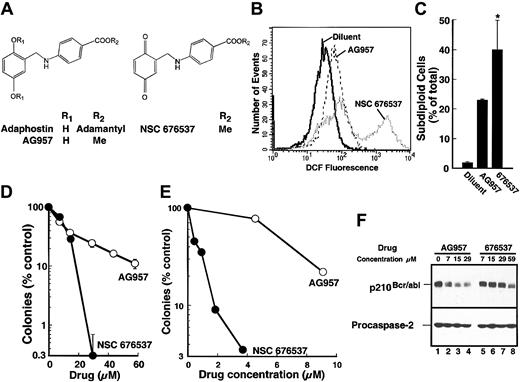
Differential effects of AG957 and its quinone derivative NSC 676537. (A) Structures of adaphostin, the parent compound AG957, and NSC 676537, which is a quinone derivative of AG957. (B) K562 cells were treated with diluent, 30 μM AG957, or 30 μM NSC 676537 for 1.5 hours, then stained with CM-H2DCFDA and examined by flow microfluorimetry to estimate peroxide levels. (C) K562 cells were treated for 24 hours with diluent, 30 μM AG957, or 30 μM NSC 676537; stained with PI; and examined by flow microfluorimetry for subdiploid cells. Error bars are mean ± SD of 3 experiments. *P < .05 relative to AG957. (D) K562 cells were incubated for 24 hours in the presence of the indicated agent, washed, and plated in 3% agar to determine clonogenic survival. (E) Peripheral blood mononuclear cells from a patient with CML were incubated with the indicated agent for 24 hours, washed, and assayed for granulocyte colonies as previously described.19 Similar results were obtained using cells from 2 additional patients with CML. (F) K562 cells were incubated in the presence of the indicated agent for 8 hours. Whole-cell lysates were probed with antibodies to c-abl or, as a loading control, procaspase-2.
Differential effects of AG957 and its quinone derivative NSC 676537. (A) Structures of adaphostin, the parent compound AG957, and NSC 676537, which is a quinone derivative of AG957. (B) K562 cells were treated with diluent, 30 μM AG957, or 30 μM NSC 676537 for 1.5 hours, then stained with CM-H2DCFDA and examined by flow microfluorimetry to estimate peroxide levels. (C) K562 cells were treated for 24 hours with diluent, 30 μM AG957, or 30 μM NSC 676537; stained with PI; and examined by flow microfluorimetry for subdiploid cells. Error bars are mean ± SD of 3 experiments. *P < .05 relative to AG957. (D) K562 cells were incubated for 24 hours in the presence of the indicated agent, washed, and plated in 3% agar to determine clonogenic survival. (E) Peripheral blood mononuclear cells from a patient with CML were incubated with the indicated agent for 24 hours, washed, and assayed for granulocyte colonies as previously described.19 Similar results were obtained using cells from 2 additional patients with CML. (F) K562 cells were incubated in the presence of the indicated agent for 8 hours. Whole-cell lysates were probed with antibodies to c-abl or, as a loading control, procaspase-2.
Antibodies recognizing the following antigens were purchased as indicated: phosphotyrosine from Upstate Biotechnology (Lake Placid, NY); c-abl from Oncogene Research (Cambridge, MA); p53 (clone 1801) from Neomarkers (Fremont, CA); phospho-Ser15 -p53, phospho-Ser20 -p53 and phospho-Thr68-Checkpoint kinase 2 (Chk2) from Cell Signaling Technology (Beverly, MA); actin from Santa Cruz Biotechnology (Santa Cruz, CA); and procaspase-2 from Pharmingen (La Jolla, CA). Anti-Chk2 was a kind gift from J. Chen (Mayo Clinic, Rochester, MN). All other materials were obtained as previously indicated.14
Cell lines
K562 cells (American Type Culture Collection, Manassas, VA) and ML-1 cells (a kind gift from Michael Kastan, St Jude Children's Hospital, Memphis, TN) were passaged in RPMI 1640 containing 10% heat-inactivated fetal bovine serum, 100 U/mL penicillin G, 100 μg/mL streptomycin, and 2 mM glutamine (medium A).
Leukemia samples
After informed consent was provided according to the Declaration of Helsinki, clinical samples were studied under the aegis of protocols approved by the institutional review boards of the Mayo Clinic and the University of Maryland Medical Center in accordance with the policies of the US Department of Health and Human Services. Peripheral blood samples were obtained from patients with untreated CML or CLL. Heparinized bone marrow aspirates were obtained from patients with newly diagnosed AML prior to institution of induction therapy. Mononuclear cells from blood or marrow were isolated on Ficoll-Hypaque gradients as described,14 washed with RPMI 1640 medium, and subjected to various assays.
Quantitation of subdiploid population
Tissue culture cells in medium A lacking or containing 24 mM NAC were treated with adaphostin for the indicated length of time. CLL specimens in medium A were incubated with diluent or 10 μM adaphostin for the indicated length of time. AML specimens in RPMI 1640 medium containing 10% (vol/vol) heat-inactivated human AB serum, 100 U/mL penicillin G, 100 μg/mL streptomycin, and 2 mM glutamine were incubated with diluent or varying adaphostin concentrations for 48 hours prior to harvest. At the completion of the incubation, samples were stained with PI and examined by flow microfluorimetry using a FACScan (Becton Dickinson, Mountain View, CA) as previously described.14 A total of 10 000 events were quantitated.
Detection of intracellular ROS
CM-H2DCFDA was used to measure intracellular peroxide levels. As previously reported,20 this agent diffuses into cells and is trapped by de-esterification. Subsequent reaction with peroxides generates intensely fluorescent 5-chloromethyl-2′,7′-dichlorofluorescein (DCF). After treatment with adaphostin or diluent for the indicated length of time, cells were sedimented at 100g, resuspended in phosphate-buffered saline (PBS) containing 10 μM CM-H2DCFDA, incubated at 37°C for 30 minutes in the dark, washed with PBS, and read on the FL1 channel of a Becton Dickinson FACScan. H2E, which is reported to more specificically detect superoxide anion,21 was substituted for CM-H2DCFDA in some experiments.
Alkaline elution
DNA single-strand breaks were examined by alkaline elution as previously described.22 In brief, ML-1 cells were labeled with 0.1 μCi/mL (0.0037 MBq) [methyl-C14]-thymidine for 24 hours. After a 1-hour incubation in radiochemical-free medium A, aliquots containing 1 × 10⁶ cells were exposed to adaphostin for 3 hours. Cells were then loaded onto Nucleopore polycarbonate filters (25 mm, 1 μm pore size), rinsed with 75 mM NaCl containing 24 mM Na4EDTA (ethylenediaminetetraacetic acid), and lysed by exposure of the filters to buffer consisting of 0.1 M glycine, 25 mM Na2EDTA (pH 10), 2% sodium dodecyl sulfate (SDS), and 0.5 mg/mL proteinase K. The filters were rinsed with 20 mM EDTA (pH 10) and eluted in the dark with 0.02 M EDTA (tetra-acid form) adjusted to pH 12.1 with tetrapropylammonium hydroxide. Eluent, filters, and the wash used to rinse the filter holders were collected, mixed with acidified scintillation fluid, and subjected to scintillation counting. To construct a standard curve, labeled cells were irradiated on ice with a 137Cs source at 0.9 Gy/min.
Immunoblotting
After treatment with drug or diluent as indicated in the figure legends, cells were sedimented at 200g for 10 minutes, washed once with ice cold RPMI 1640 medium containing 10 mM HEPES (N-2-hydroxyethylpiperazine-N′-2-ethanesulfonic acid; pH 7.4 at 4°C), solubilized in buffered 6 M guanidine hydrochloride under reducing conditions, and prepared for electrophoresis as previously described.23 Aliquots containing 50 μg protein (determined by the bicinchoninic acid method24 ) were separated on SDS-polyacrylamide gels containing 5% to 15% acrylamide gradients, electrophoretically transferred to nitrocellulose, and probed with antisera as described.25
Clonogenic assays
Aliquots containing 0.5 × 10⁶ K562 cells in 1 mL medium A were incubated with diluent or adaphostin in the absence or presence of NAC or BSO for the indicated lengths of time, sedimented at 100g for 5 minutes, diluted, and plated in gridded 35-mm plates in the medium of Pike and Robinson26 containing 0.3% (wt/vol) Bacto agar. After incubation for 10 to 14 days at 37°C, colonies containing 50 cells or more were counted on an inverted phase contrast microscope.
To compare the effects of AG957 and NSC 676537 on granulocyte colony-forming units (CFU-Gs), peripheral blood mononuclear cells (1 × 10⁶ cells/aliquot) from previously untreated patients with CML were plated in 0.3% (wt/vol) Bacto agar containing 50 ng/mL granulocyte colony-stimulating factor and increasing drug concentrations in Iscove modified Dulbecco medium containing 20% heat-inactivated fetal bovine serum. After 7 to 10 days, colonies were counted by phase contrast microscopy as previously described.19
Quantitation of glutathione levels
After treatment with BSO or adaphostin or both, 3-mL cell suspension (5 × 10⁵/mL) was incubated with 100 μM mBCl for 15 minutes at 37°C. The reaction was terminated by addition of trichloroacetic acid to a final concentration of 5% (wt/vol). After macromolecules were removed by centrifugation, the supernatant was extracted with an equal volume of CH2Cl2 to remove unreacted mBCl. Fluorescence in the aqueous phase was then quantitated with a Perkin Elmer (Shelton, CT) LS-50 fluorometer using excitation and emission wavelengths of 398 and 488 nm, respectively.27 Nonspecific fluorescence was determined by depleting cells of glutathione (GSH) with 1 mM diethylmaleate for 1 hour before adding mBCl.
High-performance liquid chromatography
After samples containing 10 nmol adaphostin in 1 mL RPMI 1640 medium were incubated for 1 hour in the absence or presence of 24 mM NAC, 50-μL aliquots were mixed with 150 μL acetonitrile and analyzed on a Beckman (Palo Alto, CA) model 125 dual pump gradient high-performance liquid chromatograph (HPLC) equipped with a model 168 diode array detector and an IBM personal computer 350 (IBM, Armonk, NY) with Beckman Gold Nouveau software. A Brownlee MPLC Newguard C18 precolumn (3.2 mm × 15 mm × 7 μm) and a Beckman Ultrasphere ODS column (4.6 mm × 250 mm × 5 μm) were pre-equilibrated for 20 minutes or more with mobile phase A (12.5 mM ammonium formate [pH 4.0] in 60% [vol/vol] acetonitrile). Separation was accomplished by eluting with a linear gradient from 100% mobile phase A to 100% mobile phase B (50% [vol/vol] acetonitrile) over 40 minutes at a flow rate of 2 mL/min. Detection was at 302 nm. Under these conditions the retention time for adaphostin was 6.5 to 6.6 minutes.
Statistics
Experiments were performed at least 3 times with similar results. Results were expressed as mean ± SD of the indicated number of replicates, and differences between groups were assessed using paired t tests.
Results
Adaphostin induces apoptosis in cells lacking Bcr/abl
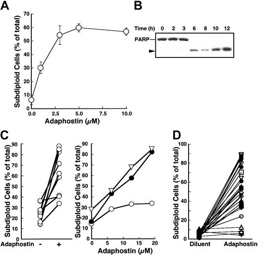
Our recent observation that adaphostin kills Bcr/abl- FDC-P1 murine myeloid cells,14 as well as previous studies showing that the parent tyrphostin AG957 kills Jurkat and HL-60 cells,16,19 led us to investigate the effects of adaphostin on additional Bcr/abl- cells. ML-1, a human AML cell line containing wild-type p53, was chosen for initial studies. Apoptotic changes were assessed using 2 different assays. Generation of extractable DNA fragments was evaluated by searching for the presence of “subdiploid” cells when flow cytometry was performed after staining with PI under hypotonic conditions.28 Caspase activation was assessed by looking for cleavage of the caspase substrate poly(adenosine diphosphate ribose) polymerase (PARP).29 As indicated in Figure 1A, adaphostin induced a dose-dependent increase in the number of “subdiploid” ML-1 cells in less than 8 hours. These changes coincided with caspase activation, as manifested by PARP cleavage to its 89-kDa signature fragment (Figure 1B). Compared to K562 cells, which were 31% apoptotic (SD 9%, n = 3) at the end of an 8-hour incubation with 10 μM adaphostin,14 ML-1 cells underwent apoptosis more rapidly and at lower adaphostin concentrations, with 59% ± 9% (n = 5) being apoptotic after an 8-hour incubation with 3 μM adaphostin.
Cells lacking Bcr/abl are susceptible to adaphostin-induced apoptosis. (A) ML-1 cells were treated with the indicated concentration of adaphostin or diluent for 8 hours, fixed, stained with PI, and examined for subdiploid cells by flow microfluorimetry. Error bars, ± 1 SD from 4 independent experiments. (B) ML-1 cells were treated with 3 μM adaphostin for the indicated times. Whole-cell lysates were subjected to immunoblotting with C-2-10 antibody, which detects full-length PARP as well as its caspase-generated 89-kDa fragment (arrow head).29 (C) Mononuclear cells isolated from bone marrows of 10 patients with newly diagnosed AML were treated with diluent (-) or 20 μM adaphostin (+) for 48 hours (left panel). Right panel indicates dose-response curve in mononuclear cells from 3 representative AML patients. (D) Lymphocytes isolated from the peripheral blood of patients with CLL were incubated in the presence of diluent or 10 μM adaphostin for 24 hours, stained with PI, and examined for subdiploid DNA content by flow microfluorimetry.
Cells lacking Bcr/abl are susceptible to adaphostin-induced apoptosis. (A) ML-1 cells were treated with the indicated concentration of adaphostin or diluent for 8 hours, fixed, stained with PI, and examined for subdiploid cells by flow microfluorimetry. Error bars, ± 1 SD from 4 independent experiments. (B) ML-1 cells were treated with 3 μM adaphostin for the indicated times. Whole-cell lysates were subjected to immunoblotting with C-2-10 antibody, which detects full-length PARP as well as its caspase-generated 89-kDa fragment (arrow head).29 (C) Mononuclear cells isolated from bone marrows of 10 patients with newly diagnosed AML were treated with diluent (-) or 20 μM adaphostin (+) for 48 hours (left panel). Right panel indicates dose-response curve in mononuclear cells from 3 representative AML patients. (D) Lymphocytes isolated from the peripheral blood of patients with CLL were incubated in the presence of diluent or 10 μM adaphostin for 24 hours, stained with PI, and examined for subdiploid DNA content by flow microfluorimetry.
To determine whether adaphostin would also induce apoptosis in clinical AML specimens, samples from 15 previously untreated patients with AML were incubated with increasing adaphostin concentrations for 48 hours. Five of these samples underwent extensive (> 80%) apoptosis in the absence of adaphostin and were, therefore, uninterpretable. Adaphostin induced at least a doubling in the percentage of apoptotic cells in 8 of the 10 remaining AML isolates (Figure 1C left panel). Dose-response curves revealed that apoptotic changes were readily induced by adaphostin concentrations as low as 6 μM (Figure 1C right panel).
To examine the activity of adaphostin activity in a more indolent neoplasm, CLL samples were treated with this agent in vitro. Adaphostin induced at least a doubling of the number of apoptotic cells in 21 of 24 (88%) CLL samples (Figure 1D). Collectively, these results indicate that Bcr/abl is not required for the toxicity of adaphostin and extend its spectrum of antileukemic activity.
Adaphostin causes DNA damage
The ML-1 cell line used in Figure 1 contains wild-type p53. Given the postulated role of this polypeptide in apoptosis induction,30,31 we next examined p53 levels in adaphostin-treated ML-1 cells. When ML-1 cells were treated with 3 μM adaphostin, p53 levels were detectably increased by 2 to 4 hours (Figure 2A). Because increased p53 levels can reflect diminished mdm2-mediated ubiquitylation as a consequence of ataxia telangiectasia mutated (ATM) or ataxia telangiectasia mutated and rad-3 related (ATR)-mediated phosphorylation on Ser15,32,33 we next examined p53 phosphorylation using an antiserum that specifically recognizes this modification. Treatment with adaphostin concentrations as low as 1 μM induced p53 phosphorylation on this residue (Figure 2B left). Similar results were obtained using antiserum that specifically recognizes p53 phosphorylated on Ser20 (Figure 2B right). These observations raised the possibility that p53 up-regulation might reflect DNA damage-induced activation of the kinases ATM, ATR, and check point kinase 2 (Chk2), which are usually responsible for phosphorylation of p53 on these residues.33 To further assess this possibility, alkaline elution was used to search for DNA single-strand breaks in adaphostin-treated ML-1 cells. Results of this analysis demonstrated dose-dependent induction of DNA damage 3 hours after addition of adaphostin (Figure 2C).
Adaphostin-induced p53 up-regulation, DNA strand breaks, and ROS production in ML-1 cells is inhibited by NAC. (A) ML-1 cells were treated with 3 μM adaphostin for up to 6 hours. Whole-cell lysates were subjected to immunoblotting with antibodies that detect p53. (B) After ML-1 cells were treated with the indicated concentrations of adaphostin for 3 hours, whole-cell lysates were probed using an antibody recognizing phosphorylation on Ser15 (left) or Ser20 (right) of p53. In the indicated lanes, the proteasome inhibitor ALLnL was included at 50 μM to stabilize p53 and ensure that the increased signal for phospho-p53 was not simply due to increased amounts of p53. (C) After ML-1 cells were treated with varying concentrations of adaphostin for 3 hours in the presence and absence of 24 mM NAC, DNA single-strand breaks were analyzed by alkaline elution.22 Elution of DNA through the filters was compared to elution after irradiation of ML-1 cells with 300 to 900 cGy ionizing radiation. (D) After a 15-minute pretreatment with 24 mM NAC (dotted line) or diluent, ML-1 cells were treated with diluent (solid line) or 3 μM adaphostin (heavy solid line or dotted line) for 90 minutes and stained with CM-H2DCFDA to assess intracellular peroxides. (E) After treatment with the indicated adaphostin concentration in the presence of 0 (-), 500 (+), or 1000 (++) U/mL catalase, ML-1 cells were fixed, stained with PI, and examined for subdiploid cells by flow microfluorimetry. Error bars, ± 1 SD from 3 independent experiments. (F) ML-1 cells were pretreated with 24 mM NAC for 15 minutes prior to addition of 3 μM adaphostin for up to 8 hours. Whole-cell lysates were subjected to immunoblotting with antibodies that detect p53 or, as a loading control, actin. (G) After ML-1 cells were treated with varying adaphostin concentrations for 8 hours in the absence or presence of 24 mM NAC, cells were stained with PI and assayed for the presence of subdiploid cells by flow microfluorimetry. (H) Adaphostin was incubated for 1 hour at 37°C in RPMI medium in the absence (-) or presence (+) of 24 mM adaphostin and subjected to HPLC. The areas of the adaphostin peaks are indicated.
Adaphostin-induced p53 up-regulation, DNA strand breaks, and ROS production in ML-1 cells is inhibited by NAC. (A) ML-1 cells were treated with 3 μM adaphostin for up to 6 hours. Whole-cell lysates were subjected to immunoblotting with antibodies that detect p53. (B) After ML-1 cells were treated with the indicated concentrations of adaphostin for 3 hours, whole-cell lysates were probed using an antibody recognizing phosphorylation on Ser15 (left) or Ser20 (right) of p53. In the indicated lanes, the proteasome inhibitor ALLnL was included at 50 μM to stabilize p53 and ensure that the increased signal for phospho-p53 was not simply due to increased amounts of p53. (C) After ML-1 cells were treated with varying concentrations of adaphostin for 3 hours in the presence and absence of 24 mM NAC, DNA single-strand breaks were analyzed by alkaline elution.22 Elution of DNA through the filters was compared to elution after irradiation of ML-1 cells with 300 to 900 cGy ionizing radiation. (D) After a 15-minute pretreatment with 24 mM NAC (dotted line) or diluent, ML-1 cells were treated with diluent (solid line) or 3 μM adaphostin (heavy solid line or dotted line) for 90 minutes and stained with CM-H2DCFDA to assess intracellular peroxides. (E) After treatment with the indicated adaphostin concentration in the presence of 0 (-), 500 (+), or 1000 (++) U/mL catalase, ML-1 cells were fixed, stained with PI, and examined for subdiploid cells by flow microfluorimetry. Error bars, ± 1 SD from 3 independent experiments. (F) ML-1 cells were pretreated with 24 mM NAC for 15 minutes prior to addition of 3 μM adaphostin for up to 8 hours. Whole-cell lysates were subjected to immunoblotting with antibodies that detect p53 or, as a loading control, actin. (G) After ML-1 cells were treated with varying adaphostin concentrations for 8 hours in the absence or presence of 24 mM NAC, cells were stained with PI and assayed for the presence of subdiploid cells by flow microfluorimetry. (H) Adaphostin was incubated for 1 hour at 37°C in RPMI medium in the absence (-) or presence (+) of 24 mM adaphostin and subjected to HPLC. The areas of the adaphostin peaks are indicated.
ROS production precedes DNA damage and apoptosis induction in ML-1 cells
Based on the presence of the readily oxidizable dihydroquinone moiety in adaphostin and its parent drug AG957, we hypothesized that these agents might be able to induce DNA damage through the generation of ROS. To test this hypothesis, adaphostin-treated ML-1 cells were stained with CM-H2DCFDA, which generates DCF after reaction with peroxides.20 Flow cytometry demonstrated that DCF fluorescence was increased to 1.67 times control (SD 0.39, n = 7, P = .009) 1.5 hours after addition of adaphostin (Figure 2D dark solid line).
If adaphostin-induced ROS were involved in DNA damage, p53 up-regulation and subsequent cell death, then treatment with antioxidants would be expected to blunt these changes. Initial experiments demonstrated that catalase failed to abolish adaphostin-induced changes (Figure 2E). These observations argue against the possibility that the ROS are generated extracellularly. In contrast, a 15-minute pretreatment with NAC, which is postulated to exert its effects by increasing intracellular GSH levels,34 markedly diminished ROS production as measured by DCF staining (Figure 2D). In addition, NAC treatment diminished the induction of DNA single-strand breaks (Figure 2C) and the up-regulation of p53 (Figure 2F), providing further support for the hypothesis that these changes occur downstream of ROS production. Moreover, NAC decreased the induction of apoptosis (Figure 2G), suggesting that the generation of ROS also contributes to the cytotoxicity of adaphostin in this Bcr/abl- cell line.
These results would be trivially explained if the sulfhydryl group of NAC directly reacted with adaphostin to deplete the active drug. To rule out this possibility, adaphostin was incubated in medium A with or without 24 mM NAC and then quantitated by HPLC. Results of this analysis indicated that NAC failed to react with adaphostin (Figure 2H).
NAC prevents adaphostin-induced loss of clonogenicity but not Bcr/abl down-regulation
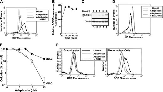
All of the antioxidant experiments described above were conducted in ML-1 cells, which do not contain Bcr/abl. To determine whether similar events occur in Bcr/abl+ cells, ROS production was examined in adaphostin-treated K562 cells. Within 2 hours after addition of adaphostin, DCF fluorescence was 1.79 times control (SD 0.6, n = 11, P = .002). ROS were increased by 75% ± 8% (n = 3) as early as 15 minutes after addition of adaphostin (Figure 3B) and were followed by a progressive increase in phosphorylation of the checkpoint kinase Chk2 (Figure 3C). As was the case in ML-1 cells, this ROS increase was blunted by NAC (Figure 3A dotted line). Conversion of H2E to ethidium, which is reported to detect superoxide,21 was also elevated within 1 hour (Figure 3D light solid line versus heavy solid line) and was 1.91 times control (SD 0.24, n = 3, P = .02). Although others have shown that caspase inhibitors diminish ROS production by camptothecin, vinblastine, and doxorubicin,35 this was not the case for adaphostin. Addition of 50 μM zVAD(OMe)-fmk, a broad-spectrum caspase inhibitor,36 did not alter superoxide generation (Figure 3D light solid line versus dotted line), placing ROS production upstream of caspase activation.
NAC inhibits adaphostin-induced ROS production and cytotoxicity in K562 cells. (A) K562 cells were treated for 90 minutes with diluent (light solid line), 10 μM adaphostin (heavy solid line), or 10 μM adaphostin in the presence of 24 mM NAC (added 15 minutes prior to adaphostin; dotted line). Peroxide production was assessed using CM-H2DCFDA. (B) K562 cells were treated for 0 to 90 minutes with 10 μM adaphostin and stained with CM-H2DCFDA. Fluorescence intensity was measured as indicated in panel A and normalized to diluent-treated cells. (C) K562 cells were treated with 10 μM adaphostin for the indicated length of time. Whole-cell lysates were subjected to SDS-polyacrylamide gel electrophoresis (SDS-PAGE) followed by blotting with serum that recognizes the activating phosphorylation of Chk2 on Thr.68 As a loading control, the same blot was probed with anti-Chk2 monoclonal antibody. (D) K562 cells were treated for 1 hour with diluent (heavy solid line), 10 μM adaphostin (light solid line), or 10 μM adaphostin after a 15-minute pretreatment with 50 μM zVAD (OMe)-fmk (dotted line). Superoxide was detected using H2E. (E) K562 cells were treated for 24 hours with the indicated concentrations of adaphostin alone (○) or together with 24 mM NAC (•). At the completion of the incubation, cells were washed and plated in 0.3% agar to allow colonies to form. Error bars, ± SD from quadruplicate samples. (F) Peripheral blood granulocytes (left panel) or mononuclear cells (right panel) from a previously untreated patient with CML were incubated for 1.5 hours in diluent (light solid line), 10 μM adaphostin (heavy solid line), or 10 μM adaphostin added 15 minutes after 24 mM NAC (dotted line). At the completion of the incubation, cells were stained with CM-H2DCFDA and examined by flow microfluorimetry to quantitate peroxide levels. Similar results were obtained in 2 additional CML patients.
NAC inhibits adaphostin-induced ROS production and cytotoxicity in K562 cells. (A) K562 cells were treated for 90 minutes with diluent (light solid line), 10 μM adaphostin (heavy solid line), or 10 μM adaphostin in the presence of 24 mM NAC (added 15 minutes prior to adaphostin; dotted line). Peroxide production was assessed using CM-H2DCFDA. (B) K562 cells were treated for 0 to 90 minutes with 10 μM adaphostin and stained with CM-H2DCFDA. Fluorescence intensity was measured as indicated in panel A and normalized to diluent-treated cells. (C) K562 cells were treated with 10 μM adaphostin for the indicated length of time. Whole-cell lysates were subjected to SDS-polyacrylamide gel electrophoresis (SDS-PAGE) followed by blotting with serum that recognizes the activating phosphorylation of Chk2 on Thr.68 As a loading control, the same blot was probed with anti-Chk2 monoclonal antibody. (D) K562 cells were treated for 1 hour with diluent (heavy solid line), 10 μM adaphostin (light solid line), or 10 μM adaphostin after a 15-minute pretreatment with 50 μM zVAD (OMe)-fmk (dotted line). Superoxide was detected using H2E. (E) K562 cells were treated for 24 hours with the indicated concentrations of adaphostin alone (○) or together with 24 mM NAC (•). At the completion of the incubation, cells were washed and plated in 0.3% agar to allow colonies to form. Error bars, ± SD from quadruplicate samples. (F) Peripheral blood granulocytes (left panel) or mononuclear cells (right panel) from a previously untreated patient with CML were incubated for 1.5 hours in diluent (light solid line), 10 μM adaphostin (heavy solid line), or 10 μM adaphostin added 15 minutes after 24 mM NAC (dotted line). At the completion of the incubation, cells were stained with CM-H2DCFDA and examined by flow microfluorimetry to quantitate peroxide levels. Similar results were obtained in 2 additional CML patients.
To further evaluate the role of ROS production on the antiproliferative effects of adaphostin, K562 cells treated with NAC or diluent were incubated for 24 hours in the presence of varying adaphostin concentrations, washed, and plated in soft agar. As indicated in Figure 3E, NAC diminished the antiproliferative effects of adaphostin, causing a 2.1-fold increase (SD 0.4, n = 3, P = .04) in the adaphostin concentration that inhibits colony formation by 50% (IC50).
In additional experiments, freshly isolated mononuclear cells and granulocytes from untreated patients with CML were stained with CM-H2DCFDA after treatment with adaphostin ex vivo. Elevated ROS levels were detected in both cell populations within 90 minutes of adaphostin exposure (Figure 3F). NAC again blunted the ROS production. These observations confirm that adaphostin-induced up-regulation of ROS is observed in clinical CML specimens as well as tissue culture cell lines.
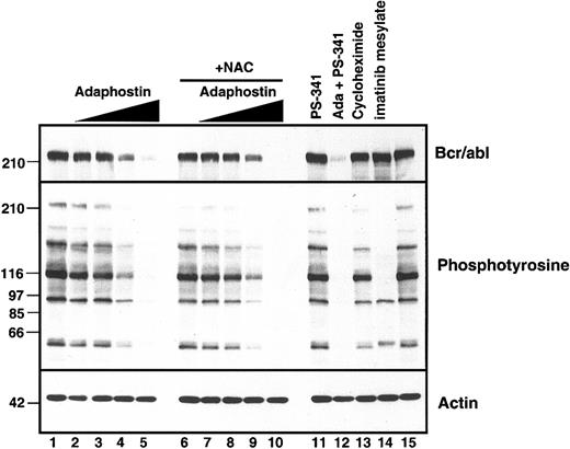
Previous experiments have shown that adaphostin and its parent drug AG957 cause p210Bcr/abl down-regulation in K562 cells within 8 hours.14,19 As is shown in lane 13 of Figure 4, this down-regulation is not explained merely by inhibition of protein synthesis. Instead, it has been suggested that cross-linking of p210Bcr/abl by the parent drug AG957 might contribute to Bcr/abl down-regulation.37 In view of these results, we examined the effect of NAC on adaphostin-induced Bcr/abl down-regulation. As indicated in Figure 4, NAC had little effect on adaphostin-induced Bcr/abl down-regulation or inhibition of tyrosine phosphorylation. Thus, adaphostin-induced ROS generation and p210Bcr/abl down-regulation are separable and distinct.
Adaphostin-induced Bcr/abl down-regulation is not affected by NAC. After 15 minutes of pretreatment with diluent (lanes 1-5) or 24 mM NAC (lanes 6-10), adaphostin was added to a final concentration of 0 (lanes 1 and 5), 1.25 (lanes 2 and 6), 2.5 (lanes 3 and 7), 5 (lanes 4 and 8), or 10 μM (lanes 5 and 10). Whole-cell lysates were prepared 8 hours later and subjected to SDS-PAGE followed by immunoblotting with antibodies that recognize c-abl (upper panel), phosphotyrosine (middle panel), or actin as a loading control (lower panel). In lanes 11-15, as controls, cells were treated with the proteasome inhibitor bortezomib (PS-341) at 50 nM in the absence or presence of 10 μM adaphostin, with 100 μM cycloheximide, or with 5 μM imatinib mesylate for 8 hours.
Adaphostin-induced Bcr/abl down-regulation is not affected by NAC. After 15 minutes of pretreatment with diluent (lanes 1-5) or 24 mM NAC (lanes 6-10), adaphostin was added to a final concentration of 0 (lanes 1 and 5), 1.25 (lanes 2 and 6), 2.5 (lanes 3 and 7), 5 (lanes 4 and 8), or 10 μM (lanes 5 and 10). Whole-cell lysates were prepared 8 hours later and subjected to SDS-PAGE followed by immunoblotting with antibodies that recognize c-abl (upper panel), phosphotyrosine (middle panel), or actin as a loading control (lower panel). In lanes 11-15, as controls, cells were treated with the proteasome inhibitor bortezomib (PS-341) at 50 nM in the absence or presence of 10 μM adaphostin, with 100 μM cycloheximide, or with 5 μM imatinib mesylate for 8 hours.
Relationship between adaphostin action and GSH levels
The hypothesis that ROS production contributes to the cytotoxicity of adaphostin suggests that (1) this agent might cause a decrease in endogenous GSH levels and (2) deliberate GSH depletion might sensitize cells to adaphostin. Both of these predictions were confirmed.
To assay for possible adaphostin-induced GSH depletion, K562 cells were treated with adaphostin for varying lengths of time and then treated with mBCl, which forms a fluorescent adduct on covalent binding to GSH. Treatment with BSO, an inhibitor of GSH synthesis, served as a positive control for these experiments. As indicated in Figure 5A, 10 μM adaphostin caused a 30% decrease (n = 3, P < .05) in GSH levels at l hour. Interestingly, GSH levels tended to recover at later time points, raising the possibility that K562 cells adapted by increased GSH synthesis. Nonetheless, these results provide independent confirmation that adaphostin induces oxidative stress.
GSH depletion potentiates adaphostin toxicity. (A) K562 cells were incubated with diluent (3 hours) or 10 μM adaphostin for the indicated length of time. Cells were then stained with 100 μM mBCl for 15 minutes and assayed for fluorescence. K562 cells incubated with 1 mM BSO for 24 hours were used as a positive control for GSH depletion. Cells incubated without mBCl provided a negative control. Error bars are mean ± SD of 3 independent experiments. *P < .02 compared to control. **P < .05 compared to control. (B) K562 cells were treated for 24 hours with diluent, 2.5 μM adaphostin, or 5 μM imatinib mesylate in the presence of 0, 100 μM, 500 μM, 1 mM, or 2 mM BSO (added 10 minutes prior to adaphostin or imatinib mesylate). At the completion of the incubation, samples were stained with PI and analyzed for flow microfluorimetry for cells containing less than 2N DNA content. Error bars are mean ± SD of 3 independent experiments. (C) K562 cells were treated for 4 hours with 1 mM BSO (•) or diluent (○). After addition of the indicated concentration of adaphostin, incubation in the presence or absence of BSO was continued for an additional 24 hours. At the completion of the incubation, cells were resuspended in drug-free medium and assayed for clonogenic survival. Error bars, ± 1 SD from quadruplicate samples.
GSH depletion potentiates adaphostin toxicity. (A) K562 cells were incubated with diluent (3 hours) or 10 μM adaphostin for the indicated length of time. Cells were then stained with 100 μM mBCl for 15 minutes and assayed for fluorescence. K562 cells incubated with 1 mM BSO for 24 hours were used as a positive control for GSH depletion. Cells incubated without mBCl provided a negative control. Error bars are mean ± SD of 3 independent experiments. *P < .02 compared to control. **P < .05 compared to control. (B) K562 cells were treated for 24 hours with diluent, 2.5 μM adaphostin, or 5 μM imatinib mesylate in the presence of 0, 100 μM, 500 μM, 1 mM, or 2 mM BSO (added 10 minutes prior to adaphostin or imatinib mesylate). At the completion of the incubation, samples were stained with PI and analyzed for flow microfluorimetry for cells containing less than 2N DNA content. Error bars are mean ± SD of 3 independent experiments. (C) K562 cells were treated for 4 hours with 1 mM BSO (•) or diluent (○). After addition of the indicated concentration of adaphostin, incubation in the presence or absence of BSO was continued for an additional 24 hours. At the completion of the incubation, cells were resuspended in drug-free medium and assayed for clonogenic survival. Error bars, ± 1 SD from quadruplicate samples.
To determine whether GSH depletion sensitizes cells to adaphostin, K562 cells were treated with adaphostin in the absence or presence of BSO. BSO by itself did not induce apoptosis or enhance the cytotoxicity of imatinib mesylate (Figure 5B). In contrast, it markedly enhanced the induction of apoptosis by low adaphostin concentrations. Further experiments demonstrated that BSO caused a 3.9-fold decrease in adaphostin IC50 (SD 1.1, n = 3, P = .05) in colony-forming assays (Figure 5C).
Effects of AG957 and its quinone derivative on ROS production and cytotoxicity
Examination of the structure of adaphostin and its parent drug AG957 (Figure 6A) suggested that the dihydroquinone moiety might be the source of the ROS. If so, then treating cells with an oxidized version of this pharmacophore would be predicted to generate more ROS and more cytotoxicity. Because the quinone analog of adaphostin was not available, this hypothesis was tested by comparing the effects of AG957 and NSC 767537 (Figure 6A). The quinone elicited a greater increase in DCF fluorescence than AG957 at equivalent concentrations of 30 μM (Figure 6B). At the same dose, the quinone induced more apoptosis in K562 cells as well (Figure 6C). Consistent with these results, K562 cells (Figure 6D) and CFU-Gs from patients with CML (Figure 6E) were more sensitive to inhibition of colony formation by the quinone than the parent agent. When the abilities of AG957 and the quinone to down-regulate p210Bcr/abl were compared, however, the quinone was less effective than AG957 (Figure 6F), again dissociating the antiproliferative effects of this class of agents from their ability to down-regulate Bcr/abl.
Discussion
In the present study, we report that adaphostin induces early up-regulation of intracellular ROS followed by DNA strand breaks, all of which precede the onset of apoptotic changes. The ability of the antioxidant NAC to inhibit these changes and GSH depletion to facilitate them provides strong evidence that ROS production plays a role in the cytotoxicity of adaphostin. Consistent with this second mechanism of action, adaphostin was observed to induce apoptosis in a variety of Bcr/abl- leukemia samples and cell lines. These results have potentially important implications for the future clinical development of this agent.
Previous studies demonstrated that adaphostin inhibits Bcr/abl kinase activity in vitro.19 Subsequent investigation, however, indicated that down-regulation of Bcr/abl-induced phosphorylation occurred much more slowly after adaphostin exposure than after treatment with imatinib mesylate.14 Paradoxically, adaphostin-treated cells died more rapidly.14 These observations, coupled with the ability of adaphostin to kill Bcr/abl- cells,14,16-18 raised the possibility that adaphostin might have a second mechanism of action. Consistent with this possibility, we demonstrated adaphostin-induced ROS generation in Bcr/abl+ K562 cells (Figure 3A) and Bcr/abl- ML-1 cells (Figure 2D). Further analysis suggested that both peroxides and superoxide might be elevated after adaphostin treatment (Figure 3A,D).
Several observations suggest that elevated ROS play a critical role in the cytotoxicity of adaphostin. First, elevated ROS appear early (Figure 3B) relative to DNA strand breaks (Figure 2C), p53 up-regulation (Figure 2A), and subsequent apoptotic changes (Figure 1 and Mow et al14 ). Second, concurrent treatment with the antioxidant NAC diminishes adaphostin-induced ROS production, DNA damage, and cytotoxicity (Figures 2, 3). Third, BSO-induced GSH depletion enhances the cytotoxicity of adaphostin (Figure 5). Fourth, NSC 76537, a quinone derived from the parent tyrphostin AG957, showed both increased ability to up-regulate ROS and enhanced antiproliferative activity (Figure 6). Collectively, these observations led to the suggestion that adaphostin causes generation of ROS, which lead to free radical-induced DNA damage followed by apoptotic biochemical changes. This proposed mechanism of action provides a potential explanation for the ability of adaphostin to kill cells lacking Bcr/abl (Figure 1).14,16-19
Continued interest in adaphostin stems, at least in part, from its ability to selectively kill myeloid progenitors from patients with CML.14,19,38 Although this selectivity appears to be at odds with a mechanism in which adaphostin up-regulates ROS, studies of Sattler et al39 provide a potential explanation for this paradox. In particular, Bcr/abl expression by itself up-regulates ROS.39 Conceivably this Bcr/abl-induced ROS elevation makes it easier for adaphostin-induced oxidative stress to exceed a critical threshold and induce apoptosis in Bcr/abl-expressing cells as compared to normal myeloid progenitors. Although further studies are required to confirm this explanation, the ability of oxidizing agents to selectively kill neoplastic cells has ample precedent. In particular, arsenic trioxide, which has also been shown to up-regulate ROS,40,41 selectively induces differentiation followed by apoptosis in progranulocytes containing the promyelocytic leukemia-retinoic-acid receptor (PML/RAR) fusion protein compared to their normal counterparts.
Interestingly, the generation of ROS and induction of cytotoxicity appear to be, at least in part, separable from 2 other aspects of adaphostin function. Although previous studies have indicated that adaphostin inhibits Bcr/abl kinase in vitro, it was difficult in the present study to detect inhibition of Bcr/abl-mediated tyrosine phosphorylation independent of Bcr/abl down-regulation (Figure 4). These results raise questions about the ability of adaphostin to inhibit Bcr/abl kinase in situ. Previous studies have also demonstrated that adaphostin induces p210Bcr/abl down-regulation.14,19 The observation that adaphostin was more potent than AG957 at down-regulating Bcr/abl and inducing cytotoxicity raised the possibility that these 2 processes might be linked. In the present study, however, Bcr/abl down-regulation is clearly a separate and distinct aspect of adaphostin function. In particular, NAC treatment diminished adaphostin-induced cytotoxicity (Figure 3E) but had little effect on adaphostin-induced Bcr/abl down-regulation (Figure 4). Because Bcr/abl conveys resistance to a wide variety of apoptosis-inducing agents,42 it is possible that this ability to down-regulate Bcr/abl contributes to the effectiveness of adaphostin as an inducer of apoptosis in Bcr/abl+ cells.
Collectively, the present results have important implications for the future study of adaphostin. The demonstration that this agent has at least 2 mechanisms of action in Bcr/abl-containing cells suggests that it might be difficult for leukemia cells to develop resistance. Consistent with this possibility, we have been unable to generate adaphostin-resistant K562 cells in vitro (S.H.K. and J.C., unpublished observations, January 2000 through March 2003). Coupled with the demonstration that this agent remains active in imatinib mesylate-resistant cells,14 these observations suggest that further study of adaphostin in Bcr/abl-derived malignancies might be reasonable. In addition, the demonstration that adaphostin induces apoptosis in AML and CLL specimens in vitro suggests that the activity of this agent against hematologic malignancies might be broader than originally envisioned. These observations should be considered in future clinical trials of this novel agent.
Prepublished online as Blood First Edition Paper, August 14, 2003; DOI 10.1182/blood-2003-02-0562.
Supported in part by National Institutes of Health grants R01 CA 85972 (S.H.K., A.T.), F32 CA93055 (J.C.), and R01 CA91542 (N.E.K.) and a predoctoral fellowship from the Mayo Foundation (J.H.). The authors have no potential financial conflicts of interest to report.
The publication costs of this article were defrayed in part by page charge payment. Therefore, and solely to indicate this fact, this article is hereby marked “advertisement” in accordance with 18 U.S.C. section 1734.
We thank Michael Kastan for the kind gift of ML-1 cells, Junjie Chen for anti-Chk2 antibody, Sherman Stinson and Keith Bible for providing advice that enabled measurement of adaphostin levels, Greg Gores for the use of his fluorometer, Jackie Greer for assistance in procuring AML specimens, Timothy Kottke and Phyllis Svingen for technical assistance, and Deb Strauss for secretarial assistance.


This feature is available to Subscribers Only
Sign In or Create an Account Close Modal