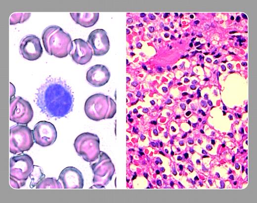
Peripheral smear (left) shows a typical hairy cell with multiple cytoplasmic projections, and violet staining nucleus quite distinct from the dense blue-black of a mature lymphocyte. Marrow biopsy (right) shows an infiltrate of hairy cells with the characteristic open, “fried egg” appearance of the cytoplasm.
Copyright © 2003 by The American Society of Hematology
2003


 Peter Maslak, Memorial Sloan-Kettering Cancer Center
This feature is available to Subscribers Only
Sign In or Create an Account Close Modal