Abstract
Monocytes from patients with sickle cell disease (SCD) are in an activated state. However, the mechanism of activation of monocytes in SCD is not known. Our studies showed that placenta growth factor (PlGF) activated monocytes and increased mRNA levels of cytokines (tumor necrosis factor-α [TNF-α] and interleukin-1β [IL-1β]) and chemokines (monocyte chemotactic protein-1 [MCP-1], IL-8, and macrophage inflammatory protein-1β [MIP-1β]) in both normal monocytes and in the THP-1 monocytic cell line. This increase in mRNA expression of cytochemokines was also reflected in monocytes derived from subjects with SCD. We studied the PlGF-mediated downstream cellular signaling events that caused increased transcription of inflammatory cytochemokines and chemotaxis of THP-1 monocytes. PlGF-mediated cytochemokine mRNA and protein expression was inhibited by PD98059 and wortmannin, inhibitors of mitogen-activated protein kinase kinase (MAPK/MEK) kinase and phosphatidylinositol-3 (PI3) kinase, respectively, but not by SB203580, a p38 kinase inhibitor. PlGF caused a time-dependent transient increase in phosphorylation of extracellular signal–regulated kinase-1/2 (ERK-1/2), which was completely inhibited by wortmannin, indicating that activation of PI3 kinase preceded MEK activation. PlGF also induced transient phosphorylation of AKT. MEK and PI3 kinase inhibitors and antibody to Flt-1 abrogated PlGF-induced chemotaxis of THP-1 monocytes. Overexpression of a dominant-negative AKT or a dominant-negative PI3 kinase p85 subunit in THP-1 monocytes attenuated the PlGF-mediated phosphorylation of ERK-1/2, cytochemokine secretion, and chemotaxis. Taken together, these data show that activation of monocytes by PlGF occurs via activation of Flt-1, which results in activation of PI3 kinase/AKT and ERK-1/2 pathways. Therefore, we propose that increased levels of PlGF in circulation play an important role in the inflammation observed in SCD via its effects on monocytes.
Introduction
The clinical manifestations of sickle cell disease (SCD) include chronic hemolytic anemia, frequent infections, and recurrent episodes of painful crises.1-7 Vascular occlusion leading to episodes of painful crises and damage to various end organs is the major cause of morbidity and mortality in SCD.1,3,6,8 Hebbel and coworkers have shown that the extent of adherence of sickle red blood cells (SS RBCs) to cultured endothelial cells appears to parallel the clinical severity of vaso-occlusive events in SCD.9 However, SCD patients with an identical defect in their β-globin genes show wide variability in the frequency and severity of vaso-occlusive crises and can remain asymptomatic for prolonged periods.2 This could be due to the epistatic effects of coinherited genes such as α-globin and γ-globin,10-13 both of which have been shown to alter disease severity.13,14 However, the effects of α- and γ-globin gene expression are not always predictable, suggesting that there must be factors other than those directly related to the RBC in the pathophysiology of vaso-occlusion in SCD.
Clinically, leukocytosis is associated with increased frequency of vaso-occlusive crisis (VOC).15,16 Moreover, a chronically elevated white cell count is a clear indicator of mortality,17 frequency of acute chest syndrome,8,18 and development of stroke19,20 in SCD. Because polymorphonuclear neutrophils (PMNs) and monocytes are activated during infection and inflammation,15,21 we hypothesize they may play a role in the initiation and/or potentiation of vaso-occlusive episodes. Treatment of individuals with SCD with hydroxyurea causes a decrease in white blood cell count and a decrease in myeloperoxidase activity, indicating that reduced leukocytosis and leukocyte activation results in improvement in the incidence of vaso-occlusive crises.22-24 A recent study25 shows that monocytes in SCD patients are in an activated state, because they express more interleukin-1β (IL-1β) and tumor necrosis factor-α (TNF-α) compared with normal monocytes. Furthermore, mononuclear cells from SCD patients can activate cultured endothelial cells, as judged by the increased expression of cell adhesion molecules and tissue factor in endothelial cells.25 However, the mechanism by which monocytes in SCD patients become activated is relatively not understood.
In the accompanying article by Perelman et al,26 beginning on page 1506, we show that plasma levels of placenta growth factor (PlGF) are higher in SCD patients compared with healthy controls and that they correlate with disease severity. The data also suggest that the higher levels of PlGF present in patients with SCD may be due to hypoxia, increased erythropoiesis, and erythropoietin concentrations due to the anemia. We26 have also shown that PlGF activates mononuclear cells (MNCs), resulting in increased gene expression of cytokines (TNF-α, IL-1β) and chemokines (monocyte chemotactic protein-1 [MCP-1], IL-8, and macrophage inflammatory protein-1β [MIP-1β]).
The present study shows that PlGF activated the monocyte fraction of the MNCs. We have examined the cellular signaling mechanism by which PlGF interaction with peripheral blood monocytes (PBMs) and THP-1 monocytes results in increased expression of proinflammatory cytochemokines. We show that interaction of PlGF with Flt-1 receptor on monocytes caused downstream activation of phosphatidylinositol-3 (PI3) kinase/AKT and extracellular signal–regulated kinase-1/2 (ERK-1/2) pathways, which increased expression of aforementioned inflammatory cytokines and chemokines. We also show that PlGF-induced activation of Src kinase, PI3 kinase/AKT, and mitogen-activated protein (MAP) kinase in monocytes promotes chemotaxis of monocytes.
Materials and methods
Media and other cell culture reagents were obtained from Invitrogen (Grand Island, NY). Genistein, PP2 (4-amino-5-(4-chlorophenyl)-7-(t-butyl) pyrazolo [3,4-d] pyrimidine), wortmannin, LY294002 (2-(4-morpholinyl)-8-phenyl-4H-1-benzopyran-4-1), PD98059 (2′-amino-3′-methoxyflavone), and actinomycin D were obtained from Calbiochem (La Jolla, CA). SB203580 and U73122 were purchased from Biomol (Plymouth Meeting, PA). 32P–uridine triphosphate (32P-UTP) was obtained from ICN Biomedical (Irvine, CA). Antibodies to AKT, phospho AKT, and phospho ERK-1/2 were purchased from Cell Signaling Technology (Beverly, MA). Antibodies to ERK-1/2 and all other secondary antibodies were purchased from Santa Cruz Biotechnology (Santa Cruz, CA). DuoSet enzyme-linked immunosorbent assay (ELISA) development system for quantitation of chemokines, antihuman vascular endothelial growth factor-receptor-1 (VEGF-R1) (Flt-1), and recombinant human placenta growth factor was obtained from R&D Systems (Minneapolis, MN). All other reagents not otherwise listed were obtained from Sigma (St Louis, MO).
Isolation of human peripheral blood monocytes
Blood was obtained from healthy volunteers and sickle cell anemia patients (HbSS) after obtaining informed consent according to a protocol approved by the institutional review committee at the University of Southern California Comprehensive Sickle Cell Center, Los Angeles County-University of Southern California Hospital. Human peripheral blood monocytes were isolated from blood collected in EDTA (ethylenediaminetetraacetic acid) as anticoagulant.27 Briefly, 10 volumes of blood sample (30 mL) were mixed with 1 volume (3 mL) of a solution composed of 6% dextran 500 in 0.9% NaCl. The tube was allowed to stand at room temperature for 45 minutes, which resulted in the sedimentation of erythrocytes. The leukocyte-rich plasma was harvested, layered over Nyco-prep media, density 1.068 g/mL (Accurate Chemical and Scientific, Westbury, NY) at a ratio of 2:1, and centrifuged at 600g for 15 minutes. The monocyte fraction was further purified according to manufacturer's instructions. Finally, these cells were allowed to adhere for 1 to 2 hours in a 60-mm Petri dish in the tissue-culture incubator. By this procedure nonadherent lymphocytes were removed. Monocytes isolated by this procedure had purity in the range of about 90% as assessed by labeling with MO2–fluorescein isothiocyanate (MO2-FITC) antibody (Coulter Diagnostics, Hialeah, FL) followed by FACScan (Becton Dickinson, San Jose, CA) analysis as previously described27 with a yield of 55% to 75%.
Cell culture
THP-1, a promonocytic cell line, was obtained from the American Type Culture Collection (ATCC) (Manassas, VA). Cells were cultured in RPMI 1640 medium containing 10% heat-inactivated fetal bovine serum.
RNase protection assay
THP-1 monocytes were treated with PlGF for various times, and total RNA was isolated from cultured cells with TriZOL reagent (Life Technologies, Bethesda, MD). Assays were performed with a custom-made Riboquant Multi-Probe RNase protection assay system (Pharmingen, San Diego, CA). In brief, the isolated RNA (10 μg) was hybridized with 32P-labeled probes overnight at 56°C followed by RNase digestion according to manufacturer's instructions. Glyceraldehyde-3-phosphate dehydrogenase (GAPDH) was used as an internal control. After digestion, the protected fragments were resolved on a 5% denaturing polyacrylamide gel and transferred to Whatman filter paper no. 3, which was dried and later exposed to x-ray film. The intensity of bands corresponding to TNF-α, MIP-1β, IL-1β, MCP-1, IL-8, and GAPDH mRNA were analyzed using a gel documentation system. Values are expressed as relative expression of mRNA normalized to housekeeping GAPDH mRNA.
Quantification of chemokines
THP-1 cells (1 × 10⁶/mL) were incubated in serum-free RPMI 1640 in the presence or absence of PlGF (250 ng/mL). At the end of the indicated periods, ranging from 1 to 24 hours, the medium was collected and cell debris removed by low-speed centrifugation at 1500 rpm for 10 minutes. Supernatant was stored at –80°C until further use. Levels of chemokines (MIP-1β, MCP-1, and IL-8) in the clarified supernatants were assayed using specific DuoSet ELISA development system (R&D Systems) according to the manufacturer's instructions.
Western blot analysis of MAPK and AKT
For Western blot analysis, THP-1 cells were incubated in RPMI 1640 medium containing 2% fetal bovine serum (FBS) for 12 to 18 hours. Medium was aspirated and replaced with fresh medium prior to treatment with PlGF. Where indicated, cells (5 × 10⁶ cells) were incubated with pharmacologic inhibitors for 30 minutes prior to PlGF treatment. At the end of the treatment, medium was aspirated and cells were lysed in 100 μL of 1× SDS sample buffer. The lysate was centrifuged at 14 000g for 20 minutes at 4°C and the supernatant heated at 95°C to 100°C for 5 minutes. An aliquot (10 μg) of supernatant was subjected to electrophoresis on a 10% sodium dodecyl sulfate–polyacrylamide gel electrophoresis (SDS-PAGE) gel followed by transfer to a nitrocellulose membrane (Bio-Rad, Hercules, CA). Activation of AKT and ERK-1/2 was assessed using 1:1000 dilutions of antiphospho-specific antibodies (Cell Signaling Technology). Horseradish peroxidase–conjugated secondary antibodies were used to develop the membrane. The protein bands were detected with Supersignal chemiluminescence substrate (Pierce Biotechnology, Rockford, IL). The intensity of bands was quantified by scanning utilizing an Alpha Imager 2000 gel documentation system (Alpha Innotech, San Leandro, CA). Blots were stripped and reprobed using a 1:1000 dilution of antibodies against the nonphosphorylated forms of AKT and ERK-1/2 to monitor protein loading.
Chemotaxis assay
Chemotaxis was assayed in 96-well plates (Neuro Probe, Gaithersburg, MD) having Transwell inserts of 5-μm pore size. Briefly, THP-1 monocytes were washed twice and resuspended in RPMI 1640 medium containing 2% serum and then loaded onto inserts at 5 × 10³ cells per 50 μL for each well; 30 μL RPMI 1640 medium containing the chemoattractants (PlGF/MCP-1) at the indicated concentrations was placed in the bottom compartment. After 2 hours of incubation at 37°C with 5% CO2, cells were scraped from the upper chamber and washed with phosphate-buffered saline (PBS) (100 μL) to remove nonmigrated cells. This was followed by addition of PBS containing 2 mM EDTA. After 15 minutes at 4°C, the plate was centrifuged at 200g in a Jouan Centrifuge CT-422 with a plate carrier. The filter was removed, and cells in the well were counted by the trypan blue exclusion method. To investigate the effect of inhibitors involved in the signal transduction, THP-1 cells were preincubated for 30 minutes with the indicated inhibitors before loading onto Transwell inserts. Each sample was tested in triplicate.
Transfection of monocytes
THP-1 cells were transfected with a plasmid expressing a dominant-negative (DN) mutant of AKT and a dominant-negative mutant PI3 kinase (DNM-Δp85), generously provided by David Ann, USC School of Pharmacy, Los Angeles. Ten micrograms of plasmid were incubated with 10 μL lipofectamine (Invitrogen) in 100 μL serum-free Dulbecco modified Eagle medium (DMEM) for 15 minutes. The liposome-DNA complexes were added to the THP-1 cell suspension (2 × 10⁶ cells per milliliter) and incubated at 37°C for 24 hours in tissue-culture incubator as described.28 These cells were then treated with PlGF to determine the activation of ERK-1/2.
Results
Expression of cytokines and chemokines in monocytes in healthy and SS patients
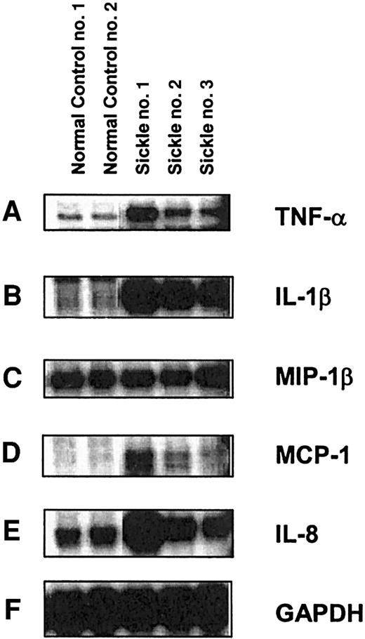
Previous studies have shown that both polymorphonuclear neutrophils (PMNs)21 and monocytes29 are in activated state in SCD, which may contribute to increased adherence of PMNs/monocytes to endothelium. Moreover, it has been shown that intracellular levels of TNF-α and IL-1β are higher in monocytes of SCD patients compared with African American and white controls.29 Thus, we examined the mRNA expression of a panel of inflammatory cytokines and chemokines by RNase protection assay (RPA) analysis in RNA isolated from peripheral blood monocytes (PBMs) from subjects with SCD and from healthy controls. As shown in Figure 1, expression of TNF-α (Figure 1A) and IL-1β (Figure 1B) was several fold higher in monocytes of SCD relative to healthy subjects. Expression of the chemokines MCP-1 (Figure 1D) and IL-8 (Figure 1E) were also several fold higher in monocytes from SCD compared with healthy controls. However, MIP-1β expression (Figure 1C) was only modestly increased by 1.2- to 1.4-fold.
RPA analysis of mRNA expression of cytokines and chemokines in monocytes derived from healthy and SCD patients. Peripheral blood monocytes were isolated from the whole blood of SCD patients and healthy individuals. RNA was isolated using TriZOL reagent, and 2 μg RNA from each sample was analyzed by RNase protection assay using a Riboquant Multi-Probe RNase protection assay system (Pharmingen). RPA was performed with riboprobes for (A) TNF-α, (B) IL-1β, (C) MIP-1β, (D) MCP-1, (E) IL-8, and (F) GAPDH.
RPA analysis of mRNA expression of cytokines and chemokines in monocytes derived from healthy and SCD patients. Peripheral blood monocytes were isolated from the whole blood of SCD patients and healthy individuals. RNA was isolated using TriZOL reagent, and 2 μg RNA from each sample was analyzed by RNase protection assay using a Riboquant Multi-Probe RNase protection assay system (Pharmingen). RPA was performed with riboprobes for (A) TNF-α, (B) IL-1β, (C) MIP-1β, (D) MCP-1, (E) IL-8, and (F) GAPDH.
Role of placenta growth factor in activation of monocytes
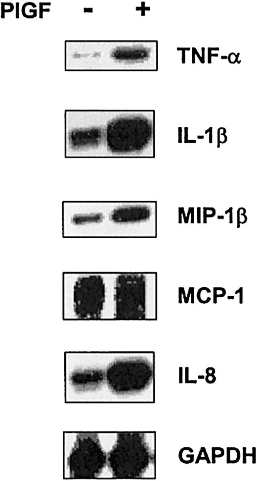
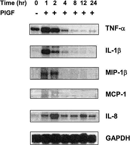
These studies indicated that monocytes in SCD were in activated state in vivo, generating cytokines and chemokines. We have also shown that PlGF, a factor produced by erythroid cells, was responsible for activation of the mononuclear cell fraction, composed of monocytes and lymphocytes.26 Furthermore, we show that plasma PlGF concentration was directly related to the incidence of vaso-occlusive events.26 Thus, we hypothesized that interaction of PlGF with the circulating monocytes may induce expression of proinflammatory cytochemokines. As shown in Figure 2, treatment of PBMs from healthy individuals with PlGF caused an increase in the mRNA expression of cytokines (TNF-α and IL-1β) and chemokines (MCP-1, IL-8, and MIP-1β) as determined using RPA analysis. Similarly, treatment of THP-1, a promonocytic cell line, with PlGF caused a time-dependent increase in the expression of the same cytokines and chemokines (Figure 3). Because both primary monocytes and the THP-1 monocytic cells exhibited a similar profile of cytochemokine expression in response to PlGF, we used THP-1 cells for subsequent studies.
Effect of PlGF on mRNA expression of cytokines and chemokines in normal peripheral blood monocytes. Peripheral blood monocytes isolated from whole blood of healthy individuals were treated with PlGF (250 ng/mL) for 2 hours. RPA was performed with riboprobes as indicated in Figure 1. Representative data are shown from 3 sets of experiments.
Effect of PlGF on mRNA expression of cytokines and chemokines in normal peripheral blood monocytes. Peripheral blood monocytes isolated from whole blood of healthy individuals were treated with PlGF (250 ng/mL) for 2 hours. RPA was performed with riboprobes as indicated in Figure 1. Representative data are shown from 3 sets of experiments.
Up-regulation of cytokine and chemokine mRNA expression in THP-1 monocytes by PlGF. THP-1 cells were treated with PlGF for time periods of 1 to 24 hours, and RNA was isolated; 10 μg RNA samples were hybridized with 32P-labeled antisense mRNA probes and digested with RNase and T1 nuclease. The protected hybridized probe fragments were resolved on 5% polyacrylamide gel. The intensity of radioactive bands in the autoradiogram was quantitated by the Alpha Imager documentation system. The relative mRNA levels were determined by normalizing band intensities of TNF-α, IL-1β, MIP-1β, MCP-1, and IL-8 with that of GAPDH housekeeping gene. The data presented are representative of 1 of 3 replicate experiments.
Up-regulation of cytokine and chemokine mRNA expression in THP-1 monocytes by PlGF. THP-1 cells were treated with PlGF for time periods of 1 to 24 hours, and RNA was isolated; 10 μg RNA samples were hybridized with 32P-labeled antisense mRNA probes and digested with RNase and T1 nuclease. The protected hybridized probe fragments were resolved on 5% polyacrylamide gel. The intensity of radioactive bands in the autoradiogram was quantitated by the Alpha Imager documentation system. The relative mRNA levels were determined by normalizing band intensities of TNF-α, IL-1β, MIP-1β, MCP-1, and IL-8 with that of GAPDH housekeeping gene. The data presented are representative of 1 of 3 replicate experiments.
Effect of PlGF treatment on the expression of cytokines and chemokines by THP-1 cells
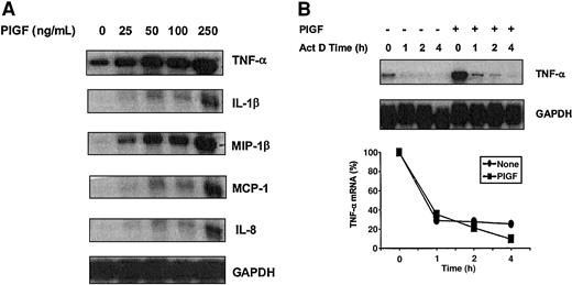
As shown in Figure 4A, there was a dose-dependent effect of PlGF (25 to 250 ng/mL) on the mRNA expression of cytokine and chemokines in THP-1 monocytes, with optimal effect at 250 ng/mL. This dose of PlGF was used in all experiments unless otherwise indicated. We then measured if increased mRNA expression of cytokines and chemokines in THP-1 monocytes was due to increased stability of mRNA. Extremely low basal levels of IL-1β, MCP-1, IL-8, and MIP-1β mRNA in THP-1 monocytes precluded determination of mRNA half-life of these proinflammatory cytochemokines in response to PlGF. We could measure TNF-α mRNA stability due to a significant basal level of expression. To measure the mRNA half-life (t1/2) of TNF-α, the transcriptional inhibitor actinomycin D was added to the culture medium 2 hours after PlGF treatment, and mRNA was analyzed at different time points. As shown in Figure 4B, the t1/2 of TNF-α mRNA in untreated THP-1 monocytes was 45 minutes, which did not change significantly upon PlGF treatment (t1/2 = about 50 minutes) in agreement with the previous results.30
Effect of PIGF dose on mRNA expression of cytochemokines and stability of TNF-α mRNA. (A) Dose-dependent increase in the expression of cytochemokines in PlGF-treated THP-1 cells: THP-1 cells were treated with different concentrations of PlGF ranging from 25 to 250 ng/mL for 2 hours. RNA was isolated by TriZOL reagent, and 10 μg RNA was used for RPA analysis. (B) Effect of PlGF on the stability of TNF-α mRNA. After the treatment of THP-1 cells with PlGF (250 ng/mL) for 2 hours, actinomycin D (10 μg/mL) was added. At the indicated times, RNA was isolated. TNF-α mRNA expression was determined by RPA as described in Figure 2. The relative mRNA levels were determined by normalizing band intensities of TNF-α with that of GAPDH housekeeping gene. Representative data are shown from 2 experiments.
Effect of PIGF dose on mRNA expression of cytochemokines and stability of TNF-α mRNA. (A) Dose-dependent increase in the expression of cytochemokines in PlGF-treated THP-1 cells: THP-1 cells were treated with different concentrations of PlGF ranging from 25 to 250 ng/mL for 2 hours. RNA was isolated by TriZOL reagent, and 10 μg RNA was used for RPA analysis. (B) Effect of PlGF on the stability of TNF-α mRNA. After the treatment of THP-1 cells with PlGF (250 ng/mL) for 2 hours, actinomycin D (10 μg/mL) was added. At the indicated times, RNA was isolated. TNF-α mRNA expression was determined by RPA as described in Figure 2. The relative mRNA levels were determined by normalizing band intensities of TNF-α with that of GAPDH housekeeping gene. Representative data are shown from 2 experiments.
Effect of cell signaling inhibitors on PlGF-induced cytokine and chemokine gene expression
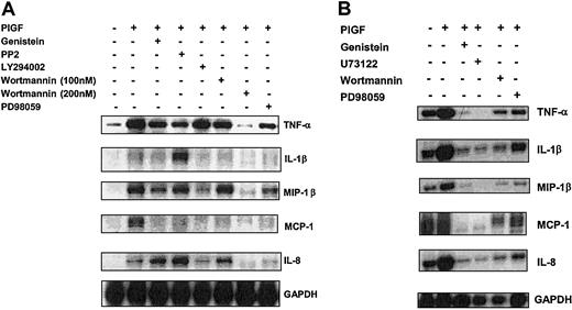
Because PlGF treatment of THP-1 monocytes caused increased gene expression of both cytokines (TNF-α and IL-1β) and chemokines (MCP-1, IL-8, and MIP-1β), we determined the effect of pharmacologic inhibitors, which have been previously shown to be specific for specific kinases in the signaling pathway. As shown in Figure 5A, pretreatment of THP-1 cells with genistein (25 μg/mL), a protein tyrosine kinase inhibitor,31 inhibited PlGF-induced mRNA expression of TNF-α (25%), IL-1β (15%), MCP-1 (35%), and MIP-1β (25%), indicating that PlGF-induced activation of protein tyrosine kinase is involved in induction of the proinflammatory cytochemokines.
Effect of signaling inhibitors on PlGF-induced cytokine and chemokine gene expression. THP-1 cells (A) and PBMs (B) were preincubated with pharmacologic inhibitors for 30 minutes, followed by treatment with PlGF (250 ng/mL) for 2 hours. RNA was isolated, and 10 μg was used for RPA analysis. The data are representative of 3 sets of experiments.
Effect of signaling inhibitors on PlGF-induced cytokine and chemokine gene expression. THP-1 cells (A) and PBMs (B) were preincubated with pharmacologic inhibitors for 30 minutes, followed by treatment with PlGF (250 ng/mL) for 2 hours. RNA was isolated, and 10 μg was used for RPA analysis. The data are representative of 3 sets of experiments.
As shown in Figure 5A, pretreatment of THP-1 monocytes with PD98059, a specific inhibitor of mitogen-activated protein kinase kinase (MAPKK, or MEK),32 attenuated the mRNA expression of cytokines (TNF-α and IL-1β) and chemokines (MCP-1, IL-8, and MIP-1β) by about 50%, indicating that activation of MAP kinase was required. Notably, SB203580, a selective p38 MAP kinase inhibitor,33 did not significantly affect the PlGF-induced expression of these cytochemokines (data not shown). Similar results were obtained with these inhibitors when peripheral blood monocytes were used (Figure 5B). These results indicate that PlGF-mediated cytochemokine induction does not involve p38 MAP kinase activation but involves MEK activation.
Role of PI3 kinase and Src kinase in PlGF-induced mRNA expression of cytochemokines
To delineate the cellular components upstream of the MAP kinases, we used LY294002 and wortmannin, PI3 kinase inhibitors,33 and PP2, a Src kinase inhibitor.34 As shown in Figure 5A, wortmannin (100 to 200 nm) reduced mRNA expression of these cytochemokines by 50% to 75% in a dose-dependent manner. However, LY294002, a weak PI3 kinase inhibitor,33 inhibited expression somewhat moderately (about 40%). These results indicate that PlGF-induced activation of PI3 kinase is required for increased expression of these cytochemokines.
PlGF-induced expression of TNF-α was inhibited by 50% in response to PP2, while expression of MIP-1β and MCP-1 was mildly affected (15% to 20%). These results indicate that activation of the Src kinase family of tyrosine kinase is required for downstream activation of mRNA of TNF-α. In contrast, the expression of IL-1β and IL-8 increased by about 50% in the presence of PP2, indicating a negative regulatory role of Src kinase on IL-1β– and IL-8–inducing pathways.
Time- and dose-dependent effect of PlGF-induced secretion of chemokines by THP-1 monocytes
We then determined if the increased transcription of cytochemokines translated to increased protein expression. The release of chemokines from THP-1 monocytes treated with PlGF (250 ng/mL) was determined over a period of 24 hours. As shown in Figure 6, PlGF treatment resulted in a time-dependent (1, 2, 4, 6, 8, and 24 hours) increase in the secretion of MCP-1, IL-8, and MIP-1β, with optimal secretion occurring at 4 to 6 hours. There was a 3-fold increase in MIP-1β secretion (481 ± 12.5 pg/mL in PlGF-treated versus 150 ± 16 pg/mL in untreated THP-1 monocytes), a 2-fold increase in IL-8 secretion (56 ± 3.0 pg/mL in PlGF-treated versus 27 ± 3 pg/mL in untreated), and a 2.5-fold increase in secretion of MCP-1 (16 ± 0.25 pg/mL in PlGF-treated versus 6.5 ± 0.15 pg/mL in untreated) in response to treatment of THP-1 monocytes with PlGF (250 ng/mL). As shown in Figure 7, PlGF exhibited an increase in the secretion of chemokines (MCP-1, IL-8, and MIP-1β) in a dose-dependent manner (25 to 250 pg/mL).
Time course of PlGF-mediated secretion of chemokines from THP-1 cells. THP-1 cells (1 × 10⁶/mL) were treated with PlGF (250 ng/mL) for time periods of 1 to 24 hours. Cell-free supernatant was collected, and 100 μL from the total 1 mL supernatant was used for determining levels of (A) MIP-1β, (B) MCP-1, and (C) IL-8 using the DuoSet ELISA from R&D Systems. Data are means ± SD of n = 3, each experiment run in duplicate.
Time course of PlGF-mediated secretion of chemokines from THP-1 cells. THP-1 cells (1 × 10⁶/mL) were treated with PlGF (250 ng/mL) for time periods of 1 to 24 hours. Cell-free supernatant was collected, and 100 μL from the total 1 mL supernatant was used for determining levels of (A) MIP-1β, (B) MCP-1, and (C) IL-8 using the DuoSet ELISA from R&D Systems. Data are means ± SD of n = 3, each experiment run in duplicate.
Effect of PlGF dose on the secretion of chemokines from THP-1 cells. THP-1 cells (1 × 10⁶/mL) were treated with PlGF (25 to 250 ng/mL) for time periods of 1 and 4 hours. The supernatant was collected and (A) MIP-1β, (B) MCP-1, and (C) IL-8 levels quantified as indicated in Figure 6. Data are expressed as means ± SD of n = 3.
Effect of PlGF dose on the secretion of chemokines from THP-1 cells. THP-1 cells (1 × 10⁶/mL) were treated with PlGF (25 to 250 ng/mL) for time periods of 1 and 4 hours. The supernatant was collected and (A) MIP-1β, (B) MCP-1, and (C) IL-8 levels quantified as indicated in Figure 6. Data are expressed as means ± SD of n = 3.
Effect of kinase inhibitors on PlGF-induced secretion of chemokines by THP-1 cells
Because PlGF-induced chemokine (MCP-1, IL-8, and MIP-1β) mRNA expression was attenuated by PI3 kinase inhibitor (wortmannin) and MAP kinase inhibitor (PD98059), we examined the effect of these inhibitors on PlGF-induced secretion of these chemokines. As shown in Figure 8, both wortmannin and PD98059 reduced release of MIP-1β and MCP-1 by about 50%. IL-8 secretion was inhibited 25% by wortmannin and 30% by PD98059. The extent of inhibition of the release of these chemokines by PI3 kinase inhibitor (wortmannin) and MAP kinase inhibitor (PD98059) was similar to the effect on transcription of these genes (Figure 5). Genistein reduced the secretion of MIP-1β and MCP-1 by 40% and 50%, respectively. In contrast, genistein had little effect on PlGF-induced secretion of IL-8. PP2, a Src kinase inhibitor, inhibited PlGF-induced secretion of MIP-1β and MCP-1 by about 25%. However, PP2 inhibitor did not significantly affect PlGF-induced secretion of IL-8 (Figure 8).
Effect of signaling inhibitors on the PlGF-induced release of chemokines in THP-1 monocytes. THP-1 monocytes (1 × 10⁶/mL) were treated with various pharmacologic inhibitors for 30 minutes. THP-1 cells were transfected with a plasmid expressing a dominant-negative (DN) mutant of AKT (DNAKT) and a dominant-negative mutant PI3 kinase (DNM-Δp85) as described in “Materials and methods.” The cells were then treated with PlGF for 4 hours, and the cell-free supernatant was collected and analyzed for the release of (A) MIP-1β, (B) MCP-1, and (C) IL-8. Data are expressed as means ± SD of n = 3.
Effect of signaling inhibitors on the PlGF-induced release of chemokines in THP-1 monocytes. THP-1 monocytes (1 × 10⁶/mL) were treated with various pharmacologic inhibitors for 30 minutes. THP-1 cells were transfected with a plasmid expressing a dominant-negative (DN) mutant of AKT (DNAKT) and a dominant-negative mutant PI3 kinase (DNM-Δp85) as described in “Materials and methods.” The cells were then treated with PlGF for 4 hours, and the cell-free supernatant was collected and analyzed for the release of (A) MIP-1β, (B) MCP-1, and (C) IL-8. Data are expressed as means ± SD of n = 3.
PlGF-induced activation of AKT in THP-1 cells
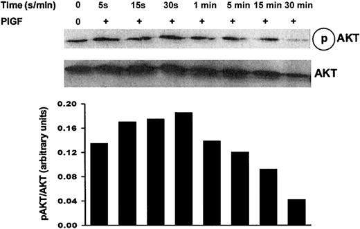
Because PI3 kinase inhibitor attenuated PlGF-induced gene expression of cytokines and chemokines, we examined the role of the serine kinase AKT, a downstream target of PI3 kinase. The full activation of AKT by growth factors requires phosphorylation on serine 473, which correlates with AKT kinase activity.35 THP-1 monocytes were stimulated with PlGF (250 ng/mL) for 5, 15 and 30 seconds, and 1, 5, 15, and 30 minutes, followed by a Western blot analysis using an antibody to AKT phosphorylated at Ser473. AKT phosphorylation increased from 5 to 30 seconds (Figure 9) and gradually declined thereafter. By 30 minutes the phosphorylation of AKT decreased below the basal level, indicating that AKT phosphorylation by PlGF is transient. Increased phosphorylation of AKT occurred without changes in its protein levels (Figure 9).
PlGF-induced AKT phosphorylation in THP-1 cells. THP-1 cells were grown overnight in RPMI medium containing 2% FBS. Cells were then starved for 2 hours in serum-free media and treated with PlGF for the indicated times. Cell lysates were analyzed for phospho AKT by Western blot using antiphospho AKT (pSer473) antibody. Equal loading of the gels was confirmed by probing with antibody specific for unphosphorylated AKT (lower panel, AKT).
PlGF-induced AKT phosphorylation in THP-1 cells. THP-1 cells were grown overnight in RPMI medium containing 2% FBS. Cells were then starved for 2 hours in serum-free media and treated with PlGF for the indicated times. Cell lysates were analyzed for phospho AKT by Western blot using antiphospho AKT (pSer473) antibody. Equal loading of the gels was confirmed by probing with antibody specific for unphosphorylated AKT (lower panel, AKT).
PlGF-induced activation of ERK-1/2 in THP-1 cells
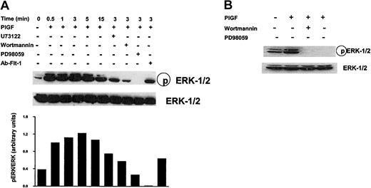
Previous studies have shown that monocytes express Flt-1 (VEGF-R1) but not Flk-1 (VEGF-R2) receptor.36 We examined whether PlGF phosphorylated ERKs and required signaling thru Flt-1. Reverse transcriptase–polymerase chain reaction (RT-PCR) analysis of THP-1 monocytes revealed the presence of Flt-1 mRNA but not flk-1 mRNA (data not shown). As shown in Figure 10A, treatment of THP-1 monocytes with PlGF (250 ng/mL) increased the phosphorylation of both ERK-1 (p44) and ERK-2 (p42). The peak phosphorylation of ERK-1/2 was observed at 3 minutes and declined thereafter. Moreover, antibody to Flt-1 inhibited PlGF-induced phosphorylation of ERK-1 and ERK-2 by 100% and 75%, respectively (Figure 10A).
PIGF-induced ERK phosphorylation in both THP-1 cells and monocytes. (A) PlGF-induced ERK-1/2 activity in THP-1 monocytes. THP-1 cells were incubated with RPMI containing 2% serum overnight. The cells were then incubated with fresh serum-free media for 2 hours and then treated with PlGF (250 ng/mL) for the indicated time periods. Where indicated THP-1 cells were preincubated with U73122 (1 μM), wortmannin (200 nM), PD98059 (10 μM), and Ab–Flt-1 (5 μg/mL) for 30 minutes before treatment with PlGF. Cell lysates were analyzed for ERK-1 and -2 activities by Western blot using antiphospho MAPK (Thr202/Tyr204) antibodies (upper panel, pERK-1/2). Blots were stripped and reprobed with antibodies specific for unphosphorylated ERK-1/2 (lower panel, ERK-1/2). The experiment shown is representative of 3 experiments. (B) PlGF-induced ERK-1/2 activity in peripheral blood monocytes. Peripheral blood monocytes (1 × 10⁶/mL) were incubated in RPMI medium containing 1% FBS for 1 hour followed by treatment with PlGF (250 ng/mL) for 3 minutes. Prior to treatment, the cells were either incubated with wortmannin (200 nM) or PD98059 (10 μM) for 30 minutes. Cell lysates were prepared, and 10 μg of the protein was subjected to 10% SDS-PAGE. The blot was probed with phospho ERK-1/2 antibody (upper panel), stripped, and reprobed with total ERK-1 antibody (lower panel) to show equal loading.
PIGF-induced ERK phosphorylation in both THP-1 cells and monocytes. (A) PlGF-induced ERK-1/2 activity in THP-1 monocytes. THP-1 cells were incubated with RPMI containing 2% serum overnight. The cells were then incubated with fresh serum-free media for 2 hours and then treated with PlGF (250 ng/mL) for the indicated time periods. Where indicated THP-1 cells were preincubated with U73122 (1 μM), wortmannin (200 nM), PD98059 (10 μM), and Ab–Flt-1 (5 μg/mL) for 30 minutes before treatment with PlGF. Cell lysates were analyzed for ERK-1 and -2 activities by Western blot using antiphospho MAPK (Thr202/Tyr204) antibodies (upper panel, pERK-1/2). Blots were stripped and reprobed with antibodies specific for unphosphorylated ERK-1/2 (lower panel, ERK-1/2). The experiment shown is representative of 3 experiments. (B) PlGF-induced ERK-1/2 activity in peripheral blood monocytes. Peripheral blood monocytes (1 × 10⁶/mL) were incubated in RPMI medium containing 1% FBS for 1 hour followed by treatment with PlGF (250 ng/mL) for 3 minutes. Prior to treatment, the cells were either incubated with wortmannin (200 nM) or PD98059 (10 μM) for 30 minutes. Cell lysates were prepared, and 10 μg of the protein was subjected to 10% SDS-PAGE. The blot was probed with phospho ERK-1/2 antibody (upper panel), stripped, and reprobed with total ERK-1 antibody (lower panel) to show equal loading.
THP-1 monocytes were then treated with specific inhibitors prior to the addition of PlGF. In the presence of wortmannin, a PI3 kinase (PI3K) inhibitor, ERK phosphorylation was completely inhibited in PlGF-stimulated monocytes. Moreover, U73122 (1 μM), a phospholipase C inhibitor,37 completely abrogated PlGF-induced ERK-1 phosphorylation and reduced the phosphorylation of ERK-2 by more than 80%. As a positive control, PD98059, an inhibitor of MEK, completely abolished ERK phosphorylation in THP-1 monocytes treated with PlGF.
These above results were then confirmed in peripheral blood monocytes. As shown in Figure 10B, PlGF-induced phosphorylation of ERK at 5 minutes (optimal phosphorylation) was attenuated by both PI3 kinase and MEK inhibitors. Taken together, these findings indicate that activation of Flt-1 receptor, phospholipase C, and PI3 kinase/AKT by PlGF is required for activation of ERK in monocytes.
Effect of overexpression of dominant-negative isoforms of PI3 kinase and AKT on PlGF-induced phosphorylation of ERK
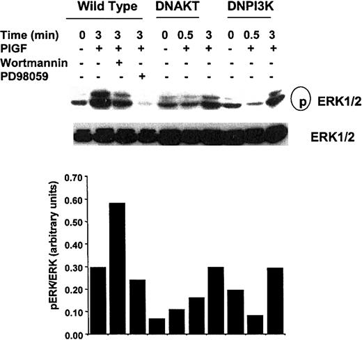
To specifically address the role of PI3 kinase/AKT in PlGF-induced modulation of ERK phosphorylation, we overexpressed a dominant-negative AKT (DNAKT) and a dominant-negative PI3 kinase p85 subunit (DNM-Δp85). As shown in Figure 11, overexpression of DNAKT in THP-1 monocytes significantly attenuated the ability of PlGF to promote phosphorylation of ERK-1/2 by 0.5 to 3 minutes. Notably, PlGF caused optimal phosphorylation of ERK at 3 minutes (Figure 10). Similarly, transfection of THP-1 monocytes with DNM-Δp85 resulted in reduced phosphorylation of ERK-1/2 in response to PlGF. These results are consistent with the data obtained with pharmacologic inhibitors and confirm that PlGF-induced phosphorylation of ERK involves activation of PI3 kinase/AKT.
Effect of transfection of dominant-negative AKT and DNM-Δp85 PI3 kinase on PlGF-mediated ERK phosphorylation in THP-1 monocytes. THP-1 cells were transfected with either DNAKT or DNM-Δp85 PI3 kinase as described in “Materials and methods.” Transfected cells were treated with PlGF for 3 minutes. Cell lysates were probed with antibody to ERKs as described in Figure 10. The data are representative of 3 experiments.
Effect of transfection of dominant-negative AKT and DNM-Δp85 PI3 kinase on PlGF-mediated ERK phosphorylation in THP-1 monocytes. THP-1 cells were transfected with either DNAKT or DNM-Δp85 PI3 kinase as described in “Materials and methods.” Transfected cells were treated with PlGF for 3 minutes. Cell lysates were probed with antibody to ERKs as described in Figure 10. The data are representative of 3 experiments.
Effect of expression of dominant-negative PI3 kinase and AKT on PlGF-induced secretion of chemokines by THP-1 monocytes
Our studies showed that wortmannin reduced PlGF-induced secretion of chemokines (Figure 8). To unequivocally establish that the effect of pharmacologic inhibitor of PI3 kinase was specific, we expressed dominant-negative PI3 kinase and AKT in THP-1 cells and studied PlGF-induced secretion of chemokines (MIP-1β, MCP-1, and IL-8). As shown in Figure 8, expression of dominant-negative PI3K (DNPI3K) in THP-1 reduced PlGF-induced secretion of IL-8 by 45%, of MCP-1 by 30%, and of MIP-1β by 30%. Similar results were obtained when THP-1 cells were transfected with DNAKT. These data are in accord with the results obtained with the use of wortmannin, a PI3 kinase inhibitor. These studies and data obtained with wortmannin, a PI3 kinase inhibitor, show that PlGF-induced signaling resulting in the secretion of chemokines involves activation of PI3 kinase.
Chemotactic response of THP-1 monocytes to PlGF
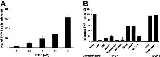
We determined whether PlGF increased monocyte chemotaxis. As shown in Figure 12A, PlGF caused a dose-dependent increase in the migration of monocytes. To determine whether the migration of THP-1 monocytes required a PlGF gradient, we performed a checkerboard analysis. As shown in Table 1, the maximal increase in the migration of monocytes occurred in response to a positive concentration gradient between the 2 compartments of the Boyden chamber, which decreased as the difference in the concentration of PlGF between lower and upper compartments declined. These results indicate that the response of THP-1 monocytes to PlGF was a result of chemotaxis and not of chemokinesis, as has been observed with VEGF-induced chemotaxis of monocytes.38 Notably, MCP-1 (100 ng/mL), a positive control in these experiments, induced chemotaxis of THP-1 monocytes to a level slightly lower than PlGF alone (Figure 12B).
Dose-dependent effect of PlGF on the chemotaxis of THP-1 monocytes. (A) THP-1 cells (5 × 10³ cells) were added to the upper compartment of a Neuro Probe chemotaxis chamber. The indicated concentrations of either PlGF and/or MCP-1 were added to the lower compartments, the chambers were incubated for 2 hours at 37°C, and then the migrated cells were stained and counted as described in the text. Results show mean ± SD of 4 independent experiments. (B) For inhibitor studies, THP-1 monocytes were pretreated with the respective pharmacologic inhibitors or antibody for 30 minutes. THP-1 cells transfected with the dominant-negative mutants of AKT and PI3 kinase were also used for the chemotaxis assay.
Dose-dependent effect of PlGF on the chemotaxis of THP-1 monocytes. (A) THP-1 cells (5 × 10³ cells) were added to the upper compartment of a Neuro Probe chemotaxis chamber. The indicated concentrations of either PlGF and/or MCP-1 were added to the lower compartments, the chambers were incubated for 2 hours at 37°C, and then the migrated cells were stained and counted as described in the text. Results show mean ± SD of 4 independent experiments. (B) For inhibitor studies, THP-1 monocytes were pretreated with the respective pharmacologic inhibitors or antibody for 30 minutes. THP-1 cells transfected with the dominant-negative mutants of AKT and PI3 kinase were also used for the chemotaxis assay.
PIGF induces THP-1 monocyte chemotaxis
Checkerboard analysis . | . | . | . | . | . | |||||
|---|---|---|---|---|---|---|---|---|---|---|
. | Upper compartment PIGF (nM) . | . | . | . | . | |||||
| Lower compartment (PIGF nM) | None | 0.5 | 1.0 | 2.5 | 5.0 | |||||
| None | 12.0 | 24.0 | 36.0 | 38.0 | 34.0 | |||||
| 0.5 | 48.0 | 145.0 | 117.0 | 46.0 | 56.0 | |||||
| 5.0 | 312.0 | 262.0 | 270.0 | 90.0 | 62.0 | |||||
Different concentrations of PIGF were added to the upper and/or lower compartments of the Neuro Probe chamber. The number of migrated cells were counted for each sample after 2 hours. Data from a representative experiment are shown.
Effect of pharmacologic inhibitors on PlGF-induced THP-1 monocyte chemotaxis
We determined whether pharmacologic inhibitors of PlGF signaling also resulted in attenuation of monocyte chemotaxis. As shown in Figure 12B, genistein (tyrosine kinase inhibitor), PP2 (Src kinase inhibitor), U73122 (phospholipase C inhibitor), wortmannin (PI3 kinase inhibitor), and PD98059 (MAP kinase inhibitor) reduced THP-1 chemotaxis in the range of 90% to 100%. Moreover, expression of dominant-negative AKT (DNAKT) and a dominant-negative PI3 kinase p85 subunit (DNM-Δp85) in THP-1 monocytes resulted in reduced (about 50%) chemotaxis in response to PlGF (Figure 12B). However, MCP-1–induced chemotaxis of THP-1 monocytes was not inhibited by wortmannin (Figure 12B). Similar results with MCP-1–induced chemotaxis have been observed in peripheral blood monocytes.39 Furthermore, we observed that antibody to Flt-1 completely abrogated PlGF-induced chemotaxis of THP-1 cells (Figure 12B). These results indicate that PlGF-induced cellular signaling plays a role in chemotaxis of monocytes.
Discussion
Patients with sickle cell disease have an abnormally high baseline leukocyte count, which is also observed in sickle transgenic mice.40,41 Moreover, it has been observed that the baseline count of leukocytes is a very strong independent risk factor for severity of disease.41 The abnormally high levels of leukocytes above the baseline reflect ongoing chronic inflammation at steady state. Recent studies show that monocytes isolated from SCD blood are in the activated state.25 Furthermore, increased levels of IL-1β and TNF-α42,43 and IL-844 have been reported in sera from patients with SCD. However, the nature of stimuli, which cause monocyte activation in SCD, is not known.
A recent study shows that erythroid cells, but not other hematopoietic cells, express both vascular endothelial growth factor-A (VEGF-A) and placenta growth factor (PlGF).45 We have observed26 that PlGF is increased in SCD plasma and correlates with disease severity. Moreover, PlGF was also shown to activate the mononuclear cell fraction.26 In the present study, we show that the monocyte fraction that expresses the Flt-1 receptor46 in the mononuclear cells is the population affected by PlGF.
We show that monocytes isolated from SCD patients are in a highly activated state as demonstrated by increased gene expression of cytokines (TNF-α and IL-1β) as well as chemokines (MCP-1, IL-8, and MIP-1β) compared with monocytes from healthy individuals. These data are consistent with studies of Belcher et al,25 who observed increased protein expression of both TNF-α and IL-1β in monocytes isolated from patients with SCD. Our data show increased gene expression of cytokines (TNF-α and IL-1β) and chemokines (MCP-1, IL-8, and MIP-1β) in peripheral blood monocytes (PBMs) treated with PlGF. We also utilized a monocyte cell line THP-1, which showed an identical pattern of gene expression in response to PlGF. Thus, we utilized the THP-1 monocytes as a model system for elucidating the signaling mechanism in response to PlGF.
Our studies show that PlGF treatment of monocytes causes a dose-dependent increase in the expression of proinflammatory cytochemokines. The increase in gene expression of these cytokines (TNF-α and IL-1β) and chemokines (MCP-1, IL-8, and MIP-1β) could have occurred as a result of increased mRNA stability. Because basal levels of IL-1β, MCP-1, IL-8, and MIP-1β mRNA in THP-1 monocytes were not detectable, one could not determine mRNA half-life of these inflammatory cytokines in response to PlGF. However, there was a basal level of expression of TNF-α mRNA, which increased several fold in response to PlGF. Therefore, half-life of TNF-α was determined. The half-life for TNF-α mRNA did not change in PlGF-treated and in untreated THP-1 monocytes. Our studies show that PlGF treatment of THP-1 monocytes causes 3-fold, 2-fold, and 1.5-fold increases in the secretion of chemokines MIP-1β, IL-8, and MCP-1, respectively, indicating a concomitant increase in the translation of the corresponding mRNA.
We evaluated the signaling pathways involved in PlGF-mediated induction of cytochemokines by using various low-molecular-weight pharmacologic inhibitors known to block specific signaling events. Specific inhibition of MEK-1/2 kinase (ERK-1/2 phosphorylating kinase) reduced mRNA expression of these cytokines in THP-1 monocytes and PBMs. In both PBMs and THP-1 monocytes, PlGF-induced expression of these cytochemokines was not inhibited by a selective p38 kinase inhibitor.33 These results are in sharp contrast to the effect of PlGF in trophoblast cells wherein PlGF causes activation of p38 kinase but not ERK-1/2 activity.47 However, PlGF has been shown to cause activation of ERK-1/2 activity in human umbilical vein endothelial cells (HUVECs).47 We also observed that PlGF caused a time-dependent increase in the phosphorylation of ERK-1/2 in THP-1 monocytes. These studies thus show that PlGF-induced signaling in monocytes involves activation of MEK kinase leading to phosphorylation of ERK-1/2.
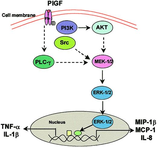
Moreover, PlGF-induced ERK-1/2 phosphorylation was inhibited by antibody to Flt-1, indicating that PlGF signaling in monocytes involves interaction with its receptor Flt-1.36 We next delineated the intermediate signaling events that occur between Flt-1 binding to PlGF and its downstream MEK activation. We show that inhibitors of PI3 kinase and phospholipase C completely inhibited PlGF-induced phosphorylation of ERKs. To confirm that PI3 kinase/AKT is upstream of ERK, we studied the effect of PlGF on the phosphorylation of AKT. These studies revealed that PlGF causes transient activation of AKT (phosphorylation of AKT at serine 473) at a very early time point (0.5 minutes) followed by a decrease at 1 minute. Furthermore, transfection of THP-1 cells with either dominant-negative AKT plasmid (DNAKT) or dominant-negative PI3 kinase p85 subunit resulted in a decrease in the phosphorylation of ERK-1/2 by PlGF. Taken together these studies show that PlGF-mediated activation of ERK-1/2 involves Flt-1, activation of phospholipase C, and PI3 kinase/AKT as illustrated in Figure 13. Because PlGF-induced cytochemokine gene expression was inhibited about 50% by wortmannin and slightly less—about 40%—by LY294002 (a weak PI3 kinase inhibitor), these results suggest that PlGF-induced signaling leading to the expression of cytokines and chemokines is PI3 kinase dependent and independent. The PI3 kinase–dependent pathway involves activation of ERK-1/2 (Figure 13), while the PI3 kinase–independent pathway may involve activation of AP-1. Further studies are required to delineate which of the transcription factors (nuclear factor–κB [NF-κB], AP-1, Egr-1, CREB-1) may play a role in the regulation of PlGF-induced cytochemokine gene expression.
Working model of PlGF-induced intracellular signal transduction cascade in monocytes. On interaction of PlGF with Flt-1 on monocytes, protein tyrosine kinase, Src kinase, PI3 kinase/AKT, and MAP kinase pathways are activated. A parallel activation of phospholipase C occurs, which activates MAP kinase.
Working model of PlGF-induced intracellular signal transduction cascade in monocytes. On interaction of PlGF with Flt-1 on monocytes, protein tyrosine kinase, Src kinase, PI3 kinase/AKT, and MAP kinase pathways are activated. A parallel activation of phospholipase C occurs, which activates MAP kinase.
Recent studies have shown that VEGF-induced signaling in endothelial cells involves activation of Src and phospholipase C-γ1 (PLC-γ1).48 Our studies showing that PP2, a Src kinase inhibitor, blocks PlGF-induced expression of TNF-α indicates that Src activation is involved in downstream activation of TNF-α. In contrast, PlGF-induced expression of IL-1β and IL-8 was 50% increased in the presence of Src kinase inhibitor PP2, indicating a negative regulatory role of Src kinase on IL-1β and IL-8 expression. Similar negative regulatory roles have been observed with VEGF, where tissue factor expression is augmented by inhibition of PI3 kinase.49 We suggest that inhibition of Src kinase augments other pathways (PI3 kinase/AKT and ERK-1/2) leading to increased expression of IL-1β and IL-8. Based on these studies one can develop potential anti-inflammatory therapeutic targets to ameliorate monocyte activation in sickle cell disease and thus prevent vaso-occlusive crises. Hebbel and colleagues25 have shown that inhibition of the transcription factor NF-κB by sulfasalazine, an anti-inflammatory agent, showed beneficial effects in a few SCD patients.
Because monocytes are activated in response to PlGF, we determined whether binding of PlGF to Flt-1 in monocytes leads to functional response. Our studies show that PlGF causes chemotaxis of THP-1 monocytes in response to PlGF gradient, in agreement with a previous report.38 Our studies demonstrate that PlGF-induced chemotaxis of THP-1 monocytes required activation of Src kinase, PI3 kinase, and MEK kinase (ERK-1/2), indicating the importance of the Src-PI3K/AKT-MAP kinase pathway in regulating the monocyte chemotaxis in response to PlGF (Figure 13). Moreover, we show that chemotaxis of monocytes mediated by PlGF gradient was completely abrogated by antibody to Flt-1, indicating the importance of PlGF receptor Flt-1 in chemotaxis.
Our studies show that PlGF causes activation of monocytes resulting in the expression of cytochemokines and suggest that PlGF may play a role in inflammation in sickle cell disease. This is in accordance with the recent studies of Luttun et al,50 who showed that anti–Flt-1, but not anti–Flk-1, reduced the mobilization of bone marrow–derived myeloid progenitors into the circulation as well as inhibited the migration of Flt-1–expressing monocytes to the sites of inflammation in the arterial wall, thereby reducing progression of atherosclerotic plaques. Moreover, these studies50 show the importance of PlGF, but not VEGF, in inflammation and atherosclerotic plaque formation. The role of PlGF in inflammation is further supported by the finding51 that targeted overexpression of PlGF-2 in the skin of mice resulted in exaggerated inflammation and edema, whereas PlGF-deficient mice show greatly reduced inflammation in the skin.
Sickle cell disease has phenotypic characteristics of an inflammatory disease41 as indicated by the observation that one finds higher than normal leukocyte counts, elevated levels of serum cytokines, and an increase in soluble intracellular adhesion molecule-1 (ICAM-1) and vascular cell adhesion molecule-1 (VCAM-1). What triggers inflammation in endothelium and the surrounding environment of circulating SS RBCs is the subject of investigation. It has been shown that interaction of sickle red blood cells, but not normal RBCs, causes activation of endothelium,52 indicating that sickle RBCs can provoke an inflammatory response. Recent studies of Kaul and Hebbel53 show that hypoxia/reoxygenation can trigger an inflammatory response in transgenic sickle mice, but not in normal mice, indicating the role of inflammation in sickle cell disease.41 We suggest that, due to increased erythropoiesis in SCD, one finds increased levels of PlGF, which may act as a trigger on monocytes to produce cytochemokines, resulting in an inflammatory response.
In conclusion, we have presented data showing that placenta growth factor causes activation of monocytes resulting in the generation of proinflammatory cytokines and chemokines. The PlGF-induced signaling involves interaction with Flt-1 receptor followed by activation of PI3 kinase/AKT and MEK kinase (ERK-1/2). The possibility of PlGF activation of other pathways, such as NF-κB, is not ruled out, because inhibitors of PI3 kinase partially (50% to 75%) inhibit cytochemokine expression. The cytochemokines released from the monocytes can activate neutrophils and endothelial cells, augmenting cell adhesion molecule expression and vascular occlusion. Our results imply that PlGF is one of the factors in the plasma responsible for the activation of monocytes in sickle cell disease. Therapeutic strategies aimed at reducing monocyte activation may be beneficial in ameliorating vaso-occlusive crises in sickle cell disease. We suggest that both PlGF and Flt-1 are potential candidates for therapeutic modulation of inflammation in sickle cell disease.
Prepublished online as Blood First Edition Paper, April 10, 2003; DOI 10.1182/blood-2002-11-3423.
Supported by National Institutes of Health grant P60-HL-48484.
The publication costs of this article were defrayed in part by page charge payment. Therefore, and solely to indicate this fact, this article is hereby marked “advertisement” in accordance with 18 U.S.C. section 1734.
We thank David Ann, USC School of Pharmacy, Los Angeles, for the generous gift of dominant-negative mutants of AKT and PI3K. We deeply appreciate the assistance of Pat Corley, RN, who obtained the blood specimens.



This feature is available to Subscribers Only
Sign In or Create an Account Close Modal