Abstract
Slow recovery of T-cell numbers and function contributes to the high incidence of life-threatening infections after cytotoxic cancer therapies. We have tested the therapeutic potential of a novel class of superagonistic CD28–specific antibodies that induce polyclonal T-cell proliferation without T-cell receptor engagement in an experimental rat model of T lymphopenia. We show that in lethally irradiated, bone marrow–reconstituted hosts, CD28 superagonist is able to dramatically accelerate repopulation by a small inoculum of mature, allotype-marked T cells. CD28-driven recovery of CD4 cells was superior to that of CD8 T cells. CD28 superagonist– expanded CD4 T cells had maintained repertoire diversity and were functional both in vitro and in vivo, suggesting that treatment with a human CD28–specific superagonist will protect T-lymphopenic patients from opportunistic infections.
Introduction
T lymphocytes are essential components of both cell-mediated and humoral immune responses to pathogens. Consequently, their depletion in the course of cytotoxic antineoplastic therapy entails life-threatening risks of opportunistic infections.1-3 The slow recovery of T-cell numbers is because of the requirement for a functional thymus to provide the highly specialized microenvironment required for the selection of a self-tolerant and self–major histocompatibility complex (MHC)–restricted T-cell repertoire. Thymic atrophy, however, sets in with puberty, leaving residual peripheral T lymphocytes as the major source of initial T-cell regeneration in adults.4,5 In this situation, “spontaneous” repopulation of the immune system with T cells from the periphery has 2 components: slow, “homeostatic” proliferation independent of foreign antigen recognition, and rapid antigen-driven clonal expansion that reduces the complexity of the T-cell repertoire and consequently the spectrum of pathogens that can be recognized.5-7 While the production of neutrophils, required for immediate defense against bacteria, is successfully boosted by granulocyte colony-stimulating factor and granulocyte-macrophage colony-stimulating factor therapy,8 no universal T-cell growth factor is available to drive rapid and uniform expansion of mature T lymphocytes spared by the cytotoxic insult.
Rapid proliferation of resting primary T cells depends on the dual engagement of the T-cell receptor (TCR) and a costimulatory receptor, of which CD28 is the most potent one characterized so far.9 Costimulation by CD28 not only drives cycling of TCR-stimulated cells, it also protects the expanding cells from apoptosis,10 thereby leading to a rapid and long-lasting numeric expansion of the clones thus addressed. Consequently, T-cell expansion by costimulation with artificial TCR and CD28 ligands is a potent approach for polyclonal unbiased T-cell expansion. Using monoclonal antibodies (mAbs) to the TCR complex and to CD28 coimmobilized on artificial surfaces, this is indeed feasable in vitro.11 In vivo, however, such strategies are hampered by the need to copresent the 2 mAbs in an immobilized form. Moreover, polyclonal activation of T cells in vivo via TCR signals results in a burst of proinflammatory and proapoptotic cytokines known as the “cytokine storm,” and hence is not suitable for generalized stimulation of T-cell recovery.12,13 Thus, the need for TCR engagement has so far hampered the exploitation of the unique mitogenic potency of CD28 for the in vivo therapy of T lymphopenia.
Monoclonal antibodies to rat CD28 can be subdivided into 2 functionally distinct groups.14 Conventional CD28-specific mAbs are potent costimulators of T-cell proliferation in vitro provided that the TCR is also engaged; such antibodies, which have no mitogenic activity when used in isolation, had previously been described for human and mouse T cells as well.15,16 In contrast, “superagonistic” CD28–specific mAbs are potent inducers of T-cell proliferation both in vitro and in vivo even in the absence of TCR engagement.14 These rat CD28–specific as well as recently identified human CD28–specific superagonists recognize a unique epitope of CD28, distinct from the binding site for conventional CD28-specific mAbs.17
As we have previously shown, the potent mitogenic effect of CD28 superagonists addresses T cells of all subclasses regardless of antigen experience or their T-cell receptor isotype.14 Furthermore, we had found that polyclonal T-cell expansion with CD28 superagonists in vivo is accompanied by the expression of anti-inflammatory cytokines, most notably of interleukin-10 (IL-10),18 rather than by the toxic cytokine storm of proinflammatory mediators induced by agents that address the TCR complex, such as superantigens19 and CD3-specific mAb,12 and that this is accounted for by a transient “overexpansion” of regulatory T cells (Treg cells) in response to the CD28 superagonist.20 Together, these results suggested that CD28 superagonists may be used to numerically expand a collapsed T-cell compartment in vivo. In the present report, we have tested this hypothesis in an experimental rat model of T lymphopenia.
Materials and methods
Animal procedures
Lewis (LEW) rats used for mixed leukocyte reactions (MLRs) and skin transplantations were bred at the institute's facilities. Unmanipulated or thymectomized 10- to 13-week-old PVG rats (Harlan Winkelmann, Borchen, Germany) were irradiated with 10 Gy from an 137Cs source and reconstituted on the same day intravenously with 5 × 10⁷ syngeneic bone marrow cells. T cells prepared from lymph nodes of PVG.RT7b rats (Harlan Winkelmann) were purified by nylon wool and/or depleted of CD4 (OX35; Serotec, Oxford, United Kingdom) or CD8 (OX8; Serotec) cells by passage over T-cell recovery columns (Cedarlane, Hornby, ON, Canada) and were intravenously injected as given. Conventional and superagonistic CD28–specific mAbs JJ319 and JJ316, respectively, and isotype control mAb (MOPC-31, γ1κ) were obtained in a low endotoxin format (BD Pharmingen, Heidelberg, Germany) and injected in 1 mL of phosphate-buffered saline (PBS) intravenously or intraperitoneally with identical results. Blood was taken from anesthesized animals by orbital puncture. Preparation of lymphoid cells for fluorescence-activated cell sorter (FACS) or functional analysis followed standard procedures.14 Immunization with 100 μg keyhole limpet hemocyanin (KLH; SIGMA, Deisenhofen, Germany) was performed either intraperitoneally with alum-absorbed protein or intramuscularly with Titermax adjuvant (Alexis, San Diego, CA). Skin grafts were performed with the kind help of Dr C. Otto, Surgical Clinic of the University of Würzburg. Syngeneic and allogeneic skin grafts (2 × 2 cm) were placed on opposite sides of the chest and examined every third day.
Labeling and flow cytometry of lymphocytes
For carboxyfluoresceine diacetate succinimidyl ester (CFSE) labeling, 107 cells were incubated in PBS containing 2 μM CFSE (MoBiTec, Göttingen, Germany) for 10 minutes (transfer experiment) or 5 minutes (in vitro experiment) and washed twice before use in PBS containing 0.1% bovine serum albumin. For cell surface marker analysis, mAbs W3/25, OX-35 (both CD4), 3.4.1 (CD8β), OX-8 (CD8α), R73 (αβTCR), CD25 (OX-39), cytotoxic T lymphocyte–associated antigen (CTLA-4; WKH203), and HIS41 (RT7b) were obtained as fluorescein isothiocyanate (FITC), phycoerythrin (PE), or biotinylated conjugates from BD Pharmingen or Serotec, and used as described.14 Intracellular cytokine staining was performed on lymph node cells from KLH-primed rats restimulated in vitro for 5 days and treated for 4 hours with phorbol myristate acetate (PMA)/ionomycin and with Brefeldin A for the last 2 hours, with minor modifications of a previously published protocol.18 IL-4 was detected with mAb OX81-PE (BD Pharmingen), and interferon-γ (IFN-γ) was detected with DB1-FITC (Serotec). Nonspecific staining background was determined by excess cold antibody blockade. Intracellular staining for CTLA-4 followed the same protocol, but without in vitro restimulation. Samples were analyzed on a FACscan (Becton Dickinson, Heidelberg, Germany), collecting logarithmically amplified fluorescence signals from live-scatter gated cells.
In vitro assays
KLH-specific antibodies were detected by enzyme-linked immunosorbent assay (ELISA) using the rat κ-chain–specific biotinylated mAb MARK-1 (Serotec) and alkaline-phosphatase–conjugated streptavidin to detect KLH-specific rat antibodies. For costimulation, lymph node T cells were stimulated with immobilized anti-TCR (clone R73) plus soluble anti-CD28 (clone JJ319) as previously described21 at an initial density of 5 × 10⁴ cells/well in a final volume of 200 μL supplemented RPMI 1640 medium. The recall response to KLH was measured with 105 splenocytes/well. For MLRs, 1 × 10⁵ purified responder lymph node T cells and 105 irradiated lymph node stimulator cells were used. The incorporation of radioactivity from a 16 h/0.5 μCi (0.0185 MBq) pulse with 3H thymidine was measured by liquid scintillation counting from day 2 to 3 (costimulation and recall response) or from day 4 to 5 (MLR). Cytokine production by pooled superficial and mesenteric lymph node cells cultured at 106 cells/mL for the times indicated with 2.5 μg/mL Concanavalin A was measured by ELISA (BD Pharmingen), following the manufacturer's instructions.
Statistical analysis
Data are expressed as mean ± SD. Statistical analyses were performed by an unpaired t test.
Results
CD28 superagonists drive multiple rounds of T-cell division in vitro and in vivo
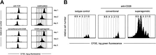
In order to compare the mitogenic activity of conventional and superagonistic anti-CD28 mAbs, we used cell division–associated dilution of the covalently cell-bound fluorescent dye CFSE. In keeping with the paradigm of costimulation, purified rat lymph node T cells showed little proliferation in response to TCR stimulation alone but underwent up to 5 cell divisions when costimulated in cell culture for 3 days with a conventional CD28-specific antibody (Figure 1A). As expected from previous results, conventional anti-CD28 by itself was unable to trigger proliferation. In contrast, the superagonistic mAb was as efficient as full TCR plus CD28-induced costimulation in driving multiple rounds of T-cell division in which virtually all input T cells participated. The mitogenic activity of the different types of CD28-specific mAbs was also compared in vivo (Figure 1B). One day after adoptive cell transfer of purified CFSE-labeled T cells into irradiated rats, the recipients received a single intravenous injection of 1 mg mAb or of an isotype-matched control antibody. Then 3 days later, an identical number of nucleated spleen cells from each group was analyzed by flow cytometry for the presence of CFSE-labeled cells. In animals treated with a mAb of irrelevant specificity or with a conventional CD28-specific mAb, few cells had progressed beyond one cell division, as is expected for the slow rate of homeostatic compared with antigen-driven proliferation.22 In contrast, the vast majority of donor cells recovered from rats treated with superagonistic anti-CD28 mAb had undergone 4 or more cell divisions, resulting in a dramatic increase in the recovery of transferred T cells. Thus, as previously reported for nonlymphopenic rats, these novel CD28-specific mAbs are potent T-cell mitogens in vivo.
Mitogenic activity of conventional and superagonistic anti-CD28 mAb. (A) In vitro proliferation of CFSE-labeled rat lymph node T cells induced by TCR stimulation alone, TCR plus CD28 costimulation, or by conventional or superagonistic CD28–specific mAb alone. (B) Cell divisions in transferred CFSE-labeled T cells after intravenous injection of conventional or superagonistic CD28–specific mAbs. Numbers indicate cell divisions of boxed populations.
Mitogenic activity of conventional and superagonistic anti-CD28 mAb. (A) In vitro proliferation of CFSE-labeled rat lymph node T cells induced by TCR stimulation alone, TCR plus CD28 costimulation, or by conventional or superagonistic CD28–specific mAb alone. (B) Cell divisions in transferred CFSE-labeled T cells after intravenous injection of conventional or superagonistic CD28–specific mAbs. Numbers indicate cell divisions of boxed populations.
Long-term T-cell repopulation by CD28 superagonist–mediated expansion of a small T-cell inoculum
In order to mimic the situation of massive T-cell depletion after cytotoxic therapy we used a model of lethal whole-body irradiation followed by transplantation of syngeneic bone marrow cells and a small number of mature resting lymph node T cells (5 × 10⁶, about 1/1000 of the normal T-cell compartment). As recipients and donors, rats of the PVG strain, which express the a-allele of the common leukocyte antigen CD45, were used. The congenic strain PVG.RT7b,which expresses the b-allele of CD45, was used as a source of mature T cells. By monitoring the expression of CD45b in the reconstituted animals, T cells derived by peripheral expansion of the mature inoculum (CD45b+) were distinguished from those derived from the transferred bone marrow cells by de novo differentiation in the thymus (CD45b–).
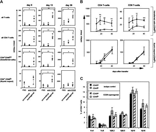
First, we compared the recovery of CD4 T cells in peripheral blood of reconstituted rats treated with 1 mg of conventional or superagonistic CD28–specific mAb on days 1 and 10 after cell transfer (Figure 2A). A dramatic increase in CD4 T cells derived from the CD45b+ inoculum was observed as early as one week after cell transfer in superagonist but not in conventional anti-CD28–treated animals. Importantly, even 5 weeks later, superagonist-treated animals contained about 20-fold higher counts of transferred CD4 T cells in their blood than PBS or conventional CD28-treated controls. Furthermore, recovery of endogenous thymus-derived CD4 T cells (CD45b–), which is initially delayed due to the requirement for thymic differentiation, was not negatively influenced by CD28 superagonist treatment. In a similar experiment, we used 5 × 10⁶ unseparated allotype-marked lymph node T cells containing approximately 80% CD4 and 20% CD8 T cells for reconstitution (Figure 2B). Both subsets were rapidly increased by CD28 superagonist treatment, with CD4 cells showing almost normal levels (around 1000 cells/μL) within 3 weeks. CD8 T cells, while clearly increased over control, were less markedly expanded. Again, recovery of endogenous T-cell subsets from bone marrow– derived cells, presumably via the thymic differentiation pathway, was delayed but unaffected by anti-CD28 therapy.
Long-term T-cell repopulation of T-lymphopenic rats by CD28 superagonist–driven expansion of a small T-cell inoculum. Lethally irradiated PVG (CD45a) rats received syngeneic bone marrow and 5 million CD45b T cells. (A) Recovery of transferred CD4+ T cells in rats after 2 injections (1 mg on days 1 and 10) of conventional or superagonistic CD28–specific mAbs. (B) Regeneration of CD4+ and CD8+ T cells in rats reconstituted with unseparated lymph node T cells and treated with superagonistic CD28–specific mAbs (•) or vehicle (PBS; ○) (1 mg on days 1 and 13). (C) Expression of TCR V segments by syngeneic (CD45a) and transferred CD4 T cells (CD45b) in spleens of rats 2 months after treatment with isotype control or CD28 superagonist. Means and SD are shown. *P < .05; **P < .01.
Long-term T-cell repopulation of T-lymphopenic rats by CD28 superagonist–driven expansion of a small T-cell inoculum. Lethally irradiated PVG (CD45a) rats received syngeneic bone marrow and 5 million CD45b T cells. (A) Recovery of transferred CD4+ T cells in rats after 2 injections (1 mg on days 1 and 10) of conventional or superagonistic CD28–specific mAbs. (B) Regeneration of CD4+ and CD8+ T cells in rats reconstituted with unseparated lymph node T cells and treated with superagonistic CD28–specific mAbs (•) or vehicle (PBS; ○) (1 mg on days 1 and 13). (C) Expression of TCR V segments by syngeneic (CD45a) and transferred CD4 T cells (CD45b) in spleens of rats 2 months after treatment with isotype control or CD28 superagonist. Means and SD are shown. *P < .05; **P < .01.
Our earlier in vitro and in vivo experiments as well as the data shown above (Figure 1) indicated that most T cells are induced to divide in response to CD28 superagonist stimulation. Nevertheless, we wanted to formally exclude the effects of a possibly contaminating superantigen that would address only T-cell subsets expressing certain Vβ segments. Accordingly, 2 months after treatment of rats that received irradiation, bone marrow transplantation, and T-cell reconstitution with either CD28 superagonist or an isotype control, we analyzed the representation of TCR V segments by flow cytometry (Figure 2C). We observed only very minor differences between T cells derived from the mature inoculum that had peripherally expanded and those that had been de novo generated in the thymus. Notably, these small deviations between thymus-derived and extrathymically expanded T cells were identical in animals treated with isotype control antibody and CD28 superagonist, ruling out a superantigen-like effect of CD28-driven T-cell expansion.
CD28 superagonist–expanded T cells are functional
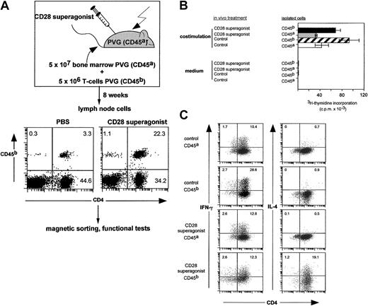
In order to assess the repopulation of peripheral lymphoid organs by the small inoculum of mature T cells expanded by CD28 superagonist stimulation, spleen and lymph node cells were prepared 2 months after initiation of the experiment. About 40% of lymph node CD4 T cells (Figure 3A) and one third of splenic CD4 T cells (not shown) were derived from the mature input cells in superagonist-treated recipients, while the equivalent figure was only about 7% in control animals. Thus, in spite of the competition imposed by the resumed thymic activity, which is known to have a negative influence on T-cell repopulation from the periphery,23 the mature T-cell inoculum expanded by CD28 superagonist therapy had persisted at high levels. In order to assess the capacity of these cells to respond to conventional TCR plus CD28 costimulation, we purified T cells derived from the mature inoculum and those derived by de novo differentiation in the thymus. Both populations derived from either superagonist-stimulated or control animals were readily induced to proliferate by a combination of plate-bound anti-TCR mAbs plus soluble conventional anti-CD28 (Figure 3B), and contained similar frequencies of IFN-γ–producing cells after activation (Figure 3C). Of note, T cells derived from the small mature inoculum introduced after irradiation 2 months earlier and expanded by CD28 superagonist therapy contained an increased frequency of IL-4–producing cells (Figure 3C), probably as a consequence of IL-4 induction by CD28 stimulation during the activation phase,18 which promoted T-helper type 2 differentiation in a fraction of the activated cells.
Functional analysis of lymph node T cells isolated from T-lymphopenic rats treated as in Figure 2B with CD28 superagonist or vehicle 2 months earlier. (A) Representation of endogenous (CD45–) and transferred (CD45b+) T cells. (B) Assessment of in vitro proliferative capacitiy to immobilized anti-TCR plus soluble anti-CD28. (C) Cytokine production by in vitro–costimulated cells.
Functional analysis of lymph node T cells isolated from T-lymphopenic rats treated as in Figure 2B with CD28 superagonist or vehicle 2 months earlier. (A) Representation of endogenous (CD45–) and transferred (CD45b+) T cells. (B) Assessment of in vitro proliferative capacitiy to immobilized anti-TCR plus soluble anti-CD28. (C) Cytokine production by in vitro–costimulated cells.
CD28-driven T-cell expansion in thymectomized recipients
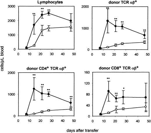
In order to evaluate to what extent restoration of the T-cell compartment from a small number of input T cells is also possible in the absence of a thymus, and to obtain animals in which the success of immune restoration could be tested in vivo without the interference of de novo differentiated T cells, we used thymectomized hosts for the following experiments. As before, rats received lethal irradiation followed by transfer of syngeneic bone marrow and 5 × 10⁶ allotype-marked lymph node T cells. A single injection of 1 mg superagonistic CD28–specific mAb, but not of an isotype control antibody, led to a rapid and dramatic recovery of circulating T-cell counts (Figure 4). Although this was followed by a slow decline, animals with CD28 superagonist treatment still had a clear advantage over those left to spontaneously recover 7 weeks later.
CD28-driven T-cell expansion in thymectomized rats. ATXBM PVG rats were reconstituted with 5 million CD45 allotype-marked T cells and injected with a single dose of 1 mg CD28 superagonist (•) or isotype control mAb (○). Peripheral blood was analyzed for total lymphocytes, donor-derived total T cells, and their CD4 and CD8 subsets as indicated. Means and SDs of 6 animals per group are shown. *P < .05; **P < .01.
CD28-driven T-cell expansion in thymectomized rats. ATXBM PVG rats were reconstituted with 5 million CD45 allotype-marked T cells and injected with a single dose of 1 mg CD28 superagonist (•) or isotype control mAb (○). Peripheral blood was analyzed for total lymphocytes, donor-derived total T cells, and their CD4 and CD8 subsets as indicated. Means and SDs of 6 animals per group are shown. *P < .05; **P < .01.
T-cell responses of rats repopulated with small T-cell numbers and treated with CD28 superagonist
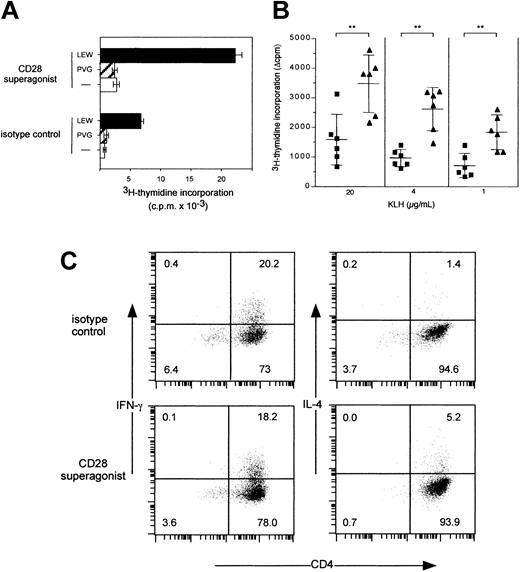
We next asked if the dramatic numeric expansion of T cells by CD28 stimulation in a highly lymphopenic setting is compatible with the maintenance of T-cell function. Initially, we compared the ability of adult-thymectomized, X-irradiated, bone marrow–reconstituted (ATXBM) PVG rats (RT1u) reconstituted 9 weeks earlier with 5 million mature T cells with or without a single injection of CD28 superagonist to respond to foreign MHC antigens by skin graft rejection in vivo and MLR in vitro. While syngeneic PVG skins healed well in both groups of animals, rejection of allogeneic LEW (RT1l) skin proceeded with similar kinetics in both isotype-treated and CD28 superagonist–treated groups, leading to complete destruction of the grafts within 2 weeks (data not shown). In vitro, both treated and control groups mounted proliferative responses to LEW allogeneic stimulator cells (Figure 5A), but lymph node cells from CD28 superagonist–treated animals were clearly superior to those from isotype-treated controls, indicating improved T-cell reactivity in the treatment group.
Functional capacitiy of T cells isolated from ATXBM PVG rats repopulated with small T-cell numbers and treated with CD28 superagonist or isotype control antibody. (A) Proliferative response to allogeneic (LEW) stimulator cells. (B) Proliferative recall response to KLH (▪ indicates isotype control; ▴, CD28 superagonist). Means and SDs are shown (n = 6). *P < .05; **P < .01. (C) Cytokine production after KLH restimulation.
Functional capacitiy of T cells isolated from ATXBM PVG rats repopulated with small T-cell numbers and treated with CD28 superagonist or isotype control antibody. (A) Proliferative response to allogeneic (LEW) stimulator cells. (B) Proliferative recall response to KLH (▪ indicates isotype control; ▴, CD28 superagonist). Means and SDs are shown (n = 6). *P < .05; **P < .01. (C) Cytokine production after KLH restimulation.
Next, we analyzed the responses to a protein model antigen, keyhole limpet hemocyanin (KLH), applied in vivo 3 weeks after T-cell reconstitution. High and similar titers of KLH-specific antibodies were detected in both treatment and control groups, indicating that even without artificial T-cell expansion, T-cell help for this highly immunogenic protein was saturating (data not shown). To quantify the presence of primed T cells, we therefore used the proliferative recall response of splenocytes prepared from these animals 4 weeks after immunization. At all concentrations of KLH tested, splenocytes from superagonist-pretreated rats responded with higher proliferation rates than those from animals treated with the isotype-matched control antibody (Figure 5B). In addition, we tested the ability of CD4 lymphoblasts generated by KLH restimulation to produce IFN-γ and IL-4 (Figure 5C). Both groups yielded comparable frequencies of cells producing IFN-γ, whereas the frequency of IL-4 producers was elevated in the superagonist-treated group. This confirmed our previous findings, shown in Figure 3C, that CD28 superagonist–driven expansion of CD4 T cells increases the T-helper type 2 compartment, while leaving T-helper type 1 function intact.
Regulatory T cells during acute CD28 stimulation and after CD28 superagonist–supported recovery from T lymphopenia
We have previously shown that the dramatic expansion of a pre-existing normal T-cell compartment by a single injection of CD28 superagonist is accompanied by a transient overrepresentation and activation of CD4+CD25+CTLA-4+ regulatory T cells (Treg cells).20 While this is the likely basis for the lack of toxicity of polyclonal T-cell activation by CD28 superagonists in vivo, an overrepresentation of Treg cells after CD28 superagonist–accelerated recovery from lymphopenia could impair overall immune responsiveness. Accordingly, we compared the frequencies of CD4+CD25+ cells in reconstituted animals treated either with the CD28 superagonist or with an isotype-matched control antibody. In all cases, coexpression of CTLA-4 was used to further confirm the Treg phenotype of the CD4+CD25+ cells analyzed (data not shown; and Lin and Hunig20 ).
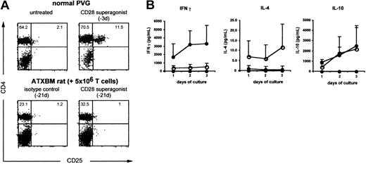
In agreement with our recent findings obtained in the LEW strain,20 the acute phase of polyclonal T-cell activation by CD28 superagonist in normal PVG rats was accompanied by a dramatic increase in the frequency of CD4+CD25+ cells (Figure 6A). In contrast, the frequency of CD25+ cells among CD4+ T cells 3 weeks after reconstitution of ATXBM rats was similar with and without CD28-assisted T-cell expansion (control: mean, 5.8 ± 2.9%; CD28 superagonist: mean, 3.3 ± 1.8%, n = 6) and to that found in normal PVG rats.
CD28-expanded T cells contain normal numbers of regulatory T cells. (A) Frequencies of CD4+CD25+ cells in lymph nodes of a normal PVG rat without (top left panel) and with (top right panel) a single injection of 1 mg of CD28 superagonist 3 days earlier, and in ATXBM rats reconstituted as in Figure 4 with (lower right panel) or without (lower left panel) CD28 stimulation. (B) Concanavalin A induced cytokine production by lymph node cells from T-cell reconstituted ATXBM rats reconstituted with (○) or without (•) CD28 stimulation. Unstimulated controls contained no detectable cytokines. Means and SDs are shown.
CD28-expanded T cells contain normal numbers of regulatory T cells. (A) Frequencies of CD4+CD25+ cells in lymph nodes of a normal PVG rat without (top left panel) and with (top right panel) a single injection of 1 mg of CD28 superagonist 3 days earlier, and in ATXBM rats reconstituted as in Figure 4 with (lower right panel) or without (lower left panel) CD28 stimulation. (B) Concanavalin A induced cytokine production by lymph node cells from T-cell reconstituted ATXBM rats reconstituted with (○) or without (•) CD28 stimulation. Unstimulated controls contained no detectable cytokines. Means and SDs are shown.
Polyclonal in vitro stimulation of lymph node cells obtained from T-lymphopenic rats 3 weeks after CD28-driven T-cell reconstitution revealed no difference in the amount of IL-10 produced (Figure 6B), supporting the notion that after the mitogenic effect of CD28 stimulation has subsided, Treg cells are not overrepresented in the expanded T-cell pool. We did, however, observe a reduction in IFN-γ and an increase in IL-4 production by lymph node cells from animals reconstituted with CD28 superagonist therapy compared with controls, indicating a bias toward a T-helper type 2 response. Importantly, as shown in Figure 5C, this does not hold true for antigen-specific T-cell responses primed in vivo with adjuvant, indicating that the ability to mount T-helper type 1 responses is not impaired after CD28-driven T-cell expansion.
Discussion
T-cell recovery in adult lymphopenic patients proceeds primarily from the periphery, takes months to years, and is characterized by oligoclonal expansions.7 Since antigen-driven proliferation is much more rapid than spontaneous, homeostatic recovery, an ideal therapeutic agent should promote expansion of all residual T cells with the speed and efficiency of antigen-driven expansion, while leaving T-cell function intact for future immune responses. Our present results show that at least with regard to CD4 T cells, CD28 superagonists fulfill these requirements. Since human CD28–specific superagonists with very similar functional properties that address the orthologous CD28 epitope in humans have also been generated,17 these monoclonal antibodies may provide effective therapeutic agents that promote rapid restoration of a collapsed T-cell compartment in T-lymphopenic patients.
So far, we have not observed toxic side effects of CD28-induced polyclonal T-cell proliferation. Our recent experiments with CD28 stimulation of normal rats suggest that this is due to a disproportionate expansion of Treg cells during this phase.20 After CD28 stimulation has subsided, however, Treg cells return to normal frequencies in this setting.20 Although the small number of T cells (5 million per animal) present in our model system for T lymphopenia at the time of CD28 superagonist injection precluded the analysis of the cytokine profile expressed during the acute phase of stimulation, it seems likely that it is also accompanied by a wave of Treg expansion, which has subsided 3 weeks after CD28-assisted T-cell reconstitution (Figure 6).
We suggest that for polyclonal in vivo expansion of T cells, mitogenic CD28 signals without TCR stimulation are advantageous not only with regard to the anti-inflammatory cytokine profile expressed during the acute phase of stimulation, but also with respect to T-cell survival. Thus, polyclonal T-cell activation induced by TCR ligands such as CD3-specific mAb or superantigens invariably results in T-cell depletion after an initial expansion phase.24,25 In contrast, the well-known antiapoptotic effects of CD28-mediated costimulation10 are even more pronounced in CD28 superagonist–induced T-cell proliferation (A. Kerstan and T.H., unpublished data, 2002) and hence are likely to contribute to the re-establishment of the T-cell compartment.
CD28 superagonist therapy is both simple and effective: a dramatic acceleration of T-cell recovery is obtained by a single antibody injection, leading to a prolonged elevation of T-cell numbers. Although the xenogeneic source of the mAb presently applied precluded repeated injections at larger intervals, we expect that if required, a species-matched CD28 superagonist can be applied several times. Indeed, T cells expanded in vivo by CD28 stimulation not only respond to costimulation (Figure 3B), but are also readily restimulated in vitro by the superagonist (data not shown).
Although both CD4 and CD8 T-cell subsets responded to CD28 stimulation in vivo, expansion of the CD4 compartment was much more pronounced than that of CD8 cells. This is in contrast to antigen-driven and homeostatic T-cell expansion (the latter of which proceeds by a CD28-independent mechanism),26 where, at least in mice, CD8 cells seem to have an edge over CD4 cells. We do not know if this will be detrimental in practice, because the expansion potential of CD8 cells upon subsequent antigen encounter may make up for a less efficient restoration during previous CD28 therapy.
In principle, application of T-cell growth factors could provide a similarly effective means of expanding the T-cell compartment. So far, however, this has been hampered by the dependence of the mitogenic activity of these cytokines on additional signals. For example, IL-2 is a potent promoter of cell cycle progression in T lymphocytes, but IL-2 reactivity depends on the induction of its receptor by antigen recognition via the TCR.27 IL-15, a closely related cytokine with similar growth factor activity and the capacity to maintain the CD8 memory pool even without further TCR stimulation, also requires TCR signals to drive naive T-cell proliferation.28-30 Finally, IL-7, a key factor for the expansion of immature lymphocyte precursors, is crucial for lymphocyte survival and for the spontaneous recovery of peripheral T cells in T-lymphopenic animals, termed “homeostatic expansion,” but the rate of cell division and hence the speed of T-cell recovery promoted by IL-7 is very low compared with the rapid clonal expansion characteristic of the antigen-driven T-cell proliferation.22,29,31,32
The most likely clinical setting in which CD28-assisted expansion of mature T cells can be exploited is after cytotoxic tumor therapy. In situations where T cells can be safely recovered before chemotherapy or radiotherapy without contaminating tumor cells (ie, with nonhematopoietic tumors), the pool of mature T cells to be addressed by the CD28 superagonist may, furthermore, be increased by reinfusion of these cells before initiating CD28-driven in vivo expansion. This form of cell therapy could be an alternative to the adoptive transfer of large numbers of T cells expanded by in vitro costimulation with anti-CD3, anti-CD28, and IL-2.33 The latter approach, which has shown first promising results in clinical settings of cancer immunotherapy,34-36 has the advantage of addressing the CD4 and CD8 subsets equally well, and recent developments indicate that CD8 T-cell expansion can be further boosted in culture by including an activating ligand for 4-1BB.37 On the other hand, in vivo expansion by CD28 superagonists is technologically much less demanding and could thus easily be made available to large numbers of patients without access to specialized facilities.
Another potential area in which CD28-driven reversal of CD4 T lymphopenia could be envisaged is AIDS. Inducing polyclonal CD4 T-cell proliferation would, however, entail the danger of activating latent HIV and providing susceptible cellular targets for the spread of infection. Accordingly, application of CD28 superagonist stimulation in HIV-infected individuals would need the shield of a very effective regimen of antiretroviral therapy. With this proviso, a potent scheme to “flush out” the virus and restore CD4 T-cell numbers can be envisaged.
In summary, the present findings suggest that CD28 superagonists provide a powerful therapeutic tool for the in vivo expansion of CD4 T cells. Possibly, for an optimal therapeutic protocol an additional component boosting CD8 expansion, such as IL-15, or superagonists addressing CD8 costimulators, such as 4-1BB, may be required. However, the central importance of CD4 T cells for the orchestration of the immune system suggests that if successful in humans, CD28 superagonist therapy may become a widely applicable and uniquely effective means of immune reconstitution.
Prepublished online as Blood First Edition Paper, May 15, 2003; DOI 10.1182/blood-2002-11-3586.
Supported by Deutsche Forschungsgemeinschaft through SFB479, Fonds der Chemischen Industrie e.V., and by TeGenero, Würzburg.
K.E. and M.R.-P. contributed equally to this work.
T.K. and T.H. declare a commercial interest in TeGenero. T.K. and T.H. are among the cofounders of TeGenero, which develops CD28 superagonists for clinical use. T.K. and T.H. also hold stock in and are scientific advisors to TeGenero.
The publication costs of this article were defrayed in part by page charge payment. Therefore, and solely to indicate this fact, this article is hereby marked “advertisement” in accordance with 18 U.S.C. section 1734.
We thank Dr Christoph Otto for help with skin grafting and Beate Geyer for expert technical assistance.

This feature is available to Subscribers Only
Sign In or Create an Account Close Modal