Abstract
Hephaestin is a membrane-bound multicopper ferroxidase necessary for iron egress from intestinal enterocytes into the circulation. Mice with sex-linked anemia (sla) have a mutant form of Hephaestin and a defect in intestinal basolateral iron transport, which results in iron deficiency and anemia. Ireg1 (SLC11A3, also known as Ferroportin1 or Mtp1) is the putative intestinal basolateral iron transporter. We compared iron levels and expression of genes involved in iron uptake and storage in sla mice and C57BL/6J mice fed iron-deficient, iron-overload, or control diets. Both iron-deficient wild-type mice and sla mice showed increased expression of Heph and Ireg1 mRNA, compared to controls, whereas only iron-deficient wild-type mice had increased expression of the brush border transporter Dmt1. Unlike iron-deficient mice, sla mouse enterocytes accumulated nonheme iron and ferritin. These results indicate that Dmt1 can be modulated by the enterocyte iron level, whereas Hephaestin and Ireg1 expression respond to systemic rather than local signals of iron status. Thus, the basolateral transport step appears to be the primary site at which the small intestine responds to alterations in body iron requirements.
Introduction
Iron is essential for normal metabolic function, and disturbances of iron homeostasis, including iron deficiency and iron overload, can have significant clinical consequences. Humans maintain iron homeostasis through regulation of intestinal iron absorption because the capacity to excrete iron is very limited.1,2 The intestinal enterocyte represents the key regulatory point for iron absorption into the body.3 Iron must cross the apical brush border of the intestinal enterocyte, translocate within the enterocyte from the apical to basolateral surfaces, and ultimately exit into the circulation. Heme and nonheme iron in the diet enter enterocytes by distinct paths but follow a convergent export route. The means by which heme is actively transported into intestinal enterocytes3 or other mammalian cells4 remains unknown, but once within enterocytes, heme oxygenase releases the iron from the heme.5 Nonheme iron uptake has been better characterized. Dcytb, a recently identified ferric reductase that resides on the apical surface of mature enterocytes, likely increases the availability of Fe++ from the dietary iron pool.6,7 Dmt1 (SLC11A2, previously Nramp2 or Dct1),8 subsequently transports Fe++ into enterocytes.9,10 Free iron from both heme and nonheme sources ends up in the same iron pool. This iron exits into the circulation through an export path likely involving at least 2 proteins: a candidate basolateral iron exporter, Ireg1 (SLC11A3, also known as Ferroportin1 or Mtp1)11-14 and a membrane-bound ferroxidase called Hephaestin (Heph, Hp). The common export path for distinct uptake systems represents a physiologic checkpoint in the control of iron absorption.15-17
The importance of Hephaestin in body iron homeostasis is illustrated by the sex-linked anemia (sla) mouse. We previously identified an in-frame deletion of 582 bases in the Heph gene in these animals,18 and affected homozygous female and hemizygous male animals possess a microcytic, hypochromic anemia caused by impaired intestinal iron transport.18-21 Iron uptake from the intestinal lumen of sla mice is normal, but the exit of iron from intestinal cells is impaired and prominent iron deposits accumulate in the enterocytes.22,23 The importance of Hp in iron absorption has been confirmed in recent studies in which sla mice were crossed with mice disrupted at the hereditary hemochromatosis locus, hfe.24 Homozygous mutant hfe/hfe mice accumulate very high levels of iron in the liver, whereas mice carrying both the hfe and sla mutations accumulate markedly less iron. Thus, the double mutant cannot export as much iron into the body for deposition in the liver.
Hp is 50% identical and 68% similar at the amino acid level to ceruloplasmin (Cp), a multicopper ferroxidase found in the plasma. Although Cp is soluble, Hp contains an N-terminal leader peptide plus 85 additional residues at the C-terminus that contain a predicted transmembrane domain and a short cytosolic tail. Cp has a ferroxidase activity that likely facilitates iron export from the reticuloendothelial system and various parenchymal cells to the plasma,25,26 and it is very likely that Hp plays a similar role in intestinal enterocytes. Heph mRNA levels are highest in intestinal tissue, which is consistent with a role in iron absorption.18,27 In addition, Hp from both an intestinal cell line and enterocytes are capable of oxidizing para-phenylenediamine (PPD), like Cp in plasma.28 These data combined with the sla phenotype suggest that Hp is a multicopper oxidase that plays a central role in whole body iron homeostasis due to its involvement in intestinal iron export at the basolateral membrane of duodenal enterocytes.
Regulation of iron transport into the mammalian body has been recently reviewed.14,29,30 Multiple regulatory pathways in the body affect iron absorption. The largest iron sinks, the erythroid mass and storage iron, influence dietary iron uptake rates from heme sources and, more dramatically, nonheme iron sources. Also, hypoxia increases iron absorption by multiple mechanisms. At the cellular level, rates of brush border iron uptake into enterocytes and basolateral iron transfer to plasma are both inversely related to body iron stores. However, the rate of basolateral iron export is considered to be a primary factor regulating iron transit across the intestinal epithelium into the body, because iron deficiency increases basolateral iron transfer rates much more than brush border iron uptake rates.14,31
Although systemic regulation of iron absorption is well established, and the recently identified duodenal iron transport molecules provide likely molecular targets for this regulation, how the small intestine responds to changes in body iron requirements is poorly understood. Because the basolateral membrane is the primary regulatory site, we hypothesized that the basolateral components, Hp and Ireg1, may be targets of this regulatory response to systemic iron deficiency. In addition to its role in identification of Hp, the sla mouse also provides a unique tool to investigate regulation of intestinal iron transport because of the combination of local intestinal iron overload with systemic iron deficiency. We therefore compared the expression of genes and proteins involved in intestinal iron uptake and storage in sla mice fed control diet with C57BL/6J mice fed control, iron-deficient, or iron-overload diets. These studies showed that Heph and Ireg1 mRNA levels respond to systemic rather than local signals of iron status.
Materials and methods
Animals and dietary treatments
The sla mice were originally obtained from the Jackson Laboratories (Bar Harbor, ME) but had been maintained on a C57BL/6J background at the Queensland Institute of Medical Research for 9 years prior to this study. Three separate diets were used. For each diet, C57BL/6J male mice were separated at weaning into 3 groups of 5 to 11 mice each. The mice were fed commercial diets obtained from Dyets (Bethlehem, PA). Mice were fed AIN-93M control diet (catalogue no. 110900, ∼ 50 ppm iron), iron-deficient diet (catalogue no. 115111, 12 ppm iron AIN-93M), or iron-overload diet (catalogue no. 115122; 2% carbonyl iron supplemented AIN-93M) for 6 months. The mice were allowed unlimited access to the diets and distilled water and were housed in cages designed to minimize coprophagia and environmental iron contamination (stainless steel grid bases used instead of bedding material and silicon stoppers in water bottles). The sla mice were fed the control AIN-93 diet for 6 months. All mouse protocols were in accordance with the National Institute of Health guidelines and approved by the Office of Lab Animal Care at the University of California, Berkeley.
Tissue preparation
At the end of the dietary treatment, mice were killed without fasting. Blood was collected by cardiac puncture and divided into aliquots for hemoglobin and plasma iron measurements and samples of liver and intestinal tissue were removed. Each piece of liver was dried by heating at 80 to 100°C for 2 hours for subsequent iron analysis. For all of our studies, we examined isolated enterocytes rather than whole gut, which contains multiple cell types (enterocytes, muscle cells, blood vessels, fibroblasts, lymphocytes, etc). For enterocyte isolation, a 6- to 8-cm segment of proximal small intestine was first rinsed with ice-cold phosphate-buffered saline (PBS), filled with ice-cold PBS containing 1.5 mM EDTA (ethylenediaminetetraacetic acid) and incubated for 10 minutes at 4°C with gentle rotation to release enterocytes. This method provides a highly enriched population of enterocytes that is substantially free of contamination by cells of the lamina propria.32 Enterocyte purity was assessed using freshly isolated cells by morphologic criteria32 and was routinely shown to be more than 95%. The enterocytes were then washed twice with ice-cold PBS and snap-frozen in liquid nitrogen for subsequent RNA, protein, enzyme, and nonheme iron analysis.
Iron status measurements
Blood hemoglobin and hematocrit levels were measured by the Mouse Pathology Laboratory at the University of California, San Francisco Cancer Center. For plasma and liver iron determination, plasma and dried liver tissue were digested in nitric acid at 80°C for 2 hours then heated at 100°C for 24 hours. Plasma and liver iron concentrations were analyzed by a Vista AX CCD Simultaneous ICP atomic emission spectrometer (ICP-AES; Varian, Palo Alto, CA) at a wavelength of 238.204 nm under optimum instrument operation conditions (power, 1 kW; plasma flow, 15.0 L/min; auxiliary flow, 1.50 L/min; nebulizer flow, 0.90 L/min). ICP-AES allows the simultaneous determination of multiple ions simultaneously with high accuracy.33,34
The nonheme iron concentration of isolated enterocytes was measured by a modification of the bathophenanthroline assay described by Torrance and Bothwell,35 which was kindly provided by Dr Robert Fleming (St Louis University School of Medicine). Data are expressed as mg Fe/g dry weight.
Northern blot analysis
Total RNA was isolated from small intestinal enterocytes using TRIzol reagent (Invitrogen, Carlsbad, CA) according to the manufacturer's protocol. Total RNA (15 μg) was separated on 1% agarose, 2.2 M formaldehyde gels and transferred to nylon membranes (Amersham, Piscataway, NJ) using standard protocols followed by UV cross-linking. The nylon membranes were hybridized overnight with a 32P-radiolabeled probe (Prime-IT II random primer labeling kit, Stratagene, La Jolla, CA; catalogue no. 300385) at 42°C in 50% formamide, 5 × saline sodium citrate (SSC), 5 × Denhardt reagent, 1% sodium dodecyl sulfate (SDS), and 100 μg/mL salmon sperm DNA. After washing to 0.1 × SSC/0.1% SDS, membranes were subjected to autoradiography. Blots were stripped with 2 mM EDTA, 0.1% SDS at 80°C for 15 minutes between hybridizations. The mouse Heph probe used corresponds to nucleotides 2068-2861 of GenBank accession no. AF082567, whereas the Ireg1 probe corresponds to nucleotides 929-1605 of GenBank accession no. AF231120. The Dmt1 probe corresponds to nucleotides 731-1240 of GenBank accession no. L33415 and detects all 4 splice forms.36 The TfR1 probe corresponds to nucleotides 1366-2221 of GenBank accession no. NM011638. The 18S rRNA band, stained with ethidium bromide, was used to normalize the quantitative densitometric analysis.
Antibodies to Hp antibodies and other proteins
To produce a rabbit polyclonal antiserum against Hp, a peptide corresponding to the 15 C-terminal amino acids (QHRQRKLRRNRRSIL) of Hp was synthesized (Howard Hughes Medical Institute-University of California, San Francisco peptide synthesis service), conjugated to keyhole limpet hemocyanin (KLH), and injected into rabbits (Animal Pharm, San Francisco, CA). Rabbits were bled at 8 weeks and the anti-Hp antiserum collected and affinity purified against the Hp peptide using the Sulfolink purification kit (Pierce, Rockford, IL). Rabbit anti-Ireg1 (CGKQLTSPKDTEPKPLEGTH) was made using the same protocol. A mouse anti-FLAG antibody was obtained from Sigma (St Louis, MO), and a rabbit antimouse ferritin (recognizes both L and H subunits) from Roche (Indianapolis, IN; catalogue no. 605022). Peroxidase-labeled antimouse or antirabbit secondary antibodies were obtained from Santa Cruz Biotechnology (Santa Cruz, CA).
Transfection of CHO cells with a Heph expression vector and Hp immunoprecipitation
Full-length mouse Heph cDNA, designated mHp, was amplified by reverse transcription–polymerase chain reaction (RT-PCR) from intestinal RNA isolated from C57BL/6J mice. Full-length mHp cDNA containing a C-terminal FLAG tag was cloned into the pcDNA3.1 vector (Invitrogen) and the resulting plasmid pmHp was transiently transfected into CHO cells using the lipofectamine method (Invitrogen). Transfected CHO cells were grown on α-minimum essential medium (α-MEM) plus 10% fetal bovine serum (FBS) medium for 48 hours, then harvested and lysed in 1% Triton X-100 in PBS plus 1 mM 4-(2-aminoethyl)benzenesulfonyl fluoride hydrochloride, 10 μM pepstatin A, and 20 μM leupeptin (Boehringer Mannheim, Mannheim, Germany) for 15 minutes at 4°C. The lysate was centrifuged at 10 000g for 20 minutes at 4°C to remove cell debris and nuclei, the supernatant was collected, and FLAG fusion protein was immunoprecipitated by rotating the supernatant overnight with 50 μL anti-FLAG M2 agarose affinity beads (Sigma) at 4°C. After 3 washes, immunoprecipitates were eluted in 40 μL SDS sample buffer for SDS–polyacrylamide gel electro-phoresis (SDS-PAGE) as described (see “Immunoblot analysis”).
Immunoblot analysis
Mouse enterocytes were lysed by extrusion through a 27-gauge needle in PBS containing 1.5% Triton X-100 and the protease inhibitors described. Lysates were centrifuged as described and the protein concentration of the supernatants determined by the Bradford assay (Bio-Rad, Hercules, CA). Samples containing 50 μg protein were denatured by boiling for 5 minutes in 2 × SDS gel loading buffer (100 mM Tris [tris(hydroxymethyl)aminomethane], pH 6.8, 4% SDS, 0.2% bromophenol blue, 20% glycerol, 200 mM 2-mercaptoethanol) electrophoretically separated by SDS-PAGE (7.5% acrylamide running gel), and transferred to nitrocellulose membranes. For Ireg1 immunoblots, the samples were not boiled.11 Blots were first incubated with blocking buffer (containing PBS, 0.1% Tween-20, and 10% nonfat dry milk), then incubated with primary antibodies in blocking solution, each for 1 hour at room temperature. Primary antibodies were used at the following concentrations: 1/500 for mouse anti-FLAG, 1/10 000 for rabbit anti-Hp, 1/5000 for rabbit anti-Ireg1, and 1/5000 for rabbit antiferritin. Blots were then washed 3 times in 0.1% PBS-T, incubated for 1 hour at room temperature with 1/20 000 diluted peroxidase-labeled antimouse or antirabbit secondary antibodies, and signals were visualized by enhanced chemiluminescence (ECL; Amersham).
Statistical analysis
In all mouse experiments, at least 5 mice were tested individually. Data were analyzed by ANOVA and P < .05 was considered statistically significant. Results are expressed as mean ± SD.
Results
Iron status of animals on different dietary regimens
To address the role of iron status in the regulation of expression of the basolateral components of iron transport, we compared mice with different systemic iron levels obtained by dietary treatment and the iron-deficient sla mouse. For each group, cohorts of at least 5 mice were maintained on iron-deficient (12 ppm iron), iron-overload (2% carbonyl iron), or control diets (40-50 ppm iron), whereas C57BL/6J-sla mice were fed the control diet. Hemoglobin and hematocrit values and plasma and liver iron concentrations in the different groups are shown in Table 1. The hemoglobin concentration and hematocrit level were significantly lower in iron-deficient mice and sla mice compared with control and iron-overload mice. Plasma and liver iron concentrations were significantly decreased in iron-deficient and sla mice compared with control mice and dramatically increased in iron-overload mice. The data show that both sla and iron-deficient mice had iron deficiency anemia and low liver iron stores. Although anemia in sla mice improves with age, our results and previous reports21,37 show persistent anemia at 6 months of age. It is important to note that the iron deficiency, as measured by liver iron stores, in sla mice is even more severe than control mice on an iron-deficient diet. These results are consistent with previous reports that also show the persistent iron deficiency in sex-linked anemia.38
Hemoglobin, hematocrit, and liver iron status in different mouse groups
. | Sla (sla/y) . | Wild-type (+/y) . | . | . | ||
|---|---|---|---|---|---|---|
. | Ctrl . | Ctrl . | Fe- . | Fe+ . | ||
| Hemoglobin level, g/dL | 8.7 ± 1.5* | 13 ± 0.7† | 8.6 ± 1.6* | 13.7 ± 0.6† | ||
| Hematocrit level, % | 31.9 ± 2.9* | 39.6 ± 6.7† | 31.0 ± 2.4* | 44.1 ± 3.9† | ||
| Plasma iron level, μg/dL | 113 ± 16* | 172 ± 19† | 119 ± 25* | 448 ± 32‡ | ||
| Hepatic iron level, μg/g | ||||||
| dry wt | 136 ± 23* | 347 ± 52† | 170 ± 30* | 3543 ± 240‡ | ||
Values represent means ± SDs, n = 5, Ctrl indicates control diet; Fe-, iron-deficient diet; and Fe+, iron-overload diet.
Values with different superscripts (*, †, ‡) are significantly different at P < .05.
Iron levels and ferritin expression in enterocytes
In contrast to the severe iron deficiency in sla mice, enterocytes from sla mice have inappropriately increased iron levels on the control diet. As shown in Figure 1A, the sla mouse had significantly higher nonheme iron concentrations in enterocytes compared with control and iron-deficient mice. The iron-overload diet led to a vast increase (∼10-fold) in enterocyte nonheme iron compared with controls. Increased enterocyte iron levels should lead to decreased iron regulatory protein (IRP) activity and increased translation of H- and L-ferritin mRNA.39 Ferritin protein levels therefore provide a physiologic end point of IRP-mediated effects and a measure of perceived cellular iron levels in the enterocytes. Consistent with the measures of nonheme iron, iron-overloaded C57BL/6J mice had very high levels of ferritin (Figure 1B). Similarly, sla mouse enterocytes had significantly higher ferritin levels than controls. Enterocytes from iron-deficient mice had significantly reduced ferritin compared with mice on the control diet. Together, these results demonstrate that the sla mouse enterocyte provides a unique model in which local iron levels are high in the enterocyte and low systemically. The dietary iron-deficient mouse has low enterocyte and liver iron levels, whereas both are high in dietary iron overload. The sla mice therefore provide a valuable tool for separating the effects of local and systemic signals of iron status on the expression of iron transport genes in the proximal small intestine.
Iron content is increased in enterocytes from sla and iron-overload mice and decreased in iron-deficient mice. (A) Nonheme iron concentration of enterocytes from sla mice fed control diet (sla) and C57BL/6J mice fed control (Ctrl), iron-deficient (Fe–), or iron-overload (Fe+) diets for 6 months. Data are expressed as micrograms Fe/g dry weight basis and each bar represents the mean ± SD of 6 determinations. Bars with different symbols are significantly different at P < .05. Enterocyte nonheme iron was significantly changed in the different groups. The enterocytes in the sla group had higher nonheme iron than the Ctrl and Fe– groups. The Fe– group had the lowest nonheme iron level. (B) Ferritin levels in enterocytes. Enterocyte lysates were probed with an antiserum directed against ferritin H and L chains. Relative ferritin levels were quantified using NIH Image and represented as relative densitometric units (RDU). Data are means ± SD as percent control from 5 independent experiments. *P < .05 versus control.
Iron content is increased in enterocytes from sla and iron-overload mice and decreased in iron-deficient mice. (A) Nonheme iron concentration of enterocytes from sla mice fed control diet (sla) and C57BL/6J mice fed control (Ctrl), iron-deficient (Fe–), or iron-overload (Fe+) diets for 6 months. Data are expressed as micrograms Fe/g dry weight basis and each bar represents the mean ± SD of 6 determinations. Bars with different symbols are significantly different at P < .05. Enterocyte nonheme iron was significantly changed in the different groups. The enterocytes in the sla group had higher nonheme iron than the Ctrl and Fe– groups. The Fe– group had the lowest nonheme iron level. (B) Ferritin levels in enterocytes. Enterocyte lysates were probed with an antiserum directed against ferritin H and L chains. Relative ferritin levels were quantified using NIH Image and represented as relative densitometric units (RDU). Data are means ± SD as percent control from 5 independent experiments. *P < .05 versus control.
Iron transport gene expression in enterocytes from sla and wild-type mice
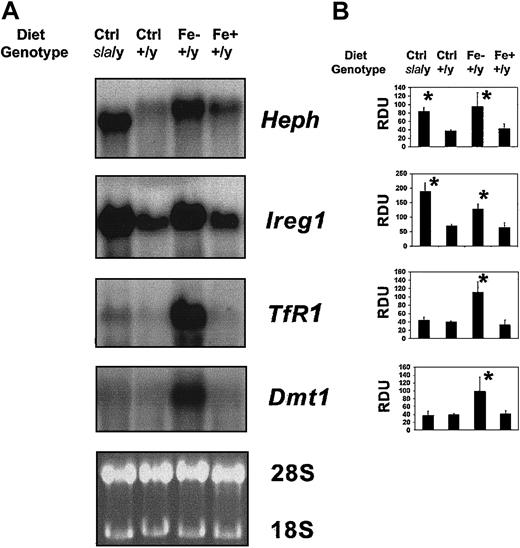
The sla mouse enterocytes had a unique RNA expression pattern as shown in Figure 2A. Quantified mRNA levels relative to 18S for each transcript from multiple independent samples are shown in Figure 2B. The sla mice fed a control diet were compared to C57BL/6J mice fed control, iron-deficient, or iron-overload diets. Transcript levels of both Heph and Ireg1 mRNA were consistently increased in both iron-deficient and sla mice and decreased in iron-overload mice. (Note that the sla enterocytes transcribe a smaller Heph mRNA band due to an in-frame deletion of 582 bases18 .) In contrast, steady-state levels of enterocyte Dmt1 and transferrin receptor (Tfr1) mRNA transcripts were dramatically increased in iron-deficient mice but not changed in sla compared to wild-type mice on an iron-sufficient diet or iron-overload diet. Thus, transcripts of genes involved in iron uptake through the apical membrane or from plasma did not change in the sla mice, but transcripts of genes involved in basolateral transit of iron to plasma were highly increased. The mRNA levels of Heph and Ireg1 are increased in the sla enterocytes despite the increased enterocyte iron levels and thus reflect systemic iron deficiency. The steady-state levels of Dmt1 and Tfr1 mRNA, in contrast to Heph and Ireg1 mRNA, appear to be modulated by the increased local iron concentration of the sla enterocyte. Both Dmt1 and Tfr1 mRNA transcripts contain iron response elements (IREs) in their 3′ untranslated region (UTR), which likely mediate the effects of varying iron status via an IRE-IRP mechanism. Ireg1 mRNA also contains a putative IRE in its 5′ UTR, but the increased mRNA levels in the face of increased intracellular iron in the sla mouse suggest that it does not have a dominant regulatory effect on Ireg1 expression in the small intestine.
Heph and Ireg1 expression is responsive to systemic rather than local iron levels. (A) Northern blot analysis of enterocytes from sla mice fed control diet (sla) and C57BL/6J mice fed control (Ctrl), iron-deficient (Fe–), or iron-overload (Fe+) diets for 6 months. Blots were sequentially hybridized to probes for Heph, Ireg1, TfR, and Dmt1. The 18S rRNA band was used as a loading control. (B) Relative mRNA levels of Heph, Ireg1, TfR1, and Dmt1 were quantified using NIH Image and densitometry values normalized to values obtained for 18S. Data are means ± SD as percent control from 5 independent experiments. *P < .05 versus control.
Heph and Ireg1 expression is responsive to systemic rather than local iron levels. (A) Northern blot analysis of enterocytes from sla mice fed control diet (sla) and C57BL/6J mice fed control (Ctrl), iron-deficient (Fe–), or iron-overload (Fe+) diets for 6 months. Blots were sequentially hybridized to probes for Heph, Ireg1, TfR, and Dmt1. The 18S rRNA band was used as a loading control. (B) Relative mRNA levels of Heph, Ireg1, TfR1, and Dmt1 were quantified using NIH Image and densitometry values normalized to values obtained for 18S. Data are means ± SD as percent control from 5 independent experiments. *P < .05 versus control.
Specificity of antiserum to Hp
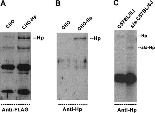
We tested the specificity of affinity-purified polyclonal antibodies to Hp in CHO cells transiently transfected with full-length FLAG-tagged Heph (Figure 3A-B). A monoclonal antibody to the FLAG epitope immunoprecipitated a relative molecular mass (Mr) 155 000 protein from mHp-transfected cells but not from the parental CHO cells (Figure 3A). The same anti-FLAG immunoprecipitated sample was probed with affinity-purified anti-Hp antibody. The anti-Hp antibody also reacted with an Mr 155 000 protein from Heph-transfected cells but did not bind any proteins from parental CHO cells (Figure 3B). This result fits closely with the predicted size of Hp.18 These data provide strong evidence that the affinity-purified antiserum detects the Hephaestin protein. We then used this antiserum for immunoprecipitations of enterocyte lysates and identified an Mr 155 000 protein in C57BL/6J mice and an Mr 130 000 truncated protein in the sla mice (Figure 3C). The sla mouse enterocytes express less Hp, so for clarity, about 5-fold more immunoprecipitate was loaded in the sla enterocyte lane (compare IgG levels). These results demonstrate that the sla mouse expresses lower amounts of a smaller protein. The sla mutation results in an in-frame 194–amino acid deletion, so these results are consistent with production of smaller and less stable protein.
Antiserum to Hp is specific and identifies Hephaestin protein in C57BL/6J and sla mice. (A-B) A full-length mouse Hp cDNA containing a C-terminal FLAG tag was transiently transfected into CHO cells (CHO-Hp). After 48 hours, clarified cell lysates were collected and used for immunoprecipitations with the monoclonal anti-FLAG antibody. Immunoprecipitates were separated by SDS-PAGE and blotted onto nitrocellulose. The blots were probed with anti-FLAG (A) or anti-Hp (B). (C) Enterocyte extracts from C57BL/6J mice and sla mice were used for immunoprecipitations with the anti-Hp antibody. Immunoprecipitates were separated by SDS-PAGE, blotted, and probed with the anti-Hp antibody. An Mr 155 000 protein is present in the C57BL/6J, whereas an Mr 130 000 protein is present in the sla mice. Because less Hp protein is present in the sla mouse, we loaded 5 times more immunoprecipitate from the sla mouse to visualize the protein.
Antiserum to Hp is specific and identifies Hephaestin protein in C57BL/6J and sla mice. (A-B) A full-length mouse Hp cDNA containing a C-terminal FLAG tag was transiently transfected into CHO cells (CHO-Hp). After 48 hours, clarified cell lysates were collected and used for immunoprecipitations with the monoclonal anti-FLAG antibody. Immunoprecipitates were separated by SDS-PAGE and blotted onto nitrocellulose. The blots were probed with anti-FLAG (A) or anti-Hp (B). (C) Enterocyte extracts from C57BL/6J mice and sla mice were used for immunoprecipitations with the anti-Hp antibody. Immunoprecipitates were separated by SDS-PAGE, blotted, and probed with the anti-Hp antibody. An Mr 155 000 protein is present in the C57BL/6J, whereas an Mr 130 000 protein is present in the sla mice. Because less Hp protein is present in the sla mouse, we loaded 5 times more immunoprecipitate from the sla mouse to visualize the protein.
Hp and Ireg1 protein levels in enterocytes from sla and wild-type mice
To determine whether the observed changes in mRNA levels resulted in changes at the protein level, we compared protein levels of Hp and Ireg1 in enterocytes from wild-type mice on the different dietary regimens and sla mice using Western blot analysis (Figure 4). The increased mRNA levels of Heph and Ireg1 in C57BL/6J mice on an iron-deficient diet compared with mice on a control diet are reflected in increased Hp and Ireg1 protein (representative blots are shown in Figures 4A and 4B, respectively). Both control and iron-overloaded enterocytes had lower Ireg1 levels, whereas sla mouse enterocytes showed as much Ireg1 as iron-deficient C57BL/6J mice. As seen by immunoprecipitation, the sla mouse enterocytes had less Hp than control mouse enterocytes. We consistently found twice as much Hp protein in iron-deficient wild-type mice compared with those on a control diet (Figure 4A).
Ireg1 protein levels are increased in sla mice and Hp and Ireg1 protein levels are increased in iron-deficient wild-type mice. Western blot analysis of enterocytes from sla mice fed control diet and C57BL/6J mice fed control (Ctrl), iron-deficient (Fe–), or iron-overload (Fe+) diets for 6 months. Enterocyte lysates were probed with antibodies directed against Hp (A) or Ireg1 (B, loading unboiled protein samples). Relative protein levels were quantified using NIH Image. Data are means ± SDs as percent control from 5 independent experiments. *P < .05 versus control. RDU indicates relative densitometric units.
Ireg1 protein levels are increased in sla mice and Hp and Ireg1 protein levels are increased in iron-deficient wild-type mice. Western blot analysis of enterocytes from sla mice fed control diet and C57BL/6J mice fed control (Ctrl), iron-deficient (Fe–), or iron-overload (Fe+) diets for 6 months. Enterocyte lysates were probed with antibodies directed against Hp (A) or Ireg1 (B, loading unboiled protein samples). Relative protein levels were quantified using NIH Image. Data are means ± SDs as percent control from 5 independent experiments. *P < .05 versus control. RDU indicates relative densitometric units.
Discussion
The availability of heritable mutants of murine iron metabolism has accelerated progress in understanding mammalian iron uptake and its regulation.40 We have undertaken physiologic and molecular analyses of the sla mutant mouse, which expresses a mutated version of the Heph gene encoding the putative multicopper ferroxidase Hephaestin.18 The sla mouse has impaired iron transit across duodenal enterocytes. The blockage appears to be at the basolateral transfer step, because enterocytes are significantly iron overloaded as shown in this study and others.41-43 As noted in previous studies, the hemoglobin levels gradually increase with age in sla mice and the difference between wild-type and sla mice narrows.21,37 However, the sla mouse continues to have significantly lower hemoglobin levels beyond 1 year of age.21,37 Despite the recovery in hemoglobin levels, sla mice continue to be severely iron deficient as shown in this and previous studies.38 In our study, sla mice have even lower liver iron levels than wild-type mice on an iron-deficient diet. The sla mouse therefore represents a unique model in which cellular iron levels are increased in the intestinal enterocytes yet systemic iron levels are low.
The enterocytes of sla mice showed a unique RNA expression pattern and demonstrated high levels of mRNA for Heph and Ireg1, similar to enterocytes from iron-deficient C57BL/6J mice. However, mRNA levels of Dmt1 and TfR1 were not significantly changed. Thus, transcripts of genes involved in iron uptake through the apical membrane or from the plasma barely increased in the sla mice, but transcripts of genes involved in basolateral transit of iron to plasma were highly increased. We would therefore suggest that Heph and Ireg1 mRNA levels are predominantly responsive to systemic levels of iron and are independent of iron levels in the enterocytes. In contrast, Dmt1 and TfR1 mRNA levels can respond to the local iron status of the enterocytes, presumably through IRP-IRE–mediated mechanisms.44,45 Our previous studies on the molecular evaluation of the mucosal block phenomenon also suggested local regulation of Dmt1 and Dcytb mRNA levels but systemic regulation of Ireg1 and Hp are highly supportive of these findings.46
Ireg1 mRNA contains a consensus IRE motif in its 5′ UTR, but there is no evidence that it plays a role in Ireg1 regulation in the intestine.13 In other genes bearing a 5′ UTR IRE, such as the ferritin genes, the binding of IRPs under low iron conditions leads to a block in translation and lower protein synthesis. However, we find the opposite direction of regulation for Ireg1 in the gut. Ireg1 protein levels are high in enterocytes from iron-deficient mice compared with iron-replete or iron-overloaded wild-type mice. Ireg1 levels remain high in sla mice even though enterocyte iron and ferritin levels are significantly higher than in iron-deficient wild-type mice. Thus, Ireg1 mRNA and Ireg1 protein levels correlate with systemic rather than local iron status. Heph mRNA levels in sla mice are similar to those in iron-deficient mice, but the smaller Hp protein is expressed in sla mice at low levels, suggesting likely instability of the sla-Hp protein containing the in-frame deletion. Dmt1 protein levels have been shown in previous studies to be increased in iron deficiency.8 Similarly, the expression of the apical ferric reductase Dcytb mRNA is also increased in iron deficiency although the mechanism is unknown.6 Therefore, iron deficiency results in increased protein expression of apical and basolateral components of iron transport, which maximizes the potential throughput of iron from dietary iron sources into the body.
How the body signals the small intestine to alter its iron transport in response to changes in body iron requirements is poorly understood, but recent evidence suggests that the liver-derived peptide Hepcidin is a strong candidate for the signaling molecule.46 Hepcidin expression is increased in liver when iron overloading occurs, and because it circulates in the plasma, it has been hypothesized to act as a negative regulator of intestinal iron absorption.47-50 Hepcidin deficiency leads to iron overload47,48 and overexpression of Hepcidin leads to severe iron deficiency49 ; we have recently found a close inverse relationship between hepatic Hepcidin expression and the expression of iron transport molecules in the duodenum.50 Hepcidin thus represents a likely candidate for the systemic signal informing the duodenum of body iron requirements and Ireg1 and Heph will therefore likely be important target genes for its mechanism of action.51
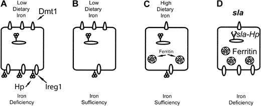
Intestinal iron transport finely balances the necessity to maximize iron import in deficiency and to minimize potential toxicity from dietary iron loads. We propose the following model for intestinal enterocyte iron transport (Figure 5) in control and sla mice. Low iron stores lead to increased expression of the components of basolateral iron transport in mature absorptive villous cells. However, Dmt1 expression in the mature enterocyte depends on cellular iron status, which reflects a combination of circulating plasma iron taken up by the crypt enterocytes via a TfR prior to differentiation and recently absorbed dietary iron. Systemic iron deficiency and an iron-deficient diet will result in maximal expression of both apical and basolateral transport systems, which maximizes the iron flux rate from diet to the circulation. However, a sudden iron load could lead to potentially toxic iron flux rates. The rapid decrease in Dmt1 expression in response to increased local iron concentrations therefore provides a protective response to high dietary iron levels. The presence of 2 layers of regulation allows for a modulated response to iron deficiency to ensure adequate iron uptake and minimize potential toxicity.
A model for local and systemic control of intestinal iron transport. (A) Low dietary iron and systemic iron deficiency lead to up-regulation of DMT1 protein levels, which increases the capacity of the enterocyte for dietary iron uptake. Similarly, increased Ireg1 and Hephaestin protein expression increases the capacity of the enterocyte for the transit of iron to plasma. (B) Low dietary iron with sufficient systemic iron stores leads to increased DMT1 expression, whereas Ireg1 and Hephaestin protein levels are decreased, which may represent a muscosal block to iron absorption. (C) High dietary iron and sufficient iron stores result in decreased levels of DMT1, Ireg1, and Hephaestin protein and minimal iron absorption. (D) In enterocytes from the sla mouse, impaired basolateral transport due to a defect in Hephaestin leads to iron accumulation. Iron accumulation in sla enterocytes leads to decreased uptake of dietary iron through decreased DMT1 mRNA and protein levels. Despite intracellular iron accumulation, Ireg1 mRNA and protein levels are increased, likely in response to systemic signals of iron deficiency.
A model for local and systemic control of intestinal iron transport. (A) Low dietary iron and systemic iron deficiency lead to up-regulation of DMT1 protein levels, which increases the capacity of the enterocyte for dietary iron uptake. Similarly, increased Ireg1 and Hephaestin protein expression increases the capacity of the enterocyte for the transit of iron to plasma. (B) Low dietary iron with sufficient systemic iron stores leads to increased DMT1 expression, whereas Ireg1 and Hephaestin protein levels are decreased, which may represent a muscosal block to iron absorption. (C) High dietary iron and sufficient iron stores result in decreased levels of DMT1, Ireg1, and Hephaestin protein and minimal iron absorption. (D) In enterocytes from the sla mouse, impaired basolateral transport due to a defect in Hephaestin leads to iron accumulation. Iron accumulation in sla enterocytes leads to decreased uptake of dietary iron through decreased DMT1 mRNA and protein levels. Despite intracellular iron accumulation, Ireg1 mRNA and protein levels are increased, likely in response to systemic signals of iron deficiency.
Prepublished online as Blood First Edition Paper, May 1, 2003; DOI 10.1182/blood-2003-02-0347.
Supported by National Institutes of Health (NIH) grants R01-DK57800, NIH R01-DK56376 and K08 DK2823 and Human Frontier Science Program RGY0328.
The publication costs of this article were defrayed in part by page charge payment. Therefore, and solely to indicate this fact, this article is hereby marked “advertisement” in accordance with 18 U.S.C. section 1734.
We thank Robert Fleming for providing the nonheme iron assay modification and also Erik Gertz and Leslie Woodhouse for help with the ICP-AES analysis. We thank Yien Kuo and Jane Gitschier for assistance in the development of the antiserum to Hephaestin.


This feature is available to Subscribers Only
Sign In or Create an Account Close Modal