Abstract
Plasma non-transferrin-bound-iron (NTBI) is believed to be responsible for catalyzing the formation of reactive radicals in the circulation of iron overloaded subjects, resulting in accumulation of oxidation products. We assessed the redox active component of NTBI in the plasma of healthy and β-thalassemic patients. The labile plasma iron (LPI) was determined with the fluorogenic dihydrorhodamine 123 by monitoring the generation of reactive radicals prompted by ascorbate but blocked by iron chelators. The assay was LPI specific since it was generated by physiologic concentrations of ascorbate, involved no sample manipulation, and was blocked by iron chelators that bind iron selectively. LPI, essentially absent from sera of healthy individuals, was present in those of β-thalassemia patients at levels (1-16 μM) that correlated significantly with those of NTBI measured as mobilizer-dependent chelatable iron or desferrioxamine chelatable iron. Oral treatment of patients with deferiprone (L1) raised plasma NTBI due to iron mobilization but did not lead to LPI appearance, indicating that L1-chelated iron in plasma was not redox active. Moreover, oral L1 treatment eliminated LPI in patients. The approach enabled the assessment of LPI susceptibility to in vivo or in vitro chelation and the potential of LPI to cause tissue damage, as found in iron overload conditions. (Blood. 2003;102:2670-2677)
Introduction
The traffic of nonheme iron, oxygen, and ascorbate in plasma represents a potential source of reactive oxygen radicals generated by reduction-oxidation cycling of iron via ascorbate and O2. Such undesirable reactions are physiologically counteracted by various protective molecules: (1) transferrin, the iron transport protein, which efficiently restricts iron's capacity for undergoing redox reactions1 ; (2) antioxidants such as vitamin E, glutathione, and, in particular, ascorbate, which, together with iron, has the dual capacity of promoting redox cycling at relatively low concentrations and acting as a powerful scavenger of radical species at higher concentrations2-5 ; and (3) metabolites with antioxidant activity such as bilirubin and urate.5 In normal conditions, the antioxidant capacity of plasma, estimated by various methods to be in the range of 1 mM,5 might greatly exceed the plasma radical-generating potential. However, in iron overload, the plasma nonheme iron concentration may rise significantly and lead to the appearance of nontransferrin bound iron (NTBI) and thereby modify the balance between pro-oxidant antioxidant activities. This seems to be the case in thalassemia, where the accumulation of plasma NTBI has been shown to correlate with the appearance of oxidation products and a decrease in plasma antioxidant capacity.6,7 However, outstanding questions have remained regarding the forms of NTBI detected in different types of clinical iron overload, particularly their redox activity, their toxic potential in plasma, and their susceptibility to chelation.
We have searched for means to determine the capacity of serum iron to engage in the formation of reactive oxidant species (ROS) in a manner that would involve minimum manipulation of the sample and be independent of exogenously added factors. The presumed source of such labile iron is assumed to be associated with NTBI, whose levels and chemical properties differ among iron-overloaded patients.8 Plasma NTBI that is accessible to chelators such as desferrioxamine (DFO), without facilitation by added mobilizing agents, is designated DCI or “directly chelatable iron”.9 DCI, when present, comprises only a fraction of the NTBI. Additional iron is chelated following addition of iron-mobilizing agents such as nitrilotriacetic acid (NTA) or oxalate, hence the name MDCI: mobilizer-dependent chelatable iron.8,10 The labile properties of those NTBI forms have yet to be assessed in terms of their capacity to engage in redox cycling. We used a method for assessing labile plasma iron (LPI) comprising 3 features: (1) the engagement of labile iron in redox cycling by using ascorbate as the redox prompting agent, (2) the detection of radical formation using an oxidant-sensitive fluorescent probe (dihydrorhodamine 123 = DHR), and (3) the application of iron-selective chelators as a means of assessing the specific involvement of labile iron in the process. Thus, the chelator-sensitive component of DHR oxidation prompted by ascorbate provides a measure for LPI in a particular biologic fluid. That measure also entails the potential antioxidant properties of the biologic fluid in question. In the present study, the component of the DHR oxidation that was blocked by iron chelators correlated quantitatively with plasma NTBI levels found in β-thalassemia patients. Oral administration of deferiprone (L1) to those patients evoked a time-dependent rise in plasma NTBI levels (DCI and MDCI) and led to a concomitant disappearance of LPI from plasma. The results indicate that massive mobilization of iron into plasma by L1 was in forms that were essentially not redox active, and, by implication, of low toxic potential.
Materials and methods
Reagents
The following materials were obtained and used without further purification: sodium ascorbate, diethylenetriaminepentaacetic acid (DTPA), ferrous ammonium sulfate, catalase, superoxide dismutase (SOD), horseradish peroxidase (Sigma, St Louis, MO), dihydrorhodamine 123 (DHR), dihydrochloride salt (Biotium, Hayward, CA), L1 (deferiprone; Apotex, Toronto, ONT, Canada), human myeloperoxidase and nitrilotriacetic acid (NTA) (Fluka, Seelze, Germany), desferrioxamine (DFO; Novartis, Basel, Switzerland), Chelex-100 chelating resin (Bio-Rad, Hercules, CA), and human serum albumin 25% and human apo-transferrin (aTf) (Kama Biological Industries, Kibbutz Beith Kama, Israel). Iron-free HEPES (N-2-hydroxyethylpiperazine-N′-2-ethanesulfonic acid)-buffered saline (HBS; HEPES 20 mM, NaCl 150 mM, pH 7.4) was obtained by treatment with 1 g 100 mL-1 Chelex-100. Residual Fe contamination was determined as 60 nM by the DHR assay, and therefore 60 nM DFO was added to the HBS to minimize the Fe contribution from HBS. Plasmalike medium (PLM) contained HEPES 20 mM, NaCl 150 mM, sodium citrate 120 μM, sodium ascorbate 40 μM, sodium phosphate dibasic 1.2 mM, sodium bicarbonate 10 mM, and human serum albumin 40 mg/mL-1 (pH 7.4).
Fe(III)/NTA complexes were formed by mixing 70 mM nitrilotriacetic acid (NTA), titrated to pH 7.0 with NaOH, with 20 mM ferrous ammonium sulfate to produce a Fe/NTA molar ratio of 1:7 and allowing the Fe(II) to oxidize to Fe(III) in ambient air for 30 minutes. Ascorbate and DHR were prepared as concentrated stocks of 5 mM in water and 50 mM in dimethyl sulfoxide (DMSO), respectively, kept frozen and thawed only once, immediately before use.
Serum samples
All of the serum samples used were from nontransfusion-dependent β-thalassemia/Hb E patients from Thailand. Ten patients participated in the short-term kinetic studies with L1. Their iron status parameters were as follows: Transferrin saturations ranged from 44% to 122%, average 90%; serum ferritins ranged from 988 to 8052 ng/mL, average 4138 ng/mL; and nonheme liver iron ranged from 6.2 to 37.1 mg/g dry weight, average 20.7 mg/g dry weight. The patients arrived at the clinic in the morning, having been instructed not to take any medications prior to arrival. Serum samples were taken just prior to administration of L1 by mouth (75 mg/kg) and subsequently at 30-minute intervals for 2 hours. All of the subjects signed a patient consent form as required by the Ethical Committee of Mahidol University, Nakornpathom, Thailand. Red blood cell (RBC) lysate: 200 μL of packed human red blood cells were washed in 10 mL HBS and resuspended in 1 mL of HBS. Lysis was achieved by 3 cycles of freeze/thaw in N2(liq) and 37°C, respectively. The lysate was centrifuged at 14 000 rpm at 5°C for 5 minutes to remove the membrane fraction. Hb concentration was determined photometrically (molar absorptivity coefficient [ϵ412]: 125 000 M-1 cm-1).
Measurement of LPI
The probe DHR is converted from its nonfluorescent to fluorescent form by various oxidants, such that the generation of reactive oxygen species can be followed as an increase in DHR fluorescence. In the LPI assay, each serum sample is tested under 2 conditions: with 40 μM ascorbate alone and with 40 μM ascorbate in the presence of 50 μM iron chelator. The difference in the rate of oxidation of DHR in the presence and absence of chelator represents the component of plasma NTBI that is redox active.
LPI assay
Quadruplicates of 20 μL serum or plasma were transferred to clear-bottom, 96-well plates (Maxisorp 96; Nunc, Rotskilde, Denmark). To 2 of the wells was added 180 μL iron-free HBS (preincubated at 37°C) containing 40 μM ascorbate and 50 μM DHR. To the 2 other wells was added 180 μL of the same solution containing 50 μM deferiprone. Immediately following reagent addition, the kinetics of fluorescence increase were followed at 37°C in a BMG Galaxy Fluostar microplate reader (BMG Lab Instruments, Offenburg, Germany) with a 485/538 nm excitation/emission filter pair, for 40 minutes, with readings every 2 minutes. The slopes (r) of DHR fluorescence intensity with time were calculated from measurements taken between 15 and 40 minutes and are given as FU minute-1 (fluorescence units per minute).
The duplicate values of r in the presence and absence of L1, rL1, and r, respectively, were averaged, and the LPI concentration (μM) was determined from calibration curves relating the difference in slopes with and without L1 against Fe concentration: LPI = Δr/rst = (r - rL1)/rst, where Δr and rst denote the L1-sensitive component of r and the calibration factor relating Δr to the Fe concentration, respectively.
Calibration curves were obtained by spiking plasmalike medium (PLM) or sera with Fe:NTA, 1:7 (mol/mol) to give final concentrations of 40 to 100 μM Fe, followed by serial dilution in PLM or in the same serum and incubation for 30 minutes at 37°C to allow binding of the Fe. Quadruplicates of 20 μL of these samples were assayed for LPI as described in the previous paragraphs. A standard curve of DHR oxidation rates versus Fe concentrations was built with data compiled from determinations done in PLM medium and sera from healthy subjects and thalassemia patients.
Measurement of DCI and MDCI components of NTBI
The concentration of directly chelatable iron (DCI) (by desferrioxamine) was performed as described previously.9 Briefly, DCI was measured by diluting serum samples in HBS buffer containing 2.5 μM fluorescein-DFO (an iron-detection probe) with and without 100 μM DFO. The iron-mediated quenching of fluorescence of the probe was used to quantify DCI, the fraction of serum iron that is directly chelatable by DFO. MDCI, mobilizer-dependent chelatable iron, was measured by diluting serum samples in HBS buffer 10 mM oxalate, 0.1 mM GaCl3, and 0.6 μM fluorescein-apo-transferrin (an iron-detection probe) with and without 2 mg/mL apo-transferrin. The iron-mediated quenching of fluorescence of the probe was used to quantify MDCI, the fraction of oxalate-mobilized serum iron bound by fluores cein-apo-transferrin.11
Results
In order to quantify labile or redox-active iron in the serum of individuals with iron overload, we devised an assay based on the promotion of redox cycling of iron by its reduction with relatively low concentrations of ascorbate (serum ascorbate is in the range of 35 to 100 μM, and the assay uses 40 μM). The products of redox cycling are detected as time-dependent oxidation of the fluorogenic probe dihydrorhodamine 123 (DHR) to the fluorescent rhodamine 123. The iron-dependent component of DHR oxidation is obtained from the inhibitory effect of specific iron chelators.
Relationships between concentration of labile iron, ascorbate, and DHR oxidation
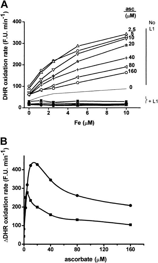
As shown in Figure 1A, mixtures of Fe(III) with ascorbate cause a substantial, Fe-dependent increase in the rate of DHR oxidation. The requirement for iron is further substantiated by the marked inhibitory effect of the iron chelator L1. Similar inhibition was found for 2 other iron chelators, desferrioxamine and apotransferrin (data not shown). In contrast to Fe, ascorbate concentration has a biphasic effect on DHR oxidation, activating total DHR oxidation rate maximally when approximately equimolar with iron and gradually becoming less active at higher concentrations. This is a characteristic feature of ascorbate, which acts as a pro-oxidant at relatively low concentrations and an antioxidant at higher concentrations.2 The relationship between chelator-sensitive DHR oxidation and ascorbate concentration is illustrated in Figure 1B, showing a maximal effect of ascorbate at ∼20 μM. We chose to use 40 μM ascorbate as a standard concentration in subsequent experiments, since this is within the range of its physiologic concentrations in serum and represents an approximate point of balance between the pro-oxidant and antioxidant activity in this system.
DHR oxidation mediated by mixtures of iron and ascorbate. (A) Samples of plasmalike medium (PLM) containing increasing concentrations of iron (as Fe(III):NTA complex) were mixed with reagent (“Materials and methods”) containing increasing concentrations of ascorbate (0-160 μM) and 50 μM DHR without (no L1) or with (+ L1) 50 μM iron chelator L1. The rate of increase in DHR fluorescence was followed and is given as arbitrary fluorescence units per time (FU × minute-1). (B) Plot of the L1-sensitive component of DHR oxidation in Graph A (“μDHR oxidation rate”) versus ascorbate concentration. Shown for comparison are 2 concentrations of Fe, 5 and 20 μM (▪ and •, respectively).
DHR oxidation mediated by mixtures of iron and ascorbate. (A) Samples of plasmalike medium (PLM) containing increasing concentrations of iron (as Fe(III):NTA complex) were mixed with reagent (“Materials and methods”) containing increasing concentrations of ascorbate (0-160 μM) and 50 μM DHR without (no L1) or with (+ L1) 50 μM iron chelator L1. The rate of increase in DHR fluorescence was followed and is given as arbitrary fluorescence units per time (FU × minute-1). (B) Plot of the L1-sensitive component of DHR oxidation in Graph A (“μDHR oxidation rate”) versus ascorbate concentration. Shown for comparison are 2 concentrations of Fe, 5 and 20 μM (▪ and •, respectively).
DHR may be oxidized by a variety of oxidants, including hypochlorous acid generated by myeloperoxidases,12 peroxynitrite anion formed by oxidation of nitric oxide13 and hydrogen peroxide in the presence of peroxidases or labile iron,14 as well as by heme, either free or protein-bound.15 Therefore, we assessed the possible contribution of peroxidases to LPI detection by adding purified horseradish peroxidase and human neutrophil myeloperoxidase to serum samples at concentrations of 100 U/mL and 1.5 U/mL, respectively. Although both enzymes increased substantially (3- to 7-fold), the rate of DHR oxidation, those activities were not mediated by labile iron since neither desferrioxamine nor L1 had any effect (data not shown). Hemoglobin, which is also a common contaminant of serum and plasma samples and a possible source of peroxidase activity,15,16 slightly increased the rate of DHR oxidation but did not contribute to the chelator-inhibitable portion of the DHR oxidation signal (not shown). However, higher concentrations of hemoglobin were found to interfere with the fluorescent signal, by light filtering effects. Similarly, heme, added in the form of hemin at concentrations up to 100 μM, failed to contribute to the chelator-inhibited LPI signal (not shown). Thus, regardless of the precise oxidizing species detected in the present assay and regardless of the possible contribution of other potential enhancers to DHR oxidation, the use of iron-selective chelators permits the specific assessment of labile forms of iron independently of other mechanisms of DHR oxidation.
We compared the relative capacity of 3 well-characterized iron chelators DFO, L1, and apo-transferrin and the nonselective metal chelator DTPA to inhibit DHR oxidation in representative sera of normal (healthy) individuals (N), hemochromatosis (HH), and thalassemia (TH) patients (Figure 2). In spite of their different chemical properties, iron-binding ratios, and affinities, DFO, L1, and apo-transferrin inhibited DHR oxidation to a similar extent. DTPA, used at 500 μM, was slightly more effective, but this could be attributed to the elimination of other contaminating metals. It is concluded that the observed effects of chelators on DHR oxidation prompted by ascorbate are specifically due to iron chelation rather than to radical scavenging.
Effect of metal chelators on ascorbate-induced DHR oxidation. Samples of sera from healthy subjects (CTRL), hemochromatotic (HH), or thalassemic (THAL) patients were assayed for DHR oxidation as described in “Materials and methods” in the presence of the iron-specific chelators desferrioxamine (DFO), L1, and apo-transferrin (aTf), as well as the general metal chelator DTPA. The concentrations of the chelators refer to their final concentrations after addition of the DHR reagent. DHR oxidation is given as in Figure 1.
Effect of metal chelators on ascorbate-induced DHR oxidation. Samples of sera from healthy subjects (CTRL), hemochromatotic (HH), or thalassemic (THAL) patients were assayed for DHR oxidation as described in “Materials and methods” in the presence of the iron-specific chelators desferrioxamine (DFO), L1, and apo-transferrin (aTf), as well as the general metal chelator DTPA. The concentrations of the chelators refer to their final concentrations after addition of the DHR reagent. DHR oxidation is given as in Figure 1.
Relationship between Tf saturation, DHR oxidation rate, and Fe concentration
Transferrin (Tf)-bound iron is known to be redox inactive,1 as confirmed in Figure 2, where excess apo-transferrin effectively inhibited the DHR oxidation. However, it was of importance to establish that this holds also for high Fe/transferrin ratios that are found in sera of individuals with high transferrin saturation levels. As shown in Figure 3 (top), for a solution of apo-transferrin, DHR oxidation prompted by ascorbate increased above background values only when the Fe binding capacity of transferrin was exceeded, for instance, at transferrin saturation of at least 100%. Therefore, transferrin-bound iron per se is not likely to be detected as labile iron in the assay. Analogously, the rate of DHR oxidation in a sample containing normal (N) sera (approximately 30% Tf saturation) or a mixture of TH sera (> 85% Tf saturation) that showed a relatively high basal level (Figure 3, bottom) rose in response to added Fe only after reaching a threshold value, which we interpret as the point exceeding Tf binding capacity. A similar titration done in PLM lacking iron-binding capacity showed no such threshold phenomenon, although the response (for instance, the rate of DHR oxidation r) to added Fe was similar to those found in either N or TH sera beyond the indicated threshold level. The inclusion of the iron chelator L1 in the medium eliminated the response to added Fe and allowed the assessment of the Fe-dependent component of DHR oxidation in both PLM and in N and TH sera. A prominent feature of DHR oxidation rates in TH is the relatively high basal level (L1-insensitive component), which is up to 10-fold higher than in N sera, although it spans a wide range.
Relationship between DHR oxidation and Fe in transferrin-containing solution, plasmalike medium, and thalassemic serum. Top: A 23-μM human apo-transferrin solution (ϵ278 = 93 000 M-1 cm-138 ) in HBS containing 10 mM Na2CO3 was reacted at room temperature for 20 minutes with Fe (Fe/NTA 1:10) to give final Fe concentrations of 0 to 82.5 μM and tested for formation of Fe-transferrin by absorption at 465 nm (right scale, filled symbols) and for DHR oxidation rate (left scale, fluorescence units/minute, FU × minute-1, open symbols) as described in “Materials and methods.” The concentration of Fe required to reach maximal A465, corresponding to 100% transferrin saturation, was 45 μM (theoretical 46 μM, based on 2 iron-binding sites per transferrin). Bottom: Fe was added as Fe/NTA to a solution of PLM (circles) or a mixture of 5 different TH sera (stars) or normal sera N (squares) supplemented (empty symbols) or not (filled symbols) with 50 μM L1. The estimated transferrin saturation values for the mixed thalassemia sera were 85% and for normal sera 28%. DHR oxidation rates were obtained as described in “Materials and methods.”
Relationship between DHR oxidation and Fe in transferrin-containing solution, plasmalike medium, and thalassemic serum. Top: A 23-μM human apo-transferrin solution (ϵ278 = 93 000 M-1 cm-138 ) in HBS containing 10 mM Na2CO3 was reacted at room temperature for 20 minutes with Fe (Fe/NTA 1:10) to give final Fe concentrations of 0 to 82.5 μM and tested for formation of Fe-transferrin by absorption at 465 nm (right scale, filled symbols) and for DHR oxidation rate (left scale, fluorescence units/minute, FU × minute-1, open symbols) as described in “Materials and methods.” The concentration of Fe required to reach maximal A465, corresponding to 100% transferrin saturation, was 45 μM (theoretical 46 μM, based on 2 iron-binding sites per transferrin). Bottom: Fe was added as Fe/NTA to a solution of PLM (circles) or a mixture of 5 different TH sera (stars) or normal sera N (squares) supplemented (empty symbols) or not (filled symbols) with 50 μM L1. The estimated transferrin saturation values for the mixed thalassemia sera were 85% and for normal sera 28%. DHR oxidation rates were obtained as described in “Materials and methods.”
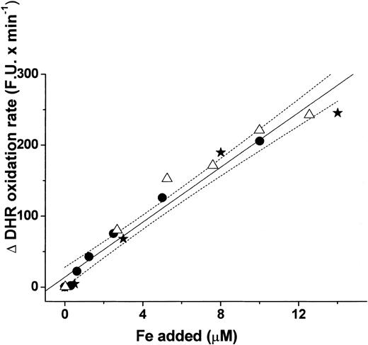
The relationship between the chelator-sensitive component of DHR oxidation rates (Δr), in fluorescence units per minute (FU minute-1) and the added Fe was linear for the different types of calibration media. The best fit for the range 0-15 μM range added Fe yielded a 19 ± 1 FU minute-1μM-1 slope (r = 0.98) with 95% confidence limits as indicated (Figure 4). For individual classes of TH and N sera (n = 5 for each class), the range of slopes was 14-20 (19 ± 2 for TH patients and 16 ± 3 for N individuals) and for PLM 18 ± 1. Since the L1-sensitive DHR oxidation rates (Δr) provide an average measure of the effective LPI concentration in a particular biologic fluid, we used rst, the slope of (Δr) versus Fe, as the standard reference value for estimating LPI.
Relationship between the rate of DHR oxidation and iron concentration in plasmalike medium (PLM), normal (N), and thalassemic (TH) sera. The different sera of (TH) and healthy (N) subjects were initially presaturated with iron by mixing with Fe/NTA (5-70 μM as in Figure 3). Fe was then added (as Fe/NTA) to them as well as to samples in PLM. Average DHR oxidation rates (fluorescence units per unit time; FU × minute-1) were obtained in parallel samples supplemented or not with 50 μM L1 (+L1 and -L1, respectively), their differences calculated and plotted against the concentration of added Fe after subtraction of the basal rate (obtained at 0 added Fe). Linear fit of the compiled data points yielded the slope of 19 ± 1 (r = 0.98) and the respective 95% confidence range (dotted line).
Relationship between the rate of DHR oxidation and iron concentration in plasmalike medium (PLM), normal (N), and thalassemic (TH) sera. The different sera of (TH) and healthy (N) subjects were initially presaturated with iron by mixing with Fe/NTA (5-70 μM as in Figure 3). Fe was then added (as Fe/NTA) to them as well as to samples in PLM. Average DHR oxidation rates (fluorescence units per unit time; FU × minute-1) were obtained in parallel samples supplemented or not with 50 μM L1 (+L1 and -L1, respectively), their differences calculated and plotted against the concentration of added Fe after subtraction of the basal rate (obtained at 0 added Fe). Linear fit of the compiled data points yielded the slope of 19 ± 1 (r = 0.98) and the respective 95% confidence range (dotted line).
Correlation between LPI and NTBI (MDCI and DCI) in thalassemia patients with iron overload
The ascorbate-prompted DHR oxidation rates (r) were determined in sera of a cohort of 57 patients with β-thalassemia both in the absence of L1 (-L1) and presence of L1 (+L1). When the respective values of DHR oxidation rates (r and rL1) were plotted against their difference, that is, Δr = (r - rL1), the resulting slopes of (-L1) and (+L1) yielded values that were not significantly different from 1 and 0, respectively (not shown). Only 10% of the samples deviated from the 95% confidence limit for both types of determinations (+L1 or -L1). This indicates that despite the increase in LPI as reflected in the r values, the basal rL1, attained by the action of L1, remained essentially constant with a slope equal to 0.08 ± 0.05, an intercept equal to35 ± 8 (r = 0.3), and a mean value of 34 ± 18. Hence, the contribution of other non-iron-related components to the DHR oxidation signal remains constant over a wide range of LPI values. The L1-insensitive component probably reflects the contribution of trace levels of various peroxidase activities such as myeloperoxidase, hemoglobin, and ceruloplasmin and shows large individual variations. We have noted that this component of the DHR oxidation signal tends to be higher in thalassemia patients than in other groups (data not shown).
The frequency of occurrence of LPI and NTBI, the latter measured as MDCI (mobilizer-chelatable iron), in thalassemia patients was examined in Figure 5. The MDCI concentrations in the sera of these patients span a wide range of values, 0 to 12.5 μM. Among the 57 sera examined, 28 showed significant levels of LPI, with values more than 0.5 μM. A linear fit of MDCI versus LPI yielded a slope equal to 1.32 ± 0.11 and an intercept equal to 0.31 ± 0.30 (r = 0.88). Therefore, in comparison to LPI, the present measurements of MDCI might underestimate NTBI by about 30%. Interestingly, even among 22 patients with relatively low MDCI levels (<0.5 μM), which are indicative of relatively low iron loads, 8 had still significant LPI levels, ranging from 0.60 to 1.7 μM. However, LPI becomes particularly prominent at relatively high levels of iron overload (MDCI values > 1.5 μM), which generally correspond to transferrin saturations higher than 70%.8
LPI in sera from thalassemic patients and its correlation with MDCI. Sera from 57 β-thalassemic patients were assayed for LPI and MDCI as described in “Materials and methods.” The values of both parameters are displayed as a histogram in the main body of the figure, and one versus the other in the inset. The best linear fit yielded the following values: slope = 1.32 ± 0.11 and intercept = 0.31 ± 0.30 (r = 0.88), with 95% confidence limits as demarcated within the dotted lines.
LPI in sera from thalassemic patients and its correlation with MDCI. Sera from 57 β-thalassemic patients were assayed for LPI and MDCI as described in “Materials and methods.” The values of both parameters are displayed as a histogram in the main body of the figure, and one versus the other in the inset. The best linear fit yielded the following values: slope = 1.32 ± 0.11 and intercept = 0.31 ± 0.30 (r = 0.88), with 95% confidence limits as demarcated within the dotted lines.
L1-mediated iron mobilization in vivo and the generation of labile plasma iron
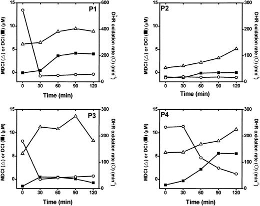
The finding that L1 effectively inhibited the iron redox activity in vitro prompted us to ask whether the same applies also in vivo to patients treated with L1. For that purpose we carried out the parallel in vivo study by analyzing DHR oxidation rates in the sera of patients receiving oral L1. A cohort of 10 thalassemic patients was monitored for 3 serum parameters immediately prior to oral L1 administration and during 2 subsequent hours: (1) DCI, desferrioxamine-chelatable iron, which measures the fraction of serum iron that is directly chelatable by DFO, using a fluoresceinated analog of DFO without added iron-mobilizing agents,9 (2) MDCI, mobilizer-dependent chelatable iron, which is measured in the presence of 10 mM oxalate as a mobilizing agent and fluorescein-apoTf as the iron-sensitive probe,11 and (3) the total rate of DHR oxidation, providing an overall measure of LPI. Figure 6 shows only 4 (of 10) cases, each representing a particular profile of time-dependent changes in DCI, MDCI, and LPI, of which the last is given in terms of DHR oxidation rates. In three fourths of patients, DCI and MDCI rose within 30 to 60 minutes of L1 administration, reaching plasma concentrations of up to 8 and 13 μM, respectively. MDCI reached relatively higher levels, apparently because of the increased iron-detection sensitivity provided by the mobilizing agent. In the same three fourths of patients the rate of DHR oxidation showed an opposite pattern to those of DCI and MDCI, namely high levels prior to L1 intake that decreased within 30 to 60 minutes of L1 intake. In one quarter of cases the initial DHR oxidation rate was almost undetectable, precluding detection of a further decrease in signal. Thus, although L1 increased the levels of serum DCI and MDCI, it not only failed to raise the LPI levels, but also actually depressed the existing LPI to undetectable levels. This is also manifested in the statistical correlation coefficient r obtained between the reciprocal of LPI and DCI or MDCI in L1-treated patients (Table 1). The r values were determined for the 4 kinetic profiles shown in Figure 6, each representing a single patient, and 6 additional profiles obtained with other patients (not shown). In 3 of 10 patients, there was no detectable LPI prior to L1 intake, and there were relatively small changes in DCI and MDCI following L1 intake. In all other 7 patients, the reciprocal correlations were significant (P < .01) between LPI and DCI (mean of 0.76 ± 0.06) and LPI and MDCI (mean of 0.74 ± 0.05), as well as the direct correlations between DCI and MDCI (0.85 ± 0.06).
Effect of oral L1 administration on serum DCI and MDCI and DHR oxidation rates. Time dependence. Serum samples from 10 β-thalassemia patients were obtained just before L1 oral administration and subsequently at 30-minute intervals. The samples were assayed for DHR oxidation rate (○, right scale), DCI (▪, left scale), and MDCI (▵, left scale) as described in “Materials and methods.” The results shown are from 4 patients, each representative of the different types of responses observed in the 10 patients.
Effect of oral L1 administration on serum DCI and MDCI and DHR oxidation rates. Time dependence. Serum samples from 10 β-thalassemia patients were obtained just before L1 oral administration and subsequently at 30-minute intervals. The samples were assayed for DHR oxidation rate (○, right scale), DCI (▪, left scale), and MDCI (▵, left scale) as described in “Materials and methods.” The results shown are from 4 patients, each representative of the different types of responses observed in the 10 patients.
Effect of oral L1 administration on serum DCI and MDCI and DHR oxidation rates
Patient no. . | LPI-DCI . | LPI-MDCI . | DCI-MDCI . |
|---|---|---|---|
| 1 | 0.658 | 0.655 | 0.994 |
| 2* | 0.104 | 0.059 | 0.787 |
| 3 | 0.842 | 0.803 | 0.858 |
| 4 | 0.968 | 0.923 | 0.897 |
| 5 | 0.646 | 0.488 | 0.929 |
| 6* | 0.181 | −0.097 | −0.139 |
| 7 | 0.504 | 0.766 | 0.57 |
| 8 | 0.905 | 0.855 | 0.983 |
| 9 | 0.825 | 0.695 | 0.579 |
| 10* | −0.45 | 0.239 | 0.669 |
| Mean (1-10) | 0.52 | 0.54 | 0.71 |
| SEM | 0.14 | 0.11 | 0.01 |
| Mean* | 0.76 | 0.74 | 0.83 |
| SEM* | 0.06 | 0.05 | 0.08 |
The cross correlation coefficients of LPI (measured as 1/DHR oxidation rate) and NTBI parameters (DCI and MDCI) measured in the plasma of 10 L1-treated thalassemia patients were calculated over the entire profile, as depicted in Figure 6 (for 4 of 10 patients). The mean correlation coefficients and their respective SEM values were calculated for all 10 profiles as well for those excluding the 3 profiles that had essentially no detectable levels of MDCI or LPI at the onset of the treatment (patients 2, 6, and 10, labeled with an asterisk).
Discussion
The propensity of iron to engage in Fenton reactions is curbed by the presence of plasma transferrin, which binds iron tightly and renders it essentially nonlabile.1,4,5 This protective property is deficient in hypotransferrinemia and is surpassed in various conditions of iron overload, giving rise to various forms of NTBI in the plasma. NTBI, unlike plasma TBI, has been assumed to be chemically labile in terms of redox activity and capacity to serve as substrate for tissue iron uptake. Indirect support for that notion has been found in the levels of plasma markers of oxidative stress in iron-overloaded thalassemia patients,6,7,17 in the deposition of labile iron in the plasma membrane of red cells,18 and in other tissues.17 However, experimental evidence for the labile nature of NTBI has rested mainly on its capacity to be chelated,8,9,11,19 a property that varies both qualitatively and quantitatively with the type of iron overload.20 Thus, while a significant fraction of the NTBI can be chelated by classic agents such as DFO applied to thalassemic plasma,9 most of the NTBI in hereditary hemochromatosis (HH) can be chelated only if it is first mobilized by agents such as NTA19 or oxalate.11 This clearly indicates the heterogeneous nature of NTBI in the different types of iron overload.8 In this work we refer to the above forms of NTBI as DCI (directly chelatable iron) and MDCI (mobilizer-dependent chelatable iron). However, while chelator accessibility of iron implies a degree of lability, it provides no indication as to its chemical reactivity.
In the present work we set out to examine directly the labile plasma iron in iron overload conditions by measuring its capacity to engage in redox cycling. The approach we used is based on a highly oxidation-sensitive probe, DHR, which is converted from the nonfluorescent dihydrorhodamine form to the fluorescent rhodamine by various oxidation reactions. DHR reacts rapidly with a variety of oxidants such as hydroxy radicals (OH·), hypochlorous acid,12 peroxynitrites,13 and hydrogen peroxide/peroxidase combinations.14,15 In order to discern the contribution of redox-active, labile iron from other potential mediators of DHR oxidation, such as peroxidases and heme-containing molecules in serum, we have used iron-specific chelators that bind and inactivate iron complexed to low-affinity ligands, but not heme-bound iron. Thus, addition of purified horseradish peroxidase or human neutrophil peroxidase to serum samples significantly increased the total DHR oxidation rate, but in a manner insensitive to the chelator L1. The blocking effect of the chelators was related to their iron-binding capacity alone because widely different types of molecules, such as DFO, L1, apo-transferrin, and DTPA (whose only common feature is iron binding), all had the same protective effect.
Redox cycling of iron requires a source of reducing equivalents for continuous regeneration of Fe(II) after its oxidation to Fe(III). In the LPI determination, this function is provided by ascorbate, which promotes redox cycling at relatively low concentrations such as those found in serum (35-100 μM).21 However, there is a dynamic equilibrium between ascorbate's action as a pro-oxidant and antioxidant that is largely dependent on its concentration relative to that of other redox active agents such as transition metals.2 While ascorbate functions in vivo as a scavenger of reactive oxygen and nitrogen species and in the regeneration of other antioxidants in biologic fluids, it also is essential for reduction of Fe(III) to Fe(II), which may be accompanied by Fenton-type side-reactions in the presence of dioxygen. This dual property of ascorbate is illustrated in Figure 1, where ascorbate-stimulated DHR oxidation reaches a maximum at equimolar iron concentrations and thereafter decreases as ascorbate-iron concentration ratios rise.
The mechanism by which ascorbate-iron combinations stimulate production of free radicals and cause damage to biomolecules has been widely studied.2,4,5,22 OH·) or other reactive radicals capable of reacting with DHR could be generated by a sequence of reactions involving Fenton chemistry: (1) reduction of Fe(III) to Fe(II) by ascorbate; (2) transfer of electrons from reduced Fe(II) to O2, giving rise to superoxide (O2·-); (3) dismutation of superoxide to H2O2, (4) transfer of electrons from Fe(II) to H2O2, giving rise to highly reactive OH · 4,5 or perferryl ions.23 Although spontaneous oxidation of DHR by atmospheric O2 occurs at a measurable rate, it is accelerated dramatically by iron-ascorbate combinations (Royall et al14 ; Figure 1). The involvement of superoxide and hydrogen peroxide is amenable to assessment via inhibition by enzymes such as superoxide dismutase and glutathione peroxidase and will be reported separately. Another possible participant in DHR oxidation is the ascorbyl radical, a long-lived product of ascorbate oxidation. Its concentration in plasma increases proportionally with ascorbate and iron concentration after plasma iron-binding capacity has been surpassed.22,24 Although ascorbyl radicals also could be generated by LPI-independent mechanisms involving ceruloplasmin, oxygen,24 or plasma peroxidases, these are not sensitive to iron chelators and therefore not likely to contribute to the LPI signal. Nonetheless, we cannot exclude the possible contribution of iron-catalyzed ascorbyl radical formation, since it is also inhibited by metal chelators such as DFO, DTPA, and apo-transferrin.22,24
Factors that can influence the LPI measurements include antioxidant and iron-binding activities of sera. Since LPI measurements are performed on intact serum or plasma, they should represent the sum of the pro-oxidant potential of the chelatable iron and the antioxidant activity of the sample. The total antioxidant activity of human plasma/serum has been estimated in the range of 1 mM5 and can be influenced by a variety of factors including diet and clinical conditions. Therefore, it is possible that sera containing similar concentrations of NTBI might have different levels of LPI, due to masking by antioxidants. In fact, thalassemia patients tend to have substantially decreased levels of plasma ascorbate and vitamin E7 and increased levels of peroxidation products such as malondialdehyde.6 This might explain to some extent the relatively high frequency of LPI in thalassemic patients: 50% of the 57 thalassemia patients examined here had substantial levels of LPI (ie, >0.5 μM), which correlated reasonably well with those of MDCI (Figure 5; Table 1). In comparison, preliminary tests of hereditary hemochromatosis patient sera with similarly high NTBI levels show only a 10% to 15% frequency of LPI (B.P.E., unpublished data, October 2002). We attribute this primarily to the chemical nature of labile iron found in thalassemia as compared to HH. Other contributing factors are the antioxidant activity and residual iron-binding activity (apo-transferrin levels) found in hemochromatosis sera as compared to thalassemic sera. A possible reflection of those properties might be seen in the relatively higher (up to 10-fold) basal levels of serum oxidative activity (ie, L1-insensitive component of DHR oxidation) found in thalassemia patients as compared to healthy or hemochromatotic patients (Figure 2).
We hypothesize that NTBI measurements based on MDCI and LPI are differentially affected by the mode and extent of iron overload, by the presence of residual serum iron-binding activity, and by the antioxidant capacity of individual sera. Apo-transferrin in sera with relatively low iron loads (eg, in hemochromatosis) will cause underestimation of both MDCI and LPI levels if NTBI is reshuffled to apo-transferrin upon “forced” chemical mobilization (as occurs in MDCI assays) or reduction (as occurs in LPI assays). In MDCI this effect is minimized by the use of transferrin-blocking agents.8,11 No such measures are taken in LPI assays. Although exogenously added apo-transferrin eliminates LPI from iron overloaded sera as efficiently as the iron chelators L1 or DFO (Figure 2), we have found that LPI is still detectable in sera with residual iron-binding capacity (ie, < 100% transferrin saturation). Conceivably, some transferrin-iron binding properties might be aberrant in such sera. In the absence of the interfering activities, a situation that is best approximated in hemosiderosis such as thalassemia, LPI can reach levels of 5 to 15 μM, which somewhat exceed the measured levels of MDCI (Figure 5, inset). Nonetheless, a close overall correlation (r = 0.88, Figure 5) was observed between LPI and MDCI levels in individual sera, indicating that in most thalassemia cases, the 2 methodologies measure the same or closely overlapping fractions of NTBI. Among the factors that might contribute to the apparent differences between the 2 measurements is the fact that ascorbate-mediated reduction of NTBI (used for LPI) involves only electron transfer, whereas oxalate-mediated mobilization of NTBI (used for MDCI) comprises a major reshuffling of the complexation state of the iron atoms. Regarding the antioxidant capacity, while it should not affect MDCI levels, it could significantly reduce those of LPI in native sera. This problem is diminished, although not necessarily eliminated, in the LPI assay by the 10-fold dilution of the serum samples in the presence of physiologic concentrations of ascorbate, thus overriding the differences in antioxidant potential inherent in the various sera.
An outstanding question in chelation therapy based on the application of deferiprone (L1) has been whether the concentrations of chelator reached in vivo are consistently high enough to prevent the formation of substoichiometric L1/Fe complexes with highly redox-active character. Such complexes could be formed during the initial iron-mobilization phase, occurring shortly after oral intake of the drug. In physiologic conditions, 3:1 L1/Fe complexes are not likely to be redox active, since all 6 of the Fe coordination points are covered by the 3 bidentate molecules of L1. In fact, the standard reduction potential of the complex Fe(L1)3,E0′ = -828 mV (relative to a standard hydrogen electrode), would render oxidation-reduction cycling of Fe kinetically unfavorable.25 However, 1:1 or 2:1 L1/Fe complexes have been predicted to be redox active and found to enhance protein fragmentation26 and chemiluminescence26,27 and to potentiate oxidative damage in iron-loaded liver cells.28 On the other hand, excess L1 inhibited linoleic acid auto-oxidation,29 benzoate hydroxylation, and ascorbate oxidation.26 We also observed that excess L1 (50 μM), such as DFO and apo-transferrin, blocks iron-mediated DHR oxidation (Figure 2). Moreover, in model cell systems (hepatocytes and macrophages) L1 treatment led to decreased lipid peroxidation activity,30,31 malondialdehyde formation,29 lactate dehydrogenase leakage,29,31 increased superoxide scavenging activity, increased protection against hypoxia-reoxygenation injury,32 and the normalization of parameters reflecting alcoholic liver injury.33 Likewise, treatment of animal models of atherosclerosis34 and colitis/ulcer35 with L1 led to an amelioration of their condition. L1 also was found to protect rat neonatal myocytes against doxorubicin-induced cytotoxicity.36 Long-term treatment of thalassemia patients with L1 or DFO brought about a decrease in the levels of cytotoxic aldehydes in plasma and repletion of red cell glutathione peroxidase activity.37
The observations made in this study are in accordance with evidence supporting the in vitro capacity of L1 to suppress the redox activity of labile iron. When L1 is given to iron-overloaded patients as part of a standard oral chelation therapy, the LPI levels in the sera of 7 of 10 thalassemia patients decreased within 30 minutes of drug intake (Figure 6). This decrease was concurrent with rises in NTBI, whether measured as DCI or as MDCI. This indicates that despite the fact that iron was mobilized into the circulation in all patients, its pro-oxidant activity was fully neutralized by L1. These results provide evidence against the likelihood that redox-active, substoichiometric L1/iron complexes are present in the circulation of L1-treated patients and indicate that L1 affects iron status both as a mobilizer of tissue iron and as an effective and safe scavenger of labile plasma iron. The major issues remaining to be to be resolved are whether (1) long-term treatment of patients with L1 leads to a reduction in the basal LPI levels, and (2) L1 in vivo chelation capacity can be further improved by ascorbate supplemented to iron-overloaded patients, as demonstrated earlier by Holfbrand and coworkers for desferrioxamine.38
Finally, the observed correlation between NTBI forms and LPI (Table 1) provides experimental support for the assumption that components of NTBI have a chemically labile character that can be revealed by physiologic concentrations of ascorbate. Since such a correlation might be masked by the presence of plasma antioxidants, LPI may be more prominent in cases where antioxidant levels decrease, such as in thalassemia7 and in compromised infants.39 The correlation also has implications for the formation of reactive oxygen species in the plasma of thalassemia patients taking high doses of ascorbate. Supplementation of these patients with ascorbate may lead to enhanced oxidant stress unless simultaneously accompanied by iron chelation. It remains to be tested to what extent LPI is also present in other pathologies in which plasma NTBI has been detected (eg, in HH, reviewed in Breuer et al8 ) and whether those LPI levels also are affected by the antioxidant properties of the plasma.
Prepublished online as Blood First Edition Paper, June 12, 2003; DOI 10.1182/blood-2003-03-0807.
Supported in part by the Incubator Program Aferrix (under which the kit for LPI detection was developed and registered) (W.B. and Z.I.C.); and the National Research Council of Thailand (Israel-Thailand joint project) (P.P.). B.P.E. is a Postdoctoral Fellow of The Hebrew University of Jerusalem supported by Apotex, Toronto, ON, Canada. Sponsored by the Israeli Ministry of Industry and Resources and The European Community 5th Framework QLRT-2001-00444.
The publication costs of this article were defrayed in part by page charge payment. Therefore, and solely to indicate this fact, this article is hereby marked “advertisement” in accordance with 18 U.S.C. section 1734.


This feature is available to Subscribers Only
Sign In or Create an Account Close Modal