Abstract
The cells of hematopoietic and vascular endothelial cell lineages are believed to share a common precursor, termed hemangioblast. However, the existence of a growth factor acting relatively specifically on hemangioblasts remains unclear. Here we report the identification of hemangiopoietin (HAPO), a novel growth factor acting on both hematopoietic and endothelial cell lineages. In vitro in the human system, recombinant human HAPO (rhHAPO) significantly stimulated the proliferation and hematopoietic and/or endothelial differentiation of human bone marrow mononuclear cells and of purified CD34+, CD133+, kinase domain receptor-positive (KDR+), or CD34+/KDR+ cell populations. In the murine system, rhHAPO stimulated the proliferation of long-term culture-initiating cells (LTC-ICs) as well as CD34+ and stem cell antigen-1 (Sca-1+) cell subsets. In vivo, subcutaneous injection of rhHAPO into normal mice resulted in a significant increase in bone marrow hematopoietic cells. Furthermore, irradiated mice injected with rhHAPO had an enhanced survival rate and accelerated hematopoiesis. Our data suggest that HAPO is a novel growth factor acting on the primitive cells of both hematopoietic and endothelial cell lineages and that HAPO may have a clinical potential in the treatment of various cytopenias and radiation injury and in the expansion of hematopoietic and endothelial stem/progenitor cells. (Blood. 2004;103:4449-4456)
Introduction
There is a long-recognized close relationship between the development of blood and endothelium, indicating that hematopoietic and endothelial cells come from hemangioblast, a transient cell stage that develops early and disappears quickly during embryonic development.1-5 The ontogenic development of hemangioblast is linked to vascular endothelial growth factor receptor 2 (kinase domain receptor [KDR])/fetal liver kinase-1 (Flk-1) because the mice lacking Flk-1 display a defect in both hematopoietic cells and vasculature.6,7 Moreover, the Flk-1+ cells isolated from differentiating embryonic stem cells generate mixed hematopoietic-endothelial colonies in unicellular culture.8 Recent studies have further suggested that in human postnatal life, AC133+, CD34+, or CD34+KDR+ cell subsets in bone marrow, peripheral blood, and cord blood also possess the functional activity of hemangioblasts, since these cells are able to differentiate into both hematopoietic and endothelial cells.9-15
A number of cytokines, including stem cell factor (SCF), thrombopoietin (TPO), vascular endothelial growth factor (VEGF), fibroblast growth factor (FGF), interleukin-3 (IL-3), fms-like tyrosine kinase 3 (FLT-3) ligand, erythropoietin (EPO), and platelet factor 4 (PF4), have been shown to promote the survival, proliferation, and/or differentiation of hematopoietic and/or endothelial cell populations.16-19 What remains to be determined is the existence of a growth factor acting relatively specifically on the primitive cells of both hematopoietic and endothelial lineages.
The present study reports the identification of hemangiopoietin (HAPO), a growth factor capable of supporting the proliferation and survival of primitive hematopioetic and endothelial cells and protecting animals from death caused by radiation injury.
Materials and methods
Bacterial strains, plasmids, reagents, and cytokines
Vector pET22b (Novagen, Madison, WI) was used to construct plasmid for the expression of HAPO in Escherichia coli host BL21 (DE3). The intermediate plasmid construct was propagated in E coli DH5α. Restriction enzymes were purchased from Takara Bio (Otsu, Shiga, Japan). The Pyrococcus furiosus (Pfu) DNA polymerase was purchased from Sangon (Shanghai, China). The primers of oligonucleotides were synthesized by BioAsia (Shanghai, China). Recombinant human SCF, EPO, granulocyte colony-stimulating factor (G-CSF), and IL-3 were kindly provided by Kirin Pharmaceuticals (Tokyo, Japan). Recombinant human granulocyte-macrophage CSF (GM-CSF) and VEGF were purchased from Sigma (St Louis, MO). Recombinant human EGF was purchased from Gibco BRL (Carlsbad, CA). The miniMACS immunomagnetic separation kits were purchased from the Miltenyi Biotech (Bergisch Gladbach, Germany).
Purification of HAPO
HAPO was initially purified from the urinary extracts of patients with aplastic anemia (AA) in our laboratory. Briefly, 55 L urine was collected from AA patients and subjected to a series of purification procedures, including ultrafiltration using an Amicon YM10 filter followed by a column of sephadex G50 to remove the salt, 3 M guanidine chlorate treatment, alcohol precipitation (60% to 90% saturation), and chromatography by WGA-Sepharose 6MB (Pharmacia, Uppsala, Sweden), reverse high-performance liquid chromatography-C18 (HPLC-C18), HPLC-Superose 6, heparine-Sepharose CL-6B, and reverse-HPLC-nucleosil C4 columns.
Purification of total RNA and RT-PCR amplification of HAPO cDNA clone
Cellular total RNA was isolated from human fetal liver according to Chomczynski's method.20 Reverse transcription-polymerase chain reaction (RT-PCR) was performed in accordance with the manufacturer's instructions. Two oligonucleotide primers of sense and antisense orientations based on the HAPO sequence with forward and reverse sequences 5′-ATGGCATGGAAAACACTTC-3′ and 5′-CGGTGGGTGCAGACTTGAT-3′ were synthesized. The HAPO RT-PCR product was confirmed by sequencing.
Molecular cloning and construction of expression plasmid pHAPO
Synthetic oligonucleotides were designed to produce an 879 bp amplified DNA fragment. Primer 1, which introduced a 5′ NcoI site, was 5′GCGGTACCATGGATGCCACCTGCAACTGT-3′. The sequence of primer 2 was 5′-GCGGGACTCGAGGGTGGGTGCAGACTTGATGG-3′. An NcoI/XhoI fragment containing the N-terminal coding sequence of HAPO was prepared by digesting the PCR-amplified fragment with NcoI and XhoI. Expression plasmid pHAPO was constructed by ligating this fragment into plasmid pET22b+, which had been previously digested with NcoI and XhoI. The insert in the plasmid was confirmed by DNA sequencing.
Expression and purification of recombinant HAPO
To characterize the biologic properties of HAPO, recombinant human HAPO (rhHAPO) was produced in our laboratory. Briefly, 50 mL cultured E coli BL21 (DE3)/pHAPO cells were used to inoculate 1000 mL of 2 × YT medium (15 g/L bacto-tryptone, 10 g/L yeast extract, 5 g/L NaCl). The cell paste was resuspended in one-fourth vol 50 mM Tris-HCl (tris(hydroxymethyl)aminomethane-HCl), pH8.0, 20% sucrose. The supernatant was loaded onto a Fast Flow (Amersham, Arlington, IL) diethylaminoethyl (DEAE) Sepharose column. The column was washed and eluted by a salt gradient from 20 mM to 120 mM NaCl, and rhHAPO activity eluted at about 100 mM NaCl. The rhHAPO fractions were pooled, adjusted to 40 mM imidazole, and loaded onto a nickel (Ni) chelating column. The column was washed and eluted by a gradient from 60 mM to 200 mM imidazole. The rhHAPO fractions, eluted at 120 mM imidazole, were pooled and dialyzed to remove the imidazole. After dialysis, the fractions were applied to a Fast Flow sulphopropyl (SP) sepharose column. The rhHAPO was eluted, and the resulting protein solution was stored at -80°C until used.
Cell isolation
Human fetal bone marrow cells were obtained from aborted fetuses of 17 to 22 weeks' gestation with informed consent. Adult bone marrow cells were obtained from consenting healthy donors. Umbilical cords were obtained from healthy, full-term placenta. Mononuclear cells (MNCs) were isolated by density gradient centrifugation over Ficoll-Hypaque. Human bone marrow CD34+, KDR+, or AC133+ cells were isolated with the use of miniMACS immunomagnetic separation kits in accordance with the manufacturer's instructions. To improve the purity, the cells in the purified fraction were applied to a second column, and the purification steps repeated. The final purity obtained was 98%. The CD34+KDR+ subpopulation was sorted from human fetal bone marrow MNCs on FACSCalibur (Becton Dickinson, San Jose, CA). Human umbilical vascular endothelial cells (HUVECs) were isolated by treating the umbilical cords with trypsin/EDTA (trypsin/ethylenediaminetetraacetic acid) and were cultured on flasks in M199 medium containing 20% fetal calf serum (FCS). On reaching 80% confluence, the HUVECs were digested and collected for further studies. Mouse bone marrow cells were obtained from the femurs of 6 to 8 weeks of age and from male BALB/c mice and performed as described previously.21
Cell culture
Clonogenic culture. Three different culture systems were applied for bioassay of rhHAPO: culture system A, hematopoietic cells; culture system B, endothelial cells; and culture system C, hematopoietic and endothelial cells. In culture system A, bone marrow MNCs were cultured in a semisolid culture consisting of 1% methylcellulose in Iscove modified Dulbecco medium (IMDM), 30% FCS, 1% bovine serum albumin (BSA), 10-4 M-mercaptoethanol, and 2 mM l-glutamine, with rhHAPO alone or rhHAPO plus a cocktail of cytokines including rhIL-3 (10 ng/mL), rhGM-CSF (10 ng/mL), and rhSCF (50 ng/mL), with or without rhEPO (3 U/mL). The cultures were incubated 37°C in a humidified atmosphere with 5% CO2. Granulocyte, monocyte, megakaryocyte, and erythroblast colony-forming units (CFU-Mix's), erythroblast burst-forming units (BFU-Es), and CFU-GMs were identified on day 14 under an inverted microscope as described previously.22 In culture system B, the CD34+KDR+ cells were cultured in a fibronectin-collagen-coated 96-well plate at concentration of 1 cell per well for 2 weeks. The M199 medium supplemented with 10 ng/mL VEGF, 10 ng/mL EGF, 100 U penicillin, 1000 U streptomycin, and 5% FCS was used for bioassay of bone marrow endothelial cells, with the addition of rhHAPO (100 ng/mL) on day 1 and day 8. In culture system C for both hematopoietic and endothelial progenitor cells, the CD34+KDR+ cells were cultured in 96-well plates at 1 cell per well in a low-glucose DMEM medium supplemented with 10 ng/mL IL-3, GM-CSF, VEGF, and EGF with or without the addition of rhHAPO on day 1 and day 8.
Liquid culture. The basic medium of liquid culture system was the IMDM supplemented with 10% FCS, 100 U/mL penicillin G, 100 μg/mL streptomycin, and 2 mM l-glutamine. The cells were cultured at various concentrations in a 24-well plate or a 96-well plate with or without rhHAPO (100 ng/mL). The cultured cells were trypsinized and counted. Viability of the cultured cells was measured by 0.4% trypan blue exclusion method at different time points.
For assay of HUVECs, the cells were seeded in 96-well plates at 5 × 10⁴ cells per well in 100 μL RPMI 1640 medium with various concentrations of rhHAPO. The MTT (3(4, 5-dimethylthiazol-2yl) 2, 5-diphenyl-tetrazolium bromide) (Sigma) assay, as described previously,19 was used to test the effect of rhHAPO on the growth of HUVECs.
Flow cytometric assay
The expression of CD34, AC133, KDR/Flk-1, c-kit, and stem cell antigen-1 (Sca-1) molecules on purified or cultured bone marrow cell populations was analyzed by means of flow cytometry. Fluorescein isothiocyanate (FITC) or phycoerythrin (PE)-conjugated anti-CD34, PE-conjugated anti-KDR (Flk-1), FITC-conjugated anti-Sca-1, and PE-conjugated anti-c-kit were provided by Becton Dickinson/PharMingen (San Diego, CA) and PE-conjugated anti-AC133 was provided by Miltenyi Biotech. Negative controls were stained with an isotype-matched FITC- or PE-conjugated immunoglobulin G (IgG), and compensation was adjusted with the use of the single-stained cell samples. Dual-color antibody marker analysis was performed by means of a FACSCalibur and CellQuest software (Becton Dickinson/PharMingen).
In vitro and in vivo study in mice
BALB/c male mice, 6 to 8 weeks of age, were used for study. Bone marrow cells were collected from femurs. In vitro colony assay of long-term culture-initiating cells (LTC-ICs) was performed as previously described.21 Briefly, the confluent marrow stroma of BALB/c mice seeded in 24-well plates developed within 3 to 4 weeks and was then irradiated at 15 Gy. These confluent layers recharged with murine bone marrow MNCs at a cell density of 1 × 10⁶/mL. Then, rhHAPO was added to the culture and incubated with cells at 37°C for 5 weeks. The liquid medium was then removed, and a methylcellulose semisolid medium, supplemented with EPO (3 U/mL), GM-CSF (10 ng/mL), G-CSF (20 ng/mL), IL-3 (20 ng/mL), and SCF (50 ng/mL), was plated directly in situ. The number of colonies was scored after 14 days of culture. Evaluation of the effect of rhHAPO in vivo in mice was performed as previously described.21
Radiation
Male BALB/c mice were exposed to gamma radiation from an experimental 137Cs γ irradiator (Gammacell 40; Atomic Energy of Canada, Kanata, ON, Canada) at 1.32 Gy/min. Exposure time was adjusted so that each animal received a 6 Gy dose.
Statistical analysis
Data are expressed as mean ± standard deviation (SD) of at least 3 independent experiments. Student t test for paired samples was applied to determine the difference. P values less than .05 were considered statistically significant.
Results
Identification, cloning, and expression of HAPO
To identify possible growth factors for hemangioblasts, we screened proteins capable of stimulating proliferation of human hematopoietic stem/progenitor cells (HSPCs) and HUVECs in vitro from the urine of patients with aplastic anemia. A polypeptide named hemangiopoietin (HAPO) was searched for on the basis of its biologic activity in positively regulating hematopoiesis and angiogenesis. After final purification procedures, approximately 500 μg protein was obtained from an initial 55 L urine, which was highly purified and showed one band on the electrophoresis gel after silver staining. The N-terminal amino acid sequence of the purified HAPO was determined by protein sequencing. On the basis of the N-terminal amino acid sequence, degenerated nucleic acid and the probes were designed to screen human fetal liver cDNA library. A specific cDNA fragment was obtained from human fetal liver cDNA library. The cDNA sequence was subjected to a basic local alignment search tool (BLAST) (National Center for Biotechnology Information, Bethesda, MD) search for sequence homologies, and a high-scoring match was obtained against human PRG4 mRNA (GenBank accession no. NM-005807), a highly glycosylated protein homologous to the articular superficial zone protein (SZP)/megakaryocyte-stimulating factor (MSF).23-25 The PRG4 gene includes 12 exons; among them, the second and third exons code 2 somatomedin B homology domains and the fourth exon codes a polypeptide with a heparin-binding domain. The amino acid sequence coded by the sixth exon includes multiple mucinlike O-linked oligosaccharide-rich repeats. The C-terminal region consisting of exon 7 to exon 12 of PRG4 is homologous to hemopexin domains of vitronectin. The cDNA clone encoding HAPO is an alternative splicing variant lacking the exon 5 of PRG4, as demonstrated by a comparative cDNA sequence analysis of HAPO and PRG4 (Figure 1A).
Sequence comparison and analysis of HAPO. Schematic presentation of the exon structure of PRG4 and expressed fragment of HAPO (A) and DNA and deduced amino acid sequence of expressed fragment of HAPO (B). The expressed cDNA fragment containing 879 bp (293 amino acids) is a truncated form of HAPO on the basis of the N-terminal amino acid and molecular weight of natural HAPO purified from the urine of patients with aplastic anemia. Recombinant human HAPO is expressed by pET-22b vector as secreted protein in periplasm. (C) Expression and purification of His-tagged terminal HAPO protein. (i) A 12% sodium dodecyl sulfate-polyacrylamide gel electrophoresis (SDS-PAGE) stained with Coomassie blue. Lane 1 indicates crude extract of E coli BL21 (DE3) from periplasm (control); lane 2, crude extract of recombinant E coli BL21 (DE3) (pET22b+/HAPO) from periplasm before induction; lane 3, crude extract of recombinant E coli BL21(DE3) (pET22b+/HAPO) from periplasm after induction; M, protein marker. (ii) A 12% SDS-PAGE stained with Coomassie blue. M indicates protein marker; lane 1, step 2-purified recombinant HAPO by metal-chelating chromatography; lane 2, step 3-purified recombinant HAPO by SP sepharose Fast Flow (FF) chromatography.
Sequence comparison and analysis of HAPO. Schematic presentation of the exon structure of PRG4 and expressed fragment of HAPO (A) and DNA and deduced amino acid sequence of expressed fragment of HAPO (B). The expressed cDNA fragment containing 879 bp (293 amino acids) is a truncated form of HAPO on the basis of the N-terminal amino acid and molecular weight of natural HAPO purified from the urine of patients with aplastic anemia. Recombinant human HAPO is expressed by pET-22b vector as secreted protein in periplasm. (C) Expression and purification of His-tagged terminal HAPO protein. (i) A 12% sodium dodecyl sulfate-polyacrylamide gel electrophoresis (SDS-PAGE) stained with Coomassie blue. Lane 1 indicates crude extract of E coli BL21 (DE3) from periplasm (control); lane 2, crude extract of recombinant E coli BL21 (DE3) (pET22b+/HAPO) from periplasm before induction; lane 3, crude extract of recombinant E coli BL21(DE3) (pET22b+/HAPO) from periplasm after induction; M, protein marker. (ii) A 12% SDS-PAGE stained with Coomassie blue. M indicates protein marker; lane 1, step 2-purified recombinant HAPO by metal-chelating chromatography; lane 2, step 3-purified recombinant HAPO by SP sepharose Fast Flow (FF) chromatography.
Using human fetal live cDNA as a template, a partial cDNA fragment of HAPO coding N-terminal region (amino acids 41 to 333, urinary metabolite) (Figure 1B) was subcloned into the pET22b+ vector and expressed in E coli BL21. The rhHAPO was efficiently secreted into periplasm and purified by a series of chromatographic columns including DEAE sepharose, Ni chelating, and SP sepharose Fast Flow columns. Purified rhHAPO has a predicted molecular mass of 31 800 Da (Figure 1C).
Effect of rhHAPO on the growth of human hematopoietic progenitor cells in vitro
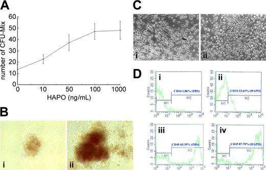
Effect on bone marrow MNCs. The rhHAPO was first tested for its effect on hematopoietic progenitor cells (HPCs) with the use of human fetal bone marrow MNCs. The rhHAPO alone stimulated the growth of CFU-Mix colonies in culture system A. The combination of rhHAPO with a cocktail of cytokines, including IL-3, GM-CSF, EPO, and SCF, resulted in an additive action on colony formation (Table 1). The stimulatory effect of rhHAPO on CFU-Mix was found to be concentration dependent, beginning at 10 ng/mL, with a maximal effect at 100 ng/mL (Figure 2A). Therefore, we used the concentration of 100 ng/mL to investigate the biologic function of rhHAPO in further experiments. In the presence of rhHAPO plus CC, the size of CFU-Mix-derived colonies was significantly increased compared with that of rhHAPO alone (Figure 2B). Further studies using liquid culture found that in the presence of rhHAPO, there were more small-size round cells grown from human fetal bone marrow cells after 4 weeks of culture (Figure 2C). When these cells were subjected to FACS analysis, a significant increase in the percentage of the CD34+ and CD45+ cells was found in the cultures with added rhHAPO. FACS analysis showed that HAPO increased percentage of CD34+ and CD45+ human fetal bone marrow mononuclear cells by 20.75% and 22.31%, respectively, as compared with control (Figure 2D).
Effect of rhHAPO on colony formation of human fetal bone marrow progenitor cells in response to cytokine stimulation
. | Colony types, no. . | . | . | . | . | ||||
|---|---|---|---|---|---|---|---|---|---|
| Additions . | CFU-M . | CFU-G . | CFU-GM . | BFU-E . | CFU-Mix . | ||||
| PBS | 3.5 ± 0.5 | 0 | 4.5 ± 0.2 | 0 | 0 | ||||
| HAPO | 3.5 ± 0.2 | 0 | 6.5 ± 2.2 | 0 | 11 ± 2.58* | ||||
| CC | 10 ± 1.41 | 0.5 ± 0.5 | 55 ± 4.5 | 6.5 ± 1.15 | 17.5 ± 1.26 | ||||
| CC + HAPO | 13 ± 1.29 | 1.5 ± 0.57 | 67.5 ± 6.5 | 8.2 ± 2.5 | 36.75 ± 6.94† | ||||
CC, the appropriate cocktail of cytokines, included rhIL-3 (10 ng/mL), rhGM-CSF (10 ng/mL), rhSCF (50 ng/mL), and rhEPO (3 U/mL). The rhHAPO was added at 100 ng/mL final concentration. Data are pooled from 3 separate experiments and expressed as mean ± SD. Bone marrow cells were plated a cell density of 1 × 10⁵/35 mm dish in a methycellulose system as described in “Materials and methods.”
CFU indicates colony-forming unit; M, macrophage; G, granulocyte; BFU-E, eruthroblast burst-forming unit; Mix, mixed colony; PBS, phosphate-buffered saline.
P < .05 versus PBS.
P < .05 versus CC group.
Biologic activity of HAPO in vitro in a human system. (A) Effect of various concentrations of rhHAPO on the growth of CFU-Mix. Human fetal marrow nonadherent cells were cultured at 1 × 10⁵/35-mm dish in methylcellulose containing growth medium with a cocktail of cytokines as described in Table 1, with or without the indicated concentrations of rhHAPO for 14 days. All cultures were performed in quadruplicate and scored with the use of an inverted microscope. Data represent mean ± SD. (B) Morphology of CFU-Mix colony in semisolid culture. The colonies stimulated by HAPO plus CC (ii) were bigger in size than those stimulated with HAPO alone (i). Original magnification × 40. (C) Morphology of human fetal bone marrow MNCs cultured in a liquid system with or without rhHAPO (100 ng/mL). Human fetal bone marrow cells were cultured without rhHAPO (i) and with rhHAPO (ii) for 4 weeks. Original magnification × 200 (D) Effect of rhHAPO on human fetal bone marrow CD34+ cells in liquid culture. Fluorescence-activated cell sorter (FACS) analysis shows an increase of the percentage of CD34+ and CD45+ cells in the presence of rhHAPO.
Biologic activity of HAPO in vitro in a human system. (A) Effect of various concentrations of rhHAPO on the growth of CFU-Mix. Human fetal marrow nonadherent cells were cultured at 1 × 10⁵/35-mm dish in methylcellulose containing growth medium with a cocktail of cytokines as described in Table 1, with or without the indicated concentrations of rhHAPO for 14 days. All cultures were performed in quadruplicate and scored with the use of an inverted microscope. Data represent mean ± SD. (B) Morphology of CFU-Mix colony in semisolid culture. The colonies stimulated by HAPO plus CC (ii) were bigger in size than those stimulated with HAPO alone (i). Original magnification × 40. (C) Morphology of human fetal bone marrow MNCs cultured in a liquid system with or without rhHAPO (100 ng/mL). Human fetal bone marrow cells were cultured without rhHAPO (i) and with rhHAPO (ii) for 4 weeks. Original magnification × 200 (D) Effect of rhHAPO on human fetal bone marrow CD34+ cells in liquid culture. Fluorescence-activated cell sorter (FACS) analysis shows an increase of the percentage of CD34+ and CD45+ cells in the presence of rhHAPO.
Effect on purified CD34+cells. To evaluate whether rhHAPO acts directly on purified hematopoietic stem cells subsets, we assayed the effect of rhHAPO on the growth of CD34+ cells purified from adult human bone marrow. Purified CD34+ marrow cells were cultured in a serum-free liquid medium containing SCF, TPO, and IL-3 with or without rhHAPO for 1 to 2 weeks. A significant increase in the number of CD34+ and AC133+ cells was obtained on day 7 and day 14. In addition, rhHAPO was able to increase the number of KDR+ cells (Table 2).
Effect of rhHAPO on the expansion of adult human bone marrow CD34+ cells
. | Purified CD34+ bone marrow cells, × 104 . | . | . | . | . | . | |||||
|---|---|---|---|---|---|---|---|---|---|---|---|
| . | Day 0 . | . | Day 7 . | . | Day 14 . | . | |||||
| Cell subset . | Control . | rhHAPO . | Control . | rhHAPO . | Control . | rhHAPO . | |||||
| CD34+ | 10 | 10 | 426 ± 4 | 526 ± 22* | 62 ± 5 | 159 ± 42* | |||||
| AC133 | 7 ± 0.1 | 7 ± 0.1 | 179 ± 2 | 288 ± 12* | 61 ± 17 | 244 ± 17* | |||||
| KDR+ | 0.4 ± 0.02 | 0.4 ± 0.02 | 0.2 ± 0.03 | 0.4 ± 0.02* | 0.11 ± 0.02 | 0.39 ± 0.01* | |||||
Purified adult bone marrow CD34+ cells were cultured at 1 × 10⁵/mL in QBSF-60 (Quality Biological, Gaithersburg, MD) serum-free medium containing a cocktail of cytokines including rhIL-3 (10 ng/mL), rhSCF (50 ng/mL), TPO (20 ng/mL), and 100 ng/mL of rhHAPO (the rhHAPO was not added in the control). Data are pooled from 3 separate experiments and expressed as mean ± SD.
P < .05 versus control.
Effect of rhHAPO on the growth of CD34+KDR+ cells in vitro
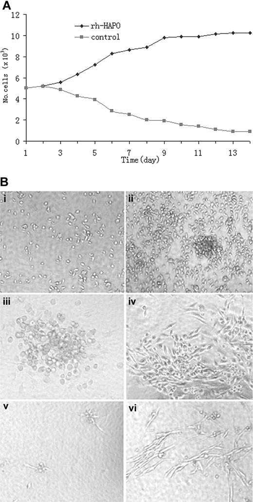
The CD34+KDR+ cell subset from human bone marrow and cord blood have been identified as the postnatal hemangioblasts with a long-term proliferative potential and hematopoietic-endothelial bilineage differentiation capacity.10 Therefore, we tested the effect of rhHAPO on the proliferation and differentiation of CD34+KDR+ cells. First, CD34+KDR+ cells were cultured at 5 × 10³ cells per well in a 24-well plate in the basic medium of liquid culture for 2 weeks. The number of viable cells in the suspension was measured by counting trypan blue-negative cells. As seen in Figure 3A, rhHAPO significantly promoted the proliferation of CD34+KDR+ cells. During culture, the number of viable cells-derived from of CD34+KDR+ cells increased in the presence of rhHAPO and declined in the absence of rhHAPO. Apart from the suspended cells, the number of adherent cells increased significantly in the presence of rhHAPO (Figure 3Bii) as compared with control culture (Figure 3Bi), indicating a direct stimulatory effect of rhHAPO on proliferation and survival of adherent and nonadherent cells derived from CD34+KDR+ cells.
Effect of rhHAPO on the growth of purified CD34+KDR+ cells. (A) Effect of rhHAPO on the growth pattern of CD34+KDR+ cells. CD34+KDR+ cells were cultured at 5 × 10³ cells per well in a 24-well plate. The number of viable cells was measured by counting trypan blue-negative cells. The log growth phase of cells was between day 3 and day 9, followed by the growth arrest phase when rhHAPO was added. Without rhHAPO, the number of CD34+KDR+ cells is decreased. (B) Hematopoietic and endothelial proliferation and differentiation of CD34+KDR+ cells in a liquid culture system. Original magnification × 200. In the absence of rhHAPO, a few cells grew in culture on day 7 (i). In contrast, significant cell proliferation with colony formation was observed in the presence of rhHAPO (ii). The suspended cells were aspirated away, and adherent cells were cultured in fresh medium with or without rhHAPO for 3 additional days. The cells grew well but displayed different morphology in the presence or absence of rhHAPO. In comparison with control culture (iii), the addition of rhHAPO into the culture significantly promoted the proliferation of endothelial-like cells (iv). When single CD34+KDR+ cells were cultured in 96-well plates in culture system C, as described in “Materials and methods,” for 2 weeks, these cells displayed different morphology in the presence or absence of rhHAPO. In comparison with control culture without rhHAPO (v), the addition of rhHAPO into the culture significantly promoted the proliferation of endothelial-like cells and formed capillary-like structures (vi).
Effect of rhHAPO on the growth of purified CD34+KDR+ cells. (A) Effect of rhHAPO on the growth pattern of CD34+KDR+ cells. CD34+KDR+ cells were cultured at 5 × 10³ cells per well in a 24-well plate. The number of viable cells was measured by counting trypan blue-negative cells. The log growth phase of cells was between day 3 and day 9, followed by the growth arrest phase when rhHAPO was added. Without rhHAPO, the number of CD34+KDR+ cells is decreased. (B) Hematopoietic and endothelial proliferation and differentiation of CD34+KDR+ cells in a liquid culture system. Original magnification × 200. In the absence of rhHAPO, a few cells grew in culture on day 7 (i). In contrast, significant cell proliferation with colony formation was observed in the presence of rhHAPO (ii). The suspended cells were aspirated away, and adherent cells were cultured in fresh medium with or without rhHAPO for 3 additional days. The cells grew well but displayed different morphology in the presence or absence of rhHAPO. In comparison with control culture (iii), the addition of rhHAPO into the culture significantly promoted the proliferation of endothelial-like cells (iv). When single CD34+KDR+ cells were cultured in 96-well plates in culture system C, as described in “Materials and methods,” for 2 weeks, these cells displayed different morphology in the presence or absence of rhHAPO. In comparison with control culture without rhHAPO (v), the addition of rhHAPO into the culture significantly promoted the proliferation of endothelial-like cells and formed capillary-like structures (vi).
When single CD34+KDR+ cells were further cultured in 96-well plate in culture system C for 2 weeks for the analysis of hematopoietic and endothelial differentiation in the presence or absence of rhHAPO, the cells grew well but displayed different morphology under the 2 conditions. In comparison with control culture without added rhHAPO (Figure 3Biii), the addition of rhHAPO into the culture significantly promoted the proliferation of endothelial-like cells (Figure 3Biv). Moreover, when single CD34+KDR+ cells were seeded in the culture system with VEGF and EGF, only a few cells grew in culture, and these cells displayed an endothelial-like morphology (Figure 3Bv). The combination of rhHAPO with VEGF and EGF promoted the formation of vascular-like structures (Figure 3Bvi), suggesting a supportive effect of rhHAPO on endothelial differentiation.
Effect of rhHAPO on endothelial cells and bone marrow cells
To determine if rhHAPO acts directly on endothelial cells, we performed the experiments using HUVECs. It was observed that rhHAPO significantly stimulated the proliferation of HUVECs. Such an action of rhHAPO was concentration dependent, with rhHAPO having a maximum effect at 100 ng/mL (Figure 4A). We then tested the effect of rhHAPO on the growth of human fetal bone marrow EPCs. Human fetal bone marrow MNCs were seeded in culture system C for 72 hours, and the adherent cells were isolated and seeded in culture system B for 7 days. FACS analysis revealed that the percentage of KDR+ and VWF+ cells was increased from 11.89% to 15.82% and from 11.74% to 21.67%, respectively (Figure 4B). When the adherent cells were further incubated in a culture system containing SCF, IL-3, TPO, and rhHAPO (Figure 4C), the formation of VWF+ endothelial cell colonies (CFU-EPCs) was significantly increased compared with control culture containing no added rhHAPO. The mean (± SD) number of colonies per culture well in rhHAPO group was 35 (± 4) colonies, compared with 21 (± 3) colonies in control group (P < .05).
Effect of HAPO on proliferation of endothelial cells. (A) The rhHAPO stimulated the proliferation of human umbilical vein endothelial cells (HUVECs) in a concentration-dependent manner. HUVECs were seeded in 96-well plates at 5 × 10⁴ cells per well in 100 μL RPMI 1640 medium with various concentrations of HAPO (0, 5, 50, 100, and 1000 ng/mL). The viability of HUVECs was analyzed by the MTT method after 72 hours. (B) Effect of rhHAPO on the growth of human fetal bone marrow endothelial cells. Human fetal bone marrow MNCs were seeded in culture system C for 72 hours, and the adherent cells were further incubated in culture system B for 7 days and then detached for FACS analysis. (C) Effect of HAPO on KDR+ human fetal bone marrow cells. Purified KDR+ cells were plated at a cell density of 1 × 10³/mL in culture system B containing a cocktail of cytokines as described, with or without rhHAPO. All cultures were performed in quadruplicate and scored after 21 days of culture. The rhHAPO significantly stimulated the formation of hematopoietic (CFU-Mix) and endothelial colonies (endothelial progenitor cell CFUs [CFU-EPCs]). Data are represented as the mean ± SD. *P < .05 versus control.
Effect of HAPO on proliferation of endothelial cells. (A) The rhHAPO stimulated the proliferation of human umbilical vein endothelial cells (HUVECs) in a concentration-dependent manner. HUVECs were seeded in 96-well plates at 5 × 10⁴ cells per well in 100 μL RPMI 1640 medium with various concentrations of HAPO (0, 5, 50, 100, and 1000 ng/mL). The viability of HUVECs was analyzed by the MTT method after 72 hours. (B) Effect of rhHAPO on the growth of human fetal bone marrow endothelial cells. Human fetal bone marrow MNCs were seeded in culture system C for 72 hours, and the adherent cells were further incubated in culture system B for 7 days and then detached for FACS analysis. (C) Effect of HAPO on KDR+ human fetal bone marrow cells. Purified KDR+ cells were plated at a cell density of 1 × 10³/mL in culture system B containing a cocktail of cytokines as described, with or without rhHAPO. All cultures were performed in quadruplicate and scored after 21 days of culture. The rhHAPO significantly stimulated the formation of hematopoietic (CFU-Mix) and endothelial colonies (endothelial progenitor cell CFUs [CFU-EPCs]). Data are represented as the mean ± SD. *P < .05 versus control.
Further studies were performed with the use of purified KDR+ human fetal bone marrow cells. In 2 representative experiments, purified KDR+ cells were plated at a cell density of 1 × 10³/mL in culture system B containing a cocktail of cytokines as described, with or without rhHAPO. All cultures were performed in quadruplicate and scored after 21 days of culture with the use of an inverted microscope. The rhHAPO significantly stimulated the formation of hematopoietic (CFU-Mix) and endothelial colonies (CFU-EPCs) (Figure 4C).
Effect of rhHAPO on the growth of murine bone marrow cells in vitro
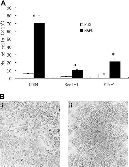
The biologic activity of rhHAPO on hematopoiesis was also studied in mice with the use of a long-term bone marrow culture system. It was found that rhHAPO stimulated the proliferation of murine bone marrow cells after 4 weeks of culture. FACS analysis revealed an increase in the numbers of various stem cell subpopulations, including CD34+, Sca-1+, and Flk-1+ cells, in response to rhHAPO stimulation. Particularly, the stimulatory effect of rhHAPO on the expansion of the CD34+ cell subset was statistically significant, as compared with control culture (Figure 5A).
Biologic activity of rhHAPO in murine system. (A) Bone marrow was obtained from BALB/c mice. The cells were seeded in 24-wells plates at a cell density of 2 × 10⁶/mL in long-term culture system with or without rhHAPO (100 ng/mL). After culture for 4 weeks, the cells were detached and subjected to FACS analysis. FACS analysis showed that rhHAPO significantly increased the number of the CD34+, Sca-1+, and flk-1+ cells as compared with control cells.*P < .05. (B) Colony formation from bone marrow long-term culture-initiating cells (LTC-ICs) in the absence (i) or presence (ii) of rhHAPO. Original magnification × 100.
Biologic activity of rhHAPO in murine system. (A) Bone marrow was obtained from BALB/c mice. The cells were seeded in 24-wells plates at a cell density of 2 × 10⁶/mL in long-term culture system with or without rhHAPO (100 ng/mL). After culture for 4 weeks, the cells were detached and subjected to FACS analysis. FACS analysis showed that rhHAPO significantly increased the number of the CD34+, Sca-1+, and flk-1+ cells as compared with control cells.*P < .05. (B) Colony formation from bone marrow long-term culture-initiating cells (LTC-ICs) in the absence (i) or presence (ii) of rhHAPO. Original magnification × 100.
To determine if rhHAPO acts on the growth of early murine stem cells, we tested the effect of rhHAPO on the proliferation of murine bone marrow long-term culture-initiating cells (LTC-ICs) as described previously.21 As seen in Figure 5Bii, rhHAPO stimulated the growth of LTC-IC-derived colonies as compared with control culture (Figure 5Bi). In 2 representative experiments, the number of LTC-IC-derived colonies in the presence of rhHAPO (10 ± 2.3/103 plated cells) was 5-fold higher than in the absence of rhHAPO (2 ± 0.8/103 plated cells).
In vivo effect of rhHAPO in normal mice
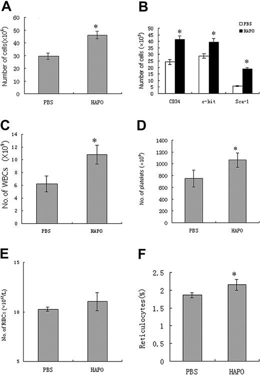
To confirm the stimulatory effect of HAPO in vivo, normal BALB/c mice received a daily subcutaneous injection of rhHAPO at 10 μg for 7 consecutive days. Figure 6A shows that on day 14 after injection of rhHAPO, the number of bone marrow nucleated cells per femur was 1.6-fold higher than in the control mice. A significant increase in the numbers of bone marrow CD34+, c-kit+, and Sca-1+ cells was observed in the mice receiving rhHAPO compared with the control mice (Figure 6B). In addition, injection of rhHAPO also resulted in an increase in circulating white blood cells (Figure 6C) and platelets (Figure 6D) in rhHAPO-treated mice. The number of circulating red blood cells had no significant change after administration of rhHAPO (Figure 6E). However, the percentage of reticulocytes slightly increased in rhHAPO-treated mice as compared with control mice (Figure 6F).
In vivo effect of rhHAPO on hematopoiesis in normal mice. The 16 mice in each group received a daily subcutaneous injection of rhHAPO at doses of 10 μg for 7 days. Shown are the effect of rhHAPO on the number of bone marrow nucleated cells (A); the numbers of the CD34+, Sca-1+, and c-kit+ cells in murine bone marrow (B); and the number of circulating white blood cells (C), platelets (D), and red blood cells (RBCs) (panel E), as well as the percentage of reticulocytes (F) on day 14 after rhHAPO administration. Data are represented as the mean ± SD. *P < .05 versus control.
In vivo effect of rhHAPO on hematopoiesis in normal mice. The 16 mice in each group received a daily subcutaneous injection of rhHAPO at doses of 10 μg for 7 days. Shown are the effect of rhHAPO on the number of bone marrow nucleated cells (A); the numbers of the CD34+, Sca-1+, and c-kit+ cells in murine bone marrow (B); and the number of circulating white blood cells (C), platelets (D), and red blood cells (RBCs) (panel E), as well as the percentage of reticulocytes (F) on day 14 after rhHAPO administration. Data are represented as the mean ± SD. *P < .05 versus control.
Radioprotective effect of rhHAPO in mice
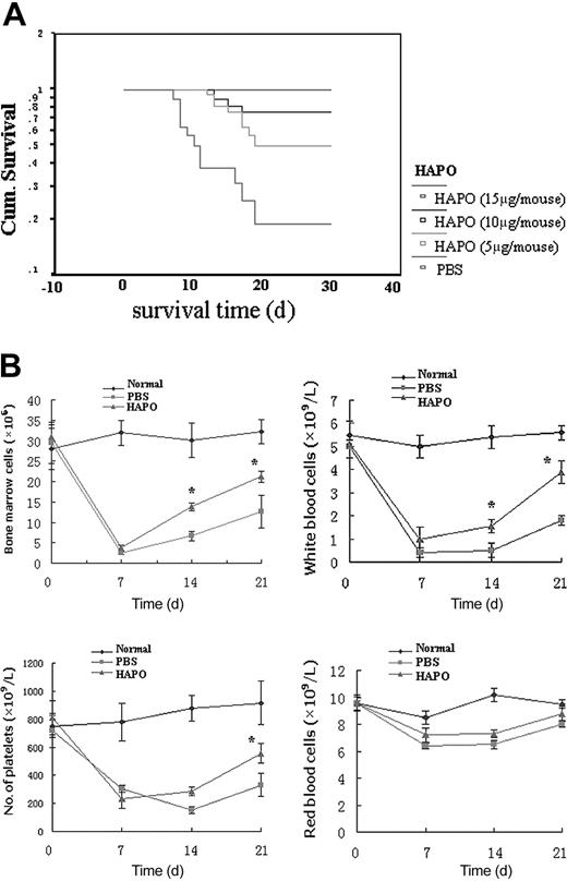
On the basis of the observation that rhHAPO stimulated normal hematopoiesis in vivo, the effect of rhHAPO on the recovery of radiation injury was further studied with the use of irradiated mice. The mice were treated with total body irradiation of 6 Gy and then received a daily subcutaneous injection of rhHAPO for 7 consecutive days. The irradiated mice receiving no rhHAPO started to die on day 6, and only 7% of mice survived for as long as 20 days after irradiation. In contrast, the survival rate of the irradiated mice receiving rhHAPO was significantly increased. Interestingly, the radioprotection of rhHAPO was found to be dose dependent. A high dose of rhHAPO (15 μg) completely protected animals from death caused by irradiation (Figure 7A).
Effect of HAPO on the survival rate of irradiated mice. (A) Survival curves of BALB/c mice after irradiation with 6.0 Gy (93% lethal dose). The 16 mice in each group received a daily subcutaneous injection of rhHAPO at doses of 5 μg, 10 μg, or 15μg for 14 days. The control mice received the same volume of PBS injected according to the same schedule. The irradiated mice started to die after 6 days. The administration of rhHAPO increased the survival rate in a dose-dependent manner, with 100% protection at a dose of 15 μg/d. (B) Quantitative changes of bone marrow and peripheral blood cells of the mice after irradiation. The 16 mice in each group received a daily subcutaneous injection of rhHAPO (10 μg) or PBS. At each time point, 3 mice in each group were randomly sampled and were subjected to examination for the content of the bone marrow CD34+ cells from the murine femurs (i) and the quantitative changes in the number of white blood cells (ii), platelets (iii), and red blood cells (iv) in the blood of the mice after irradiation. Data are represented as the mean ± SD. *P < .05 versus control.
Effect of HAPO on the survival rate of irradiated mice. (A) Survival curves of BALB/c mice after irradiation with 6.0 Gy (93% lethal dose). The 16 mice in each group received a daily subcutaneous injection of rhHAPO at doses of 5 μg, 10 μg, or 15μg for 14 days. The control mice received the same volume of PBS injected according to the same schedule. The irradiated mice started to die after 6 days. The administration of rhHAPO increased the survival rate in a dose-dependent manner, with 100% protection at a dose of 15 μg/d. (B) Quantitative changes of bone marrow and peripheral blood cells of the mice after irradiation. The 16 mice in each group received a daily subcutaneous injection of rhHAPO (10 μg) or PBS. At each time point, 3 mice in each group were randomly sampled and were subjected to examination for the content of the bone marrow CD34+ cells from the murine femurs (i) and the quantitative changes in the number of white blood cells (ii), platelets (iii), and red blood cells (iv) in the blood of the mice after irradiation. Data are represented as the mean ± SD. *P < .05 versus control.
Further study on the recovery of hematopoiesis in irradiated mice injected with rhHAPO found a significant increase in the degree of bone marrow nucleated cells on days 14 and 21 after irradiation (Figure 7Bi). The mice injected with rhHAPO also had an increased number of white blood cells (Figure 7Bii) and platelets (Figure 7iii) on days 14 and 21 after irradiation as compared with control mice.
Discussion
Aplastic anemia is an elusive disease, not only having a cytopenia and marrow hypoplasia but also containing key information on hematopoietic stem cell regeneration and microenvironmental evolution.26,27 Therefore, we initiated a search for a novel growth factor from the urine of patients with aplastic anemia. In the present work, we report the successful identification and purification of HAPO, a growth factor acting on the primitive cells of both hematopoietic and endothelial cell lineages. Sequence analysis indicates that the cDNA clone of HAPO, isolated from human fetal liver cDNA library, is an alternative splicing variant lacking exon 5 of PRG 4.23-25 To characterize the biologic activities of HAPO, a partial cDNA fragment of HAPO was expressed in E coli BL21, and highly purified rhHAPO was obtained for various in vitro and in vivo biologic assays.
We first demonstrated a stimulatory effect of rhHAPO on human hematopoiesis using different human culture systems. The rhHAPO stimulated the proliferation of human hematopoietic progenitor cells. Cloning efficacy analysis indicates that rhHAPO had a stimulatory effect on CFU-Mix but not on CFU-GM, CFU-G, CFU-M, and BFU-E, suggesting a predominant action of rhHAPO on human primitive hematopoietic stem/progenitor cells.
We then wanted to know if rhHAPO acts on the level of hemangioblast. It has been suggested that hemangioblast is a transient cell stage during embryonic development.3,4 Therefore, it is difficult to obtain a sufficient number of purified hemangioblasts from embryonic tissue for cytokine bioassay. However, previous observations have indicated that adults maintain a reservoir of hemangioblasts. For example, AC133+ human bone marrow and peripheral blood cells have the capacity to differentiate into endothelial cells. In addition, postnatal CD34+ cells and CD34+KDR+ cells can also generate hematopoietic and endothelial progeny in different in vitro and in vivo assays.8-11,14 On the basis of these observations, we tested the effect of rhHAPO on these stem cell subsets using culture systems both for hematopoietic-endothelial progenitor cells and for endothelial cells. The rhHAPO was found to directly stimulate the proliferation and differentiation of purified CD34+, KDR+, or CD34+KDR+ cells into both hematopoietic and endothelial cells The results obtained clearly show a predominantly stimulating action of rhHAPO at the level of the primitive cells of both hematopoietic and endothelial cells.
To determine if rhHAPO also acts in mice, similar experiments were conducted in the murine system. The rhHAPO was found to enhance proliferation of murine hematopoietic stem/progenitor cell populations including CD34+, Sca-1+, and Flk-1+ cells. Moreover, rhHAPO stimulated the growth of murine bone marrow LTC-ICs in liquid culture. These results demonstrate once again a stimulating action of rhHAPO on an early stage of hematopoietic stem cells.
Subsequent experiments were conducted to evaluate the clinical potential of rhHAPO in vivo in normal and irradiated mice. Administration of rhHAPO in normal mice resulted in a significant increase in bone marrow hematopoietic stem/progenitor cells, together with a slight increase of white blood cells and platelets in blood. These data indicate a positive action of rhHAPO on hematopoiesis in vivo and imply a potential of this factor in hemoprotection. We therefore performed in vivo studies on radio-protection of rhHAPO. The results obtained demonstrate that rhHAPO significantly protected animals from death caused by radiation damage. The irradiated mice injected with rhHAPO had an enhanced survival rate. Such a radioprotective effect of rhHAPO was dose dependent, starting at a rhHAPO dose of 55 μg. An injection of a higher dose of rhHAPO (15 μg) resulted in a 100% survival rate of the irradiated mice. Comparative analysis of control and experimental groups of mice revealed that the mechanism of action of rhHAPO that protects mice from irradiation was related to an accelerated hematopoiesis in response to rhHAPO stimulation.
We have noted that, in both in vitro and in vivo studies, a higher dose was required for rhHAPO to be effective compared with that necessary for other cytokines, including SCF, TPO, G-CSF, and FLT-3 ligand.28-30 This phenomenon suggests that HAPO may act on a relatively rare cell population and that therefore a larger amount of HAPO is needed for activation of the targeted cells. Another possibility is that HAPO may be a unique growth factor because the normal serum level of HAPO (56.42 ± 14.26 ng/mL) measured in our laboratory (ranging from 18.3 ng/mL to 91.1 ng/mL; n = 46; data not shown), by means of a sandwich enzyme-linked immunosorbent assay (ELISA), was found to be relatively higher than that of SCF, TPO, G-CSF, or FLT-3 ligand in normal serum. In addition, we must point out that the rhHAPO used in the study was preserved in a buffer without any exogenous protein as protector. This preservation may not be the best one and may cause a loss of some activity of rhHAPO. Full characterization of biologic activities, pathophysiological significance, and clinical potential of HAPO will require further studies in both animal and human systems, including preparation of highly pure and active rhHAPO and identification of HAPO receptor and signal transduction pathway as well as its modulating action on cell apoptosis and DNA repair.
In summary, the present study reports the identification and partial biologic characterization of HAPO and suggests that this cytokine possesses an important clinical potential for the management of various cytopenias and radiation injury and for the expansion of hematopoietic and endothelial stem/progenitor cells. The results obtained from in vitro studies suggest that HAPO is the growth factor acting on the primitive cells of both hematopoietic and endothelial cell lineages. In vivo in mice, although short-term radioprotection by rhHAPO has been demonstrated, further studies are needed to confirm if rhHAPO acts directly on primitive hematopoietic cells as observed in vivo, with the use of the competitive transplation assay.
Prepublished online as Blood First Edition Paper, February 19, 2004; DOI 10.1182/blood-2003-06-1825.
Supported by grants of 863 (2001AA215311, 2002AA223354) and 973 (001CB5101) projects from the Ministry of Science and Technology of China; a grant from the China Medical Board of New York (no. 01-748); and a grant from the Tianjin Committee of Science and Technology (003119811) (Z.C.H.).
The publication costs of this article were defrayed in part by page charge payment. Therefore, and solely to indicate this fact, this article is hereby marked “advertisement” in accordance with 18 U.S.C. section 1734.
The authors sincerely thank Drs Hsiang-fu Kung and Zhengyu Wang for their suggestions and critical review of the manuscript.


![Figure 4. Effect of HAPO on proliferation of endothelial cells. (A) The rhHAPO stimulated the proliferation of human umbilical vein endothelial cells (HUVECs) in a concentration-dependent manner. HUVECs were seeded in 96-well plates at 5 × 10⁴ cells per well in 100 μL RPMI 1640 medium with various concentrations of HAPO (0, 5, 50, 100, and 1000 ng/mL). The viability of HUVECs was analyzed by the MTT method after 72 hours. (B) Effect of rhHAPO on the growth of human fetal bone marrow endothelial cells. Human fetal bone marrow MNCs were seeded in culture system C for 72 hours, and the adherent cells were further incubated in culture system B for 7 days and then detached for FACS analysis. (C) Effect of HAPO on KDR+ human fetal bone marrow cells. Purified KDR+ cells were plated at a cell density of 1 × 10³/mL in culture system B containing a cocktail of cytokines as described, with or without rhHAPO. All cultures were performed in quadruplicate and scored after 21 days of culture. The rhHAPO significantly stimulated the formation of hematopoietic (CFU-Mix) and endothelial colonies (endothelial progenitor cell CFUs [CFU-EPCs]). Data are represented as the mean ± SD. *P < .05 versus control.](https://ash.silverchair-cdn.com/ash/content_public/journal/blood/103/12/10.1182_blood-2003-06-1825/5/m_zh80120462690004.jpeg?Expires=1769098021&Signature=KGYeKh-JcxhNidJrNdIh7qpQS79W4OXYUWbOrzLytJtwUghIHpjUJqfmgrDzlkNCvQkbjwVmGum2SBueEF5DdsjXmTX29oeLeNUxLcD8Vy8sHPRpBuVlgBVjyc3KdtnXzuT76wjB2cRnOd4PArcGCXcj6iKpfo4yF6bRHFb2IFd9Rq4KjFLf7eKnW5KBVh4sikxU8i6iAaRgAR92KSpb8GIjrFcV6c1qdbhYCMIQ~IUW4mBmxD8dR42bgFAC3Jfr1Ap5pCOw~oTlyJsWob3RjmNSkwuqwXx4bMfIp3qFCkTH-tnEzOTA00shQH5I7XK9wpILuE4a-ECt5yOI~vOB0Q__&Key-Pair-Id=APKAIE5G5CRDK6RD3PGA)
This feature is available to Subscribers Only
Sign In or Create an Account Close Modal