Abstract
Interleukin-6 (IL-6) is a critical factor in the regulation of stromal function and hematopoiesis. In vivo bromodeoxyuridine incorporation analysis indicates that the percentage of Lin-Sca-1+ hematopoietic progenitors undergoing DNA synthesis is diminished in IL-6-deficient (IL-6-/-) bone marrow (BM) compared with wild-type BM. Reduced proliferation of IL-6-/- BM progenitors is also observed in IL-6-/- long-term BM cultures, which show defective hematopoietic support as measured by production of total cells, granulocyte macrophage-colony-forming units (CFU-GMs), and erythroid burst-forming units (BFU-Es). Seeding experiments of wild-type and IL-6-/- BM cells on irradiated wild-type or IL-6-deficient stroma indicate that the hematopoietic defect can be attributed to the stromal and not to the hematopoietic component. In IL-6-/- BM, stromal mesenchymal precursors, fibroblast CFUs (CFU-Fs), and stroma-initiating cells (SICs) are reduced to almost 50% of the wild-type BM value. Moreover, IL-6-/- stromata show increased CD34 and CD49e expression and reduced expression of the membrane antigens vascular cell adhesion molecule-1 (VCAM-1), Sca-1, CD49f, and Thy1. These data strongly suggest that IL-6 is an in vivo growth factor for mesenchymal precursors, which are in part implicated in the reduced longevity of the long-term repopulating stem cell compartment of IL-6-/- mice. (Blood. 2004;103:3349-3354)
Introduction
Interleukin-6 (IL-6) is a multifunctional cytokine with a major role in inflammatory responses and is the primary inducer of acute-phase proteins.1 IL-6 also regulates the proliferation and differentiation of hematopoietic cells from several lineages at different maturation stages.1 In synergy with IL-3, IL-6 stimulates proliferation of granulocyte-macrophage, megakaryocyte, and erythroid progenitors.2-5 This cooperation has also been shown in several in vivo model systems designed to evaluate the effect of IL-6 on the stem cell compartment.3,4,6 The specific contribution of IL-6 to homeostasis in the lymphohematopoietic system has been evaluated in the IL-6-/- mouse model, which showed a clear reduction in the number of the earliest clonogenic precursors and in the function of long-term repopulating stem cells (LTRSCs).7 IL-6-/- mice show a decrease in absolute numbers of spleen colony-forming units (CFU-Ss) at day 12 and of pre-CFU-S hematopoietic progenitors, as well as a deficit in the LTRSC compartment.7 In addition to the effects in early hematopoiesis, IL-6-/- mice show defects in T- and B-cell function8 and in acute-phase responses.9 These results strongly suggest an important in vivo role for IL-6 in the survival and self-renewal of hematopoietic stem cells (HSCs) and early progenitors.
IL-6 is produced by a variety of cells including T and B cells, macrophages, fibroblasts, bone marrow stromal cells, and mesenchymal cells.10-12 The adherent stromal cell layer in long-term bone marrow cultures (LTBMCs) is necessary for balanced proliferation and differentiation of hematopoietic progenitors.13 IL-6 is produced at low levels in LTBMCs, and appears to participate in regulating their hematopoietic supportive activity. Anti-IL-6 antibodies decrease the production of mature myeloid cells and colony-forming progenitor cells in LTBMCs.14,15 Addition of high IL-6 concentrations to LTBMCs leads to a general increase in total cell, progenitor, and multipotential cell numbers.14 IL-6 is constitutively expressed by stromal layers; addition of serum,16 IL-1α, or tumor necrosis factor α (TNFα)17 to LTBMCs increases the amount of secreted IL-6. IL-6 affects stromal cell proliferation and production of extracellular matrix, and down-regulates collagen I expression.17
Mesenchymal stem cells (MSCs) are a subset of nonhematopoietic cells found in the bone marrow (BM) stroma. These precursors for nonhematopoietic tissues were identified by Friedenstein et al18 by their ability to form colonies when plated in serum-containing medium, and were termed colony-forming unit fibroblasts (CFU-Fs).19,20 They were subsequently denominated marrow stromal cells,21 and finally, mesenchymal stem cells.22 Human marrow-derived MSCs are of interest, since they are easily expanded in culture and can differentiate terminally into several lineages including bone, cartilage, tendon, nerve, and hematopoietic-supportive stromal cells.23 A recently described, rare MSC subpopulation, the multipotent adult progenitor cells (MAPCs), contributes to most somatic tissues.24,25 IL-6 is produced by MSCs as well as by other BM stromal cells.26,27 MSCs do not express the IL-6 receptor and do not respond to IL-611 ; however, the IL-6-rich environment in BM stroma can induce osteoblastic differentiation of marrow-derived mesenchymal progenitors in the presence of glucocorticoids and IL-6.11
Taking into consideration the role of IL-6 in hematopoietic progenitor and MSC regulation, we analyzed whether the hematopoietic deficiency observed in IL-6-/- mice can be attributed to a defect in the hematopoietic supportive activity of IL-6-/- BM stromal cells, and the extent to which development and maintenance of the MSC compartment is affected by the absence of IL-6.
Materials and methods
Mice
IL-6-deficient9 mice and wild-type littermates on a pure C57BL/6 background were bred in microisolator cages in the barrier facility at the Centro Nacional de Biotecnología under standard pathogen-free protocols. The mice used for these experiments were 8 to 12 weeks old.
Bone marrow cell isolation
BM cells were flushed from femurs and tibiae into Hanks balanced salt solution containing 2% fetal bovine serum and 0.02% sodium azide (HSA), using a 25-gauge needle. Low-density mononuclear cells (LDMNCs) were purified by centrifugation (400g, 15 minutes) in a 3-density gradient (1.090 g/mL, 1.080 g/mL, and 1.050 g/mL) prepared by mixing NycoPrep 1.077A (1.077 g/mL) and OptiPrep (1.320 g/mL) (Axis-Shield, Oslo, Norway). LDMNCs were resuspended in HSA (107 cells/mL) and incubated (30 minutes, 4°C) with purified rat antimouse monoclonal antibodies (0.5 μg/106 cells) anti-CD4, -CD8, -B220, -Gr-1, -Mac-1, and -TER-119 (all from BD PharMingen, San Diego, CA). Cells were washed with HSA, resuspended in ice-cold HSA (107 cells/mL) and incubated (8-10 beads/cell) with Dynabeads M-450 sheep antirat immunoglobulin G (IgG) (Dynal Biotech, Oslo, Norway), for 45 minutes at 4°C. Beads were separated with a magnet; the remaining medium contained the Lin- fraction.
Long-term bone marrow cultures
Primary stromata were obtained by flushing BM cells from one tibia and one femur of wild-type or IL-6-/- mice directly into a 25-cm2 flask with 10 mL Myelocult M5300 medium (StemCell Technologies, Vancouver, BC, Canada) supplemented with 10-6 M hydrocortisone, and cultured at 32°C. Half of the medium was changed weekly for up to 5 weeks, and total cell number, granulocyte macrophage-colony-forming units (CFU-GMs), and erythroid burst-forming units (BFU-Es) were evaluated weekly. Duplicate cultures were trypsinized each week to detach the stromal cell layer, and total hematopoietic cells, CFU-GMs, and BFU-Es were analyzed in the stromal layer. Murine IL-6 (200 ng/mL; Biosource, Camarillo, CA) was added weekly to some cultures.
For seeding experiments, wild-type and IL-6-/- LTBMCs were established as above and irradiated (17 Gy) 4 weeks later; 3 days after irradiation, cultures were washed and seeded with 5 × 10⁵ wild-type or IL-6-/- Lin- BM cells in 10 mL Myelocult M5300, supplemented as above. In some seeding experiments, IL-6-/- LTBMCs were established in the presence of 5 ng/mL or 20 ng/mL IL-6. The Lin- cells used in seeding experiments were free of CFU-Fs, and the irradiated stromata were unable to produce hematopoietic cells.
Clonogenic assays
CFU-GMs were analyzed (105 cells/mL) in MethoCult M3534 medium (Stem-Cell Technologies). Erythroid burst-forming units (BFU-Es) were tested (105 cells/mL) in MethoCult M3234 (StemCell Technologies) supplemented with 6 U/mL erythropoietin (StemCell Technologies), 10 ng/mL murine IL-3 (Biosource), and 50 ng/mL murine stem cell factor (Biosource). Cells were added in a volume of 300 μL, mixed, and l-mL duplicates dispensed into 35-mm plates (Falcon, Plymouth, United Kingdom). Cultures were incubated (37°C) and colonies scored on day 7 for CFU-GMs and day 12 for BFU-Es.
Cobblestone area-forming cell (CAFC) assay
Wild-type and IL-6-/- LTBMCs were established as described in “Long-term bone marrow cultures” in gelatin-coated 96-well microtiter plates and irradiated (17 Gy). Three days after irradiation, cultures were washed and seeded with 200 μL freshly prepared wild-type or IL-6-/- BM cell suspensions at 6 serial 2-fold dilutions (from 625 to 20 000 cells/well); each cell concentration was monitored in 16 replicate wells. Cells were cultured in Myelocult M5300 supplemented with 10-6 M hydrocortisone (32°C, 3 weeks) with weekly exchange of half the medium. The appearance of cobblestone areas (colonies growing beneath the stromal layer, consisting of at least 5 small, nonrefractile cells) was evaluated weekly. Individual wells were scored for the presence of cobblestone areas. CAFC frequency was determined in limiting dilution analysis using L-Calc statistical software (StemCell Technologies).
CFU-F and stroma-initiating cell (SIC) assays
The CFU-F assay was performed using MesenCult Basal Medium and MSC stimulatory supplements (StemCell Technologies), according to the manufacturer's protocols. Wild-type and IL-6-/- BM cells were grown in treated tissue-culture plates (Falcon) for 14 days, and the CFU-F number was determined by fixing cultures in ice-cold methanol for 5 minutes, Giemsa staining for 5 minutes, then counting colonies under the optical microscope.
SICs were assayed as described.28 Briefly, BM cells were resuspended in RPMI 1640 medium supplemented with 15% fetal calf serum (FCS) and 5 mM 2-mercaptoethanol, then incubated on plastic dishes (4 hours, 37°C) to deplete adherent cells. Nonadherent cells were subsequently seeded into 10-cm dishes (Falcon; 107 cells/dish) in complete medium. Cultures were incubated for 2 weeks with a weekly medium change, then fixed, Giemsa-stained, and counted as for CFU-Fs.
Fluorescence activated cell sorting (FACS) analysis
Cells were harvested by brief trypsinization, washed in ice-cold blocking buffer (phosphate-buffered saline with 0.5% bovine serum albumin), and incubated for 30 minutes in ice-cold blocking buffer containing the specific fluorescein isothiocyanate (FITC)- or phycoerythrin (PE)-labeled antibody. In all experiments, appropriate FITC- or PE-labeled nonimmune isotypes were used as negative controls. All monoclonal antibodies (mAbs) were purchased from BD Pharmingen. For each analysis, 10 000 events were recorded. Appropriate gating was performed using the forward versus side scatter dot plot; dead cells were gated out by propidium iodide staining. Stained cells were analyzed on a Coulter EPICS XL flow cytometer (Beckman Coulter, Fullerton, CA).
Results
Long-term bone marrow cultures
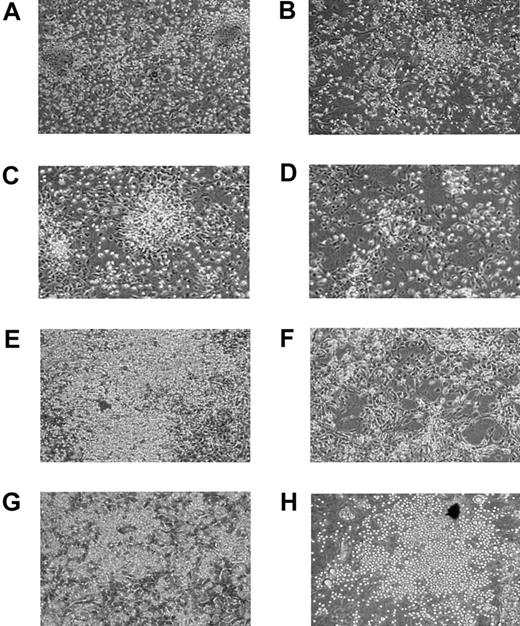
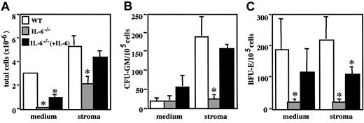
To analyze the in vitro hematopoietic supportive activity of IL-6-/- BM stroma, wild-type and IL-6-/- LTBMCs were established and monitored weekly for up to 5 weeks. Microscopic observation of wild-type LTBMCs revealed that hematopoietic foci were apparent during the first 2 weeks of culture; during the third week, large cobblestone areas were observed, and after 4 weeks a confluent adherent layer had been established (Figure 1). On the contrary, IL-6-/- LTBMCs showed a clear delay in stromal layer development and in the onset of hematopoietic activity; cobblestone areas were present after 4 weeks of culture, but the stromal layer was not yet confluent (Figure 1). Hematopoietic cell generation kinetics (total cell numbers and relative CFU-GM and BFU-E content) were scored weekly, showing a substantial reduction in the hematopoietic supportive activity of IL-6-/- LTBMCs compared with equivalent wild-type cultures. This deficiency was observed both in the cell fraction released from the stromal layer to the medium and in cells that remained attached to the stroma (Figure 2A). At 5 weeks after culture initiation, the average hematopoietic cell numbers for wild-type and IL-6-/- cultures were, respectively, 36 × 105 ± 20.6 × 10⁵ and 0.57 × 10⁵ ± 0.08 × 10⁵ cells in culture medium and 50.4 × 10⁵ ± 16.8 × 10⁵ and 20.4 × 10⁵ ± 6.8 × 10⁵ cells in the stromal layer. Relative CFU-GM and BFU-E content was also analyzed in the cells collected at each time interval (Figure 2B-C). Wild-type cultures exhibited peak CFU-GM production at week 4, whereas peak CFU-GM production was observed one week earlier in IL-6-/- cultures, suggesting an imbalance in IL-6-/- cultures in the ratio between hematopoietic progenitor proliferation and their differentiation to the granulocyte/macrophage lineage. Mean CFU-GM values for wild-type and IL-6-/- cultures at week 4 were 363 ± 132 and 72 ± 29 CFU-GMs/105 cells in conditioned medium and 295 ± 100 and 87 ± 31 CFU-GMs/105 cells in the stromal layer, respectively. Mean BFU-E values for wild-type and IL-6-/- cultures at week 5 were 188 ± 98 and 18 ± 12 BFU-Es/105 cells in conditioned medium and 218 ± 74 and 25 ± 5 BFU-Es/105 cells in the stromal layer, respectively.
Photomicrographs of wild-type and IL-6-/- LTBMCs. Cultures were established from wild-type (A, C, E, G) or IL-6-/- (B, D, F, H) BM and were maintained in Myelocult M5300 medium containing 10-6 M hydrocortisone at 32°C, with weekly exchange of half of the medium. Photomicrographs were obtained 1 (A-B), 2 (C-D), 3 (E-F), or 4 (G-H) weeks after initiation of cultures. Original magnification × 100.
Photomicrographs of wild-type and IL-6-/- LTBMCs. Cultures were established from wild-type (A, C, E, G) or IL-6-/- (B, D, F, H) BM and were maintained in Myelocult M5300 medium containing 10-6 M hydrocortisone at 32°C, with weekly exchange of half of the medium. Photomicrographs were obtained 1 (A-B), 2 (C-D), 3 (E-F), or 4 (G-H) weeks after initiation of cultures. Original magnification × 100.
Production of hematopoietic cells, CFU-GMs, and BFU-Es by wild-type and IL-6-/- LTBMCs. Cultures were established and maintained as in Figure 1. Total hematopoietic cells (A), CFU-GMs (B), and BFU-Es (C) in culture medium and in the stromal layer were evaluated at weekly intervals. Results are expressed as the mean ± standard deviation (SD) of 4 experiments. *P < .01, for wild-type versus IL-6-/- cultures.
Production of hematopoietic cells, CFU-GMs, and BFU-Es by wild-type and IL-6-/- LTBMCs. Cultures were established and maintained as in Figure 1. Total hematopoietic cells (A), CFU-GMs (B), and BFU-Es (C) in culture medium and in the stromal layer were evaluated at weekly intervals. Results are expressed as the mean ± standard deviation (SD) of 4 experiments. *P < .01, for wild-type versus IL-6-/- cultures.
IL-6 has been identified as one of the main soluble factors in serum, and is necessary for efficient establishment of LTMBCs.15 Although culture medium used for this analysis was calibrated for optimal growth, we analyzed whether exogenous IL-6 addition could overcome the defective hematopoietic support observed in IL-6-/- LTBMCs. We established IL-6-/- cultures for 5 weeks, using several concentrations of IL-6. Even at a supraoptimal IL-6 concentration (200 ng/mL) (Figure 3A), the defective hematopoietic support was only partially restored (30% and 82% of wild-type culture values for cells in medium or on stroma, respectively). Relative CFU-GM and BFU-E content was also only partially normalized (Figure 3B-C). These results show that IL-6 addition is insufficient to restore full hematopoietic supportive activity in IL-6-/- LTBMCs, and suggest that this deficiency can be attributed in part to defective BM stroma development in the absence of IL-6.
Partial restoration of the IL-6-/- LTBMC hematopoietic support defect by exogenous IL-6. Wild-type and IL-6-/- cultures were established and maintained as in Figure 1. Parallel IL-6-/- cultures were maintained in 200 ng/mL IL-6, with weekly cytokine additions. Five weeks after initiation, cultures were analyzed for production of total hematopoietic cells (A), CFU-GMs (B), and BFU-Es (C) in culture medium and in the stromal layer. Results are expressed as the mean ± SD of 4 experiments. *P < .01, for wild-type versus IL-6-/- cultures.
Partial restoration of the IL-6-/- LTBMC hematopoietic support defect by exogenous IL-6. Wild-type and IL-6-/- cultures were established and maintained as in Figure 1. Parallel IL-6-/- cultures were maintained in 200 ng/mL IL-6, with weekly cytokine additions. Five weeks after initiation, cultures were analyzed for production of total hematopoietic cells (A), CFU-GMs (B), and BFU-Es (C) in culture medium and in the stromal layer. Results are expressed as the mean ± SD of 4 experiments. *P < .01, for wild-type versus IL-6-/- cultures.
Hematopoietic support by irradiated stroma
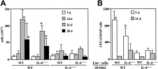
To determine whether the reduced hematopoietic cell proliferation in IL-6-/- LTBMCs can be attributed to a defect in hematopoietic progenitors or to deficient stromal support, we compared the ability of irradiated wild-type and IL-6-/- stromal layers to support proliferation and differentiation of wild-type and IL-6-/- Lin- hematopoietic populations. Total cell and relative CFU-GM numbers were monitored in the conditioned medium of irradiated, reseeded cultures. Hematopoietic and CFU-GM cell proliferation on irradiated IL-6-/- stroma was significantly reduced compared with irradiated wild-type stroma, irrespective of the seeded Lin- BM cell genotype (Figure 4). IL-6-/- Lin- and wild-type Lin- BM cell proliferation on wild-type stroma was comparable.
Kinetics of Lin- wild-type and IL-6-/- BM cell proliferation and CFU-GM production on irradiated wild-type and IL-6-/- stromal layers. Wild-type and IL-6-/- LTBMCs were established and maintained as in Figure 1. After 4 weeks, cultures were irradiated (17 Gy); 3 days after irradiation, cultures were seeded with 5 × 10⁵ Lin- wild-type or IL-6-/- BM cells. No differences were observed in CFU-GM number between Lin- wild-type and IL-6-/- BM seeded cells. Half of the medium was changed weekly, and production of total hematopoietic cells (A) and CFU-GMs (B) in culture medium was evaluated. Results are expressed as the mean ± SD of 4 experiments. *P < .01, for wild-type stroma versus IL-6-/- stroma seeded with the same type of BM cells.
Kinetics of Lin- wild-type and IL-6-/- BM cell proliferation and CFU-GM production on irradiated wild-type and IL-6-/- stromal layers. Wild-type and IL-6-/- LTBMCs were established and maintained as in Figure 1. After 4 weeks, cultures were irradiated (17 Gy); 3 days after irradiation, cultures were seeded with 5 × 10⁵ Lin- wild-type or IL-6-/- BM cells. No differences were observed in CFU-GM number between Lin- wild-type and IL-6-/- BM seeded cells. Half of the medium was changed weekly, and production of total hematopoietic cells (A) and CFU-GMs (B) in culture medium was evaluated. Results are expressed as the mean ± SD of 4 experiments. *P < .01, for wild-type stroma versus IL-6-/- stroma seeded with the same type of BM cells.
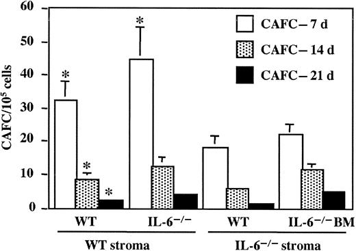
The effect of IL-6 deletion on the hematopoietic supportive activity of BM stroma was evaluated using the cobblestone area-forming cell (CAFC) assay. The CAFC subsets were quantified at weekly intervals in wild-type and IL-6-/- BM cells, using wild-type or IL-6-/- stroma. Similar to the irradiated LTBMCs (Figure 4), IL-6-/- stroma was deficient in supporting growth of wild-type CAFC-day 7 (56% reduction compared with wild-type stroma), CAFC-day 14 (67% reduction), and CAFC-day 21 (40% reduction) (Figure 5). Quantification of CAFC subsets on a single stroma type showed that CAFC numbers for IL-6-/- BM were similar to or slightly increased compared with those of wild-type BM, at all time points tested (Figure 5).
Frequency of CAFC on days 7, 14, and 21 in wild-type and IL-6-/- BM, analyzed on wild-type or IL-6-/- supportive stroma. Wild-type and IL-6-/- LTBMCs were established and maintained for 4 weeks as in Figure 1, in gelatin-coated 96-well microtiter plates, and irradiated (17 Gy). Three days after irradiation, cultures were seeded with wild-type or IL-6-/- BM cell suspensions at 6 serial 2-fold dilutions (from 625 to 20 000 cells/well) and were monitored in 16 replicate wells. The appearance of cobblestone areas was evaluated weekly, and individual wells were scored for the presence of cobblestone area. CAFC frequency was determined by limiting dilution analysis. *P < .01, for wild-type stroma versus IL-6-/- stroma seeded with the same type of BM cells.
Frequency of CAFC on days 7, 14, and 21 in wild-type and IL-6-/- BM, analyzed on wild-type or IL-6-/- supportive stroma. Wild-type and IL-6-/- LTBMCs were established and maintained for 4 weeks as in Figure 1, in gelatin-coated 96-well microtiter plates, and irradiated (17 Gy). Three days after irradiation, cultures were seeded with wild-type or IL-6-/- BM cell suspensions at 6 serial 2-fold dilutions (from 625 to 20 000 cells/well) and were monitored in 16 replicate wells. The appearance of cobblestone areas was evaluated weekly, and individual wells were scored for the presence of cobblestone area. CAFC frequency was determined by limiting dilution analysis. *P < .01, for wild-type stroma versus IL-6-/- stroma seeded with the same type of BM cells.
Exogeneous IL-6 does not fully restore hematopoietic supportive capacity of IL-6-/- LTBMCs (Figure 3). Seeding experiments were carried out using irradiated IL-6-/- stroma cultured in 5 ng/mL to 20 ng/mL IL-6, a concentration similar to that in wild-type LTBMCs after medium exchange (∼2.5 ng/mL).15 Two weeks after seeding irradiated IL-6-/- stroma with wild-type Lin- BM cells, the number of CFU-GMs and BFU-Es released to medium was analyzed. Hematopoietic support by irradiated IL-6-/- stroma established in the presence of IL-6 was not restored (not shown).
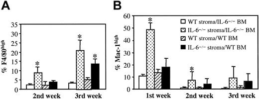
To determine whether wild-type and IL-6-/- stroma differ in supporting the proliferation of a specific hematopoietic lineage, we analyzed the expression of several lineage markers in cells released to the medium by irradiated, reseeded stroma. For both wild-type and IL-6-/- BM cells, IL-6-/- stroma showed increased Lin- BM cell differentiation into populations with high expression of macrophage markers Mac-1 and F4/80 (Figure 6), with a more pronounced effect in IL-6-/- BM cells. This suggests that, despite its reduced capacity to support hematopoiesis, IL-6-/- stroma preferentially induces differentiation to populations expressing macrophage development markers.
FACS analysis of F4/80high and Mac-1high cells in culture medium of irradiated wild-type or IL-6-/- stroma seeded with Lin- wild-type or IL-6-/- BM cells. LTBMCs were established, irradiated, and seeded as in Figure 4. F4/80high (A) and Mac-1high (B) cells were analyzed weekly in culture medium. Results are expressed as the mean ± SD of 6 experiments. *P < .01, for wild-type stroma versus IL-6-/- stroma.
FACS analysis of F4/80high and Mac-1high cells in culture medium of irradiated wild-type or IL-6-/- stroma seeded with Lin- wild-type or IL-6-/- BM cells. LTBMCs were established, irradiated, and seeded as in Figure 4. F4/80high (A) and Mac-1high (B) cells were analyzed weekly in culture medium. Results are expressed as the mean ± SD of 6 experiments. *P < .01, for wild-type stroma versus IL-6-/- stroma.
Fibroblastic precursor assays and FACS analysis of stroma
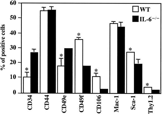
To determine whether the hematopoietic supportive deficiency of IL-6-/- stroma could be attributed to differences in the cell subpopulations that constitute BM stroma, we analyzed BM fibroblastic cell precursors in 2 clonal studies, the CFU-F and the SIC assay. These assays evaluate precursors at distinct differentiation stages within the fibroblastic/mesenchymal cell compartment.29 In IL-6-/- BM, fibroblastic/mesenchymal precursors are reduced to nearly 50% of the value for wild-type BM stroma (Table 1). Cells in IL-6-/- and wild-type stromata cultured in conditions that do not support hematopoiesis were further analyzed by FACS for antigens expressed by stromal cells and mesenchymal precursors (Figure 7). Wild-type stroma expressed a significantly higher level of vascular cell adhesion molecule-1 (VCAM-1; CD106), stem cell antigen-1 (Sca-1) (Ly-6A/E), integrin subunit α6 (CD49f), and Thy1.2 (CD90.2), and significantly lower levels of CD34 and integrin α5 (CD49e) than IL-6-/- stroma. No differences were observed in CD44 and Mac-1 expression in either stroma type (Figure 7). These differences in antigen expression levels were also observed when we analyzed CD45- BM cells, a population devoid of hematopoietic cells. Moreover, IL-6-/- CD45- BM cells expressed lower β1 integrin (CD29) and endoglin (CD105) levels compared with wild-type CD45- BM cells (not shown). The results indicate that the in vivo development of BM stroma in the absence of IL-6 alters the cellular composition of the hematopoietic stromal compartment, suggesting that IL-6 is an important in vivo growth factor for mesenchymal precursors in bone marrow.
CFU-F and SIC numbers in wild-type and IL-6−/− BM
. | CFU-Fs/106 cells . | SICs/107 cells . |
|---|---|---|
| Wild-type BM | 23.7 ± 11.5 | 25.9 ± 5.9* |
| IL-6−/− BM | 14.3 ± 10.2 | 13.3 ± 6.6 |
. | CFU-Fs/106 cells . | SICs/107 cells . |
|---|---|---|
| Wild-type BM | 23.7 ± 11.5 | 25.9 ± 5.9* |
| IL-6−/− BM | 14.3 ± 10.2 | 13.3 ± 6.6 |
Results are expressed as the mean ± SD for duplicate analysis of 5 animals.
P < .02 for wild-type versus IL-6−/− cells.
FACS analysis of wild-type and IL-6-/- stromata. Wild-type and IL-6-/- BM cells were cultured for 2 weeks in Dulbecco modified Eagle medium plus 10% FCS at 37°C, with weekly medium exchange. Cells were harvested by brief trypsinization and FACS analyzed as described (“Materials and methods”). Results are expressed as the mean ± SD of 4 experiments. *P < .01, for wild-type versus IL-6-/- stromal cells.
FACS analysis of wild-type and IL-6-/- stromata. Wild-type and IL-6-/- BM cells were cultured for 2 weeks in Dulbecco modified Eagle medium plus 10% FCS at 37°C, with weekly medium exchange. Cells were harvested by brief trypsinization and FACS analyzed as described (“Materials and methods”). Results are expressed as the mean ± SD of 4 experiments. *P < .01, for wild-type versus IL-6-/- stromal cells.
Discussion
IL-6-deficient mice show reduced longevity of the LTRSC compartment. Under steady-state conditions, the IL-6 deficiency is not limiting and can be compensated for by other cytokines; after induction of anemia and/or myeloablation, however, the late stages of hematopoietic differentiation are impaired in IL-6-/- mice.7 Here we analyzed the contribution of IL-6-/- BM stroma to this deficiency. The microenvironment provided by the LTBMC stromal component supports and regulates hematopoietic cell growth, self-renewal, and differentiation.30,31 We observed that IL-6-/- LTBMCs have a reduced capacity to support CFU-GM and BFU-E proliferation and a diminished number of cobblestone areas, which represent foci of active hematopoietic proliferation. The reduced hematopoietic supportive activity of IL-6-/- LTBMCs coincides with the observation that addition of anti-IL-6 antibodies to LTBMCs results in reduction of nonadherent myeloid cell numbers, whereas addition of exogenous IL-6 produces a dramatic increase in these populations.14,15
IL-6 is produced by MSCs11 and by other BM stromal cells16,26,27 ; through cytokine production, stromal cells may thus stimulate hematopoiesis in LTBMCs. Stromal formation in LTBMCs is IL-6 concentration-dependent15 ; reduced stromal growth was observed in cultures established in horse serum with low IL-6 concentrations (170 mU/mL, equivalent to approximately 15 pg/mL), but IL-6 addition (10 U/mL) did not improve the morphology of these cultures.15 High IL-6 concentrations inhibit SV40-transformed BM stromal cell proliferation, while enhancing anchorage-independent growth.17 Our data indicate that in IL-6-/- LTBMCs, establishment of the adherent stromal layer is delayed compared with wild-type cultures. Addition of supraoptimal IL-6 concentrations (200 ng/mL) partially restores hematopoietic activity in IL-6-/- LTBMCs without clearly improving the stromal component of these cultures.
The proliferative disadvantage of IL-6-/- cells in LTBMCs can be attributed mainly to the stromal compartment, as evidenced by our seeding experiments on irradiated stroma. IL-6-/- Lin- BM cells proliferate on irradiated wild-type stroma. Stromal cell IL-6 production appears to be sufficient to sustain proliferation of committed hematopoietic progenitors such as the CFU-GMs and BFU-Es evaluated here; other cytokines, such as IL-3, may synergize with stromal IL-6 and stimulate hematopoietic cell proliferation.32 Surprisingly, neither LTBMC hematopoietic cell-produced IL-6 nor IL-6 in the sera used are able to stimulate proliferation of Lin- BM cells when they are seeded on IL-6-deficient stroma. This supports the idea that local cytokine production by stromal cells may be important in maintaining hematopoietic progenitor proliferation, or that IL-6-/- stroma may have an intrinsic modification in cell composition that influence cultures decisively. Hematopoietic supportive deficiency of IL-6-/- stromal cells is also indicated by their reduced ability to sustain proliferation of distinct CAFC subsets with varying degrees of primitiveness. The hematopoietic supportive ability of IL-6-/- LTBMCs cannot be restored by optimal exogenous IL-6 concentrations, strongly suggesting an intrinsic defect in the stromal component. Further evidence for such a defect is the finding that the BM stromal cell clonal precursors CFU-F and SIC are significantly reduced in IL-6-/- BM (∼50% of wild-type values).
The LTBMC adherent layer is composed mainly of macrophages and myofibroblasts and a minor endothelial-like cell population.33 Human LTBMC stromal cells are mesenchymal cells that follow a vascular smooth muscle cell differentiation pathway.34,35 Detailed analysis of extracellular matrix and cytoskeletal protein expression in a variety of murine stromal cell lines led to the proposal of a model describing the sequential transition from immature mesenchymal to vascular smooth muscle-like stromal cells.35 This model reflects the broad heterogeneity of the stroma and the need for cooperation among distinct stromal cell types for maintenance of hematopoiesis. Our analysis of membrane proteins in wild-type and IL-6-/- stromal cultures showed increased expression in wild-type stroma of VCAM-1 and Sca-1, considered mesenchymal cell markers.35,36 VCAM-1 expression has been linked to the hematopoietic supportive capacity of immortalized murine BM stromal cell lines.29 Sca-1 expression was also correlated with the ability of a murine BM stromal cell line to support proliferation of 5-fluorouracil (5-FU)-treated BM cells.37 The reduced expression of β1 and α6 integrins in IL-6-/- CD45- BM cells suggests lower expression of the laminin receptor VLA-6 (α6β1) in IL-6-/- BM stromal cells. Laminin is strongly adhesive for multipotent hematopoietic FDCP-mix cells, in an interaction mediated by α6β1 integrin.38 On the other hand, β1 integrin expression down-regulates the proapoptotic protein Bim,39 increasing survival of hematopoietic progenitors.40 The reduced expression of membrane antigens observed in IL-6-/- BM stroma may account for its reduced hematopoietic supportive ability. Several cytokines stimulate BM fibroblastic cell colony formation in the SIC assay,41 as well as proliferation of BM stromal fibroblasts.42 Our data showed that genetic IL-6 deficiency causes a reduction in BM stromal precursor numbers and alters the proportion of LTBMC cell populations, thereby reducing the ability of IL-6-/- stroma to support proliferation of pluripotent and committed hematopoietic progenitors. This phenotype, similar to that observed in the hematopoietic compartment,17 indicates that IL-6 could be an in vivo growth factor for mesenchymal precursors implicated in the reduced longevity of the long-term repopulating stem cell compartment in IL-6-/- mice.
Prepublished online as Blood First Edition Paper, December 30, 2003; DOI 10.1182/blood-2003-10-3438.
Supported in part by Plan Nacional de Salud y Farmacia, CICYT (SAF2001-2262), and Plan Nacional de Genómica (GEN2001-4856-C13-02) to A.B. The Department of Immunology and Oncology was founded and supported by the Spanish Council for Scientific Research (CSIC) and by Pfizer.
The publication costs of this article were defrayed in part by page charge payment. Therefore, and solely to indicate this fact, this article is hereby marked “advertisement” in accordance with 18 U.S.C. section 1734.
We thank Asunción García for excellent technical assistance and Catherine Mark for editorial support.


This feature is available to Subscribers Only
Sign In or Create an Account Close Modal