Abstract
We compared the angiogenic potential of bone marrow plasma and the expression of vascular endothelial growth factor (VEGF), basic fibroblast growth factor (bFGF), and their receptors on plasma cells from patients with monoclonal gammopathy of undetermined significance (MGUS), smoldering multiple myeloma (SMM), and newly diagnosed multiple myeloma (NMM). Cytokine and cytokine-receptor expression was studied by bone marrow immunohistochemistry, quantitative reverse transcription-polymerase chain reaction (RT-PCR) on sorted plasma cells, and quantitative Western blot analysis. Bone marrow angiogenic potential was studied using a human in vitro angiogenesis assay. The expression levels of VEGF, bFGF, and their receptors were similar among MGUS, SMM, and NMM. Sixty-one percent of NMM samples stimulated angiogenesis in the in vitro angiogenesis assay compared with SMM (0%) and MGUS (7%) (P < .001). Importantly, 63% of MGUS samples inhibited angiogenesis compared with SMM (43%) and NMM (4%) (P < .001). The inhibitory activity was heat stable, not overcome by the addition of VEGF, and corresponded to a molecular weight below 10 kd by size-exclusion chromatography. Our results suggest that increasing angiogenesis from MGUS to NMM is, at least in part, explained by increasing tumor burden rather than increased expression of VEGF/bFGF by individual plasma cells. The active inhibition of angiogenesis in MGUS is lost with progression, and the angiogenic switch from MGUS to NMM may involve a loss of inhibitory activity. (Blood. 2004; 104:1159-1165)
Introduction
Multiple myeloma (MM) is characterized by a clonal proliferation of immunoglobulin-secreting plasma cells in the bone marrow, resulting in a variety of clinical manifestations including osteolytic bone lesions, anemia, hypercalcemia, and renal failure. It accounts for approximately 10% of all hematologic malignancies and nearly 1% of all malignancies. Estimates in the United States alone are that 15 270 new patients will receive diagnoses of myeloma in 2004 and that 11 070 people will die of the disease in the same period.1 For patients treated with conventional chemotherapy, the median survival time from diagnosis is 3 to 4 years.2 Survival time improves in patients who are able to undergo high-dose therapy and stem cell rescue.3-5 However, most patients eventually experience relapses. Better understanding of the disease biology will enable development of therapeutic agents targeted at disrupting critical biologic pathways.
Angiogenesis, or the formation of new blood vessels from existing blood vessels (in contrast to vasculogenesis or de novo formation of blood vessels), occurs physiologically during normal growth, tissue healing, and regeneration. Angiogenesis plays an important role in the development and spread of tumors, a concept that was introduced more than 3 decades ago.6 Increasing levels of tumor angiogenesis have been associated with poor prognosis for a variety of hematologic malignancies and solid tumors. New vessel formation in the bone marrow seems to play an important role in the pathophysiology of myeloma,7-10 leukemias,11,12 and myelofibrosis.13 Increased bone marrow microvessel density (MVD) in patients with myeloma appears to be an important prognostic factor.7,9,10,14
Various cytokines have been invoked as responsible for driving the process of neovascularization in solid tumors and hematologic malignancies.15 Malignant plasma cells can secrete various cytokines, including vascular endothelial growth factor (VEGF), basic fibroblast growth factor (bFGF), and hepatocyte growth factor (HGF), all known proangiogenic factors.16-20 Studies have shown that myeloma cells are capable of secreting VEGF in response to interleukin-6 (IL-6) stimulation; in turn, microvascular endothelial cells and marrow stromal cells secrete IL-6, a potent growth factor for malignant plasma cells, in response to VEGF stimulation.18,21
We have previously demonstrated a gradual increase in degree of bone marrow angiogenesis along the disease spectrum from MGUS to smoldering MM (SMM) to newly diagnosed MM (NMM) and relapsed MM (RMM).15 The biology behind this progression is unclear, and differences in angiogenic cytokine expression and bone marrow angiogenic potential among the various stages of the disease are not fully understood. In this study, we examined the expression of VEGF, bFGF, and their receptors by plasma cells in patients with MGUS, SMM, and symptomatic myeloma at the protein level and at gene expression to understand the biologic basis for this progression of bone marrow angiogenesis. We also studied the stimulus for angiogenesis in the microenvironment by evaluating the angiogenic potential of bone marrow plasma using a human in vitro angiogenesis assay.
Patients, materials, and methods
All patients gave written informed consent for research use of bone marrow and serum specimens. Approval for the study was obtained from the Mayo Institutional Review Board in accordance with federal regulations and the Declaration of Helsinki. Paraffin-embedded bone marrow biopsy samples were used for immunohistochemical stains. Bone marrow aspirate was used to obtain sorted plasma cells for reverse transcription-polymerase chain reaction (RT-PCR) and quantitative Western blot studies and to obtain plasma for experiments to assess angiogenic potential. Normal bone marrow plasma cells were isolated from marrow (All Cells LLC, Berkeley, CA). Normal bone marrow plasma was obtained from patients undergoing bone marrow biopsy for clinical indications who were eventually classified as having normal bone marrow.
Immunohistochemistry for VEGF, bFGF, and their receptors
Bone marrow core biopsy samples were obtained from patients with diagnoses of MGUS, SMM, or NMM. VEGF, bFGF, VEGF receptor 1 (VEGFR1) (flt-1), VEGFR2 (flk-1/KDR), fibroblast growth factor receptor 2 (FGFR2), and FGFR3 immunohistochemical staining of the bone marrow was performed using a labeled streptavidin-biotin peroxidase method on a Ventana ES automated immunohistochemistry stainer (Ventana Medical Systems, Tucson, AZ) using buffers and detection reagents supplied by the manufacturer.17,22 After deparaffinization, heat-induced epitope retrieval was performed using EDTA (ethylenediaminetetraacetic acid; pH 8.0) for bFGF, FGFR2, and FGFR3 and using citrate (pH 6.0) for VEGF, VEGFR1, and VEGFR2 for 30 minutes in a steamer. Slides were cooled for 5 minutes, rinsed, and put on the ES stainer. Primary antibodies (Santa Cruz Biotechnology, Santa Cruz, CA) were used at a dilution of 1:50 for VEGF, bFGF, and VEGFR2, 1:40 for FGFR2, 1:25 for VEGFR1, and 1:20 for FGFR3. Each slide was incubated with the respective primary antibody for 30 minutes. The amino ethyl carbazole (AEC) detection kit (Ventana Medical Systems, Tucson, AZ) was used for antigen visualization, and sections were counterstained with a light hematoxylin. Cytokine expression was graded as previously described17 : - indicates no staining; +, weak staining (0%-30% plasma cells positive); ++, weak to moderate staining (31%-60% plasma cells positive); and +++, strong staining (more than 60% plasma cells positive). Data on VEGF expression in NMM were obtained from a previous study.17
Slides were then read, and a visual estimate of the plasma cells staining for each cytokine and its receptors was made as a percentage of all nucleated cells. One reviewer (S.K.), who was blinded to the clinical characteristics of the patients, reviewed all slides. Using a separate section from each biopsy sample stained with hematoxylin and eosin, an estimate was made of the plasma cell percentage in each bone marrow biopsy. The percentage of plasma cells expressing the cytokine or the receptor of interest was then estimated as a ratio of these 2 values. Instances in which the ratio exceeded 1 were scored as 100% plasma cell expression of the protein of interest.
Quantitative RT-PCR on sorted plasma cells
Plasma cells from patients with MGUS (n = 10), SMM (n = 10), and NMM (n = 7) were studied for VEGF expression using quantitative RT-PCR. Similarly, bFGF expression was also studied in the 3 groups (MGUS [n = 10], SMM [n = 10], and NMM [n = 20]). Plasma cells from bone marrow aspirates were isolated using magnetic cell-sorting (MACS) separation columns with antibodies against CD138, as described by the manufacturer17 (Miltenyi Biotec, Bergisch Gladbach, Germany). These cells were further analyzed using Wright-Giemsa staining, and a purity of more than 96% was confirmed. Total RNA was isolated from these samples using an RNeasy Kit (Qiagen, Hilden, Germany), as described by the manufacturer. RT-PCR was performed as a multiplex using the Titan One Tube RT-PCR System with 15 cycles of amplification, according to the manufacturer's instructions (Roche Diagnostics GmbH, Mannheim Germany). On completion of the RT-PCR, the samples were treated with ExoSAP-IT to remove excess primer and deoxyribonucleotides (dNTPs; USB, Cleveland, OH), as described by the manufacturer. A portion of the RT-PCR product was used in the Fast Start DNA Master SYBR Green I kit. This reaction was run on the Light-Cycler (Roche Diagnostics GmbH) until a sample reached plateau stage. PCR products were run on a 1% agarose gel, and relative quantification was performed by spot densitometry comparisons with GAPDH (glyceraldehyde-3-phosphate dehydrogenase) bands using AlphaEaseFC software. Expression was quantified as a ratio of cytokine to GAPDH mRNA expression. Primer sequences were as follows: GAPDH forward, ACC ACA GTC CAT GCC ATC AC; GAPDH reverse, TCC ACC ACC CTG TTG CTT GTA; VEGF forward, GCC CAC TGA GGA GTC CAA CAT C; VEGF reverse, TTT TTG CAG GAA CAT TTA CAC G; bFGF forward, AGT CTT CGC CAG GTC ATT GAG; bFGF reverse, AGG AGA CAC AGC GGT TCG AG. Melting curve analysis of the PCR products was performed to confirm the presence of a single, specific amplicon. Precautions were taken to avoid contamination, and negative controls were run with each step. Samples and PCR reactions were prepared in different areas.
Quantitative Western blot analyses of VEGF, bFGF, VEGFR1, and VEGFR2
Patient or normal plasma cell lysates were prepared from CD138+ bead-isolated cells. Protein concentration was determined using the BCA protein assay system (Pierce, Rockford, IL). bFGF and VEGF were immunoprecipitated using 100 μg total protein; VEGFR1 and VEGFR2 were immunoprecipitated using 200 μg total protein. Cell lysates were incubated with primary antibody (5 μg anti-VEGF [Abcam, Cambridge, MA]; 5 μg anti-bFGF [BD Biosciences, San Jose, CA]; 10 μg anti-VEGFR1 and anti-VEGFR2 [gift from Imclone, New York, NY]) for a minimum of 6 hours at 4°C with constant mixing. Forty microliters goat-antimouse immunoglobulin G (IgG) (H+L) Sepharose 4B (Zymed, San Francisco, CA) was added to each, and the samples were returned to 4°C and mixed overnight. After centrifugation (2500g for 2 minutes), the supernatant was discarded, and the pellet was washed twice with 500 μL cold lysis buffer. After the last spin, any remaining fluid was aspirated, and the pellet was resuspended in 40 μL 2 × sample buffer. Samples were incubated at 70°C for 20 minutes and spun at 10 000g for 2 minutes, and 30 μL of each sample was loaded onto a 15% acrylamide/bis gel (bFGF and VEGF) or a 7.5% acrylamide/bis gel (VEGFR1 and VEGFR2). Molecular-weight markers (Rainbow Markers, Amersham Biosciences, Piscataway, NJ; SigmaMarker, Sigma, St Louis, MO) were included on each gel.
Gels were run for a minimum of 6.5 hours. Proteins were transferred to nitrocellulose (Schleicher & Schuell Biosciences, Keene, NH) for 6 hours at 4°C. Nitrocellulose sheets were blocked with TSM buffer (10% dry milk in 0.15 M NaCl, 10 mM Tris HCl, 1 mM sodium azide, pH 7.4) overnight. The blocking buffer was removed, and the nitrocellulose was incubated with the appropriate primary antibody (anti-bFGF 1:500, anti-VEGF 1:200, anti-VEGFR1 1:200, and anti-VEGFR2 1:150) overnight. Peroxidase-labeled goat-antimouse antibody (KPL, Gaithersburg, MD) was added and incubated for at least 1 hour at room temperature. Proteins were detected using chemiluminescence (ECL detection system; Amersham Biosciences, Piscataway, NJ), and the exposed films were developed using an X-Omat 2004 processor (Kodak, Rochester, NY).
Spot densitometry was performed on the bands of interest using AlphaEaseFC software on the AlphaImager optical system (Alpha Innotech, San Leandro, CA). Using the densitometry values, quantitative comparisons among patient samples were made using the ratios of the bFGF, VEGF, VEGFR1, and VEGFR2 bands to mouse immunoglobulin bands from the primary antibody used in the immunoprecipitation step.
In vitro angiogenesis assay
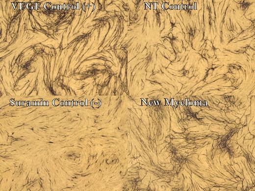
Angiogenic potential of the bone marrow plasma was studied using a human in vitro angiogenesis assay (Angiokit; TCS Cellworks, Buckinghamshire, United Kingdom).23-25 This system uses human endothelial cells cocultured with human fibroblasts and myoblasts in a 24-well plate containing optimized media supplied by the manufacturer. Endothelial cells, which initially form small islands in the culture matrix, subsequently begin to proliferate and then migrate through the matrix to form tubular structures. By the end of 2 weeks, they merge to form a network of anastomosing tubules closely resembling a capillary bed. Each 24-well plate has 6 control wells and 18 test wells. Two control wells are treated with VEGF (positive controls), resulting in an extensive network of branching vessels over a period of 10 to 12 days. Two control wells are treated with suramin (negative controls) in which there is near total inhibition of angiogenesis. Finally, 2 control wells receive no additional treatment (NT controls), and these wells show a moderate network of branching tubules that is significantly less than that of VEGF-treated wells (Figure 1).
Human in vitro angiogenesis assay. Photomicrographs demonstrate tubule formation in the human in vitro angiogenesis assay. (top left panel) Stimulation of tubule formation with the addition of VEGF. (bottom left panel) Inhibition of tubule formation with the addition of suramin. (top right panel) No treatment (NT) control without added cytokines or inhibitors. (bottom right panel) Stimulation of tubule formation by plasma of a myeloma patient. Pictures obtained at 12.5 × 0.04 (magnification/aperture).
Human in vitro angiogenesis assay. Photomicrographs demonstrate tubule formation in the human in vitro angiogenesis assay. (top left panel) Stimulation of tubule formation with the addition of VEGF. (bottom left panel) Inhibition of tubule formation with the addition of suramin. (top right panel) No treatment (NT) control without added cytokines or inhibitors. (bottom right panel) Stimulation of tubule formation by plasma of a myeloma patient. Pictures obtained at 12.5 × 0.04 (magnification/aperture).
Ten microliters bone marrow plasma (made cell free by centrifugation) from patients with MGUS, SMM, and NMM were used in the test wells. To obtain bone marrow plasma, fresh marrow aspirate (20-40 mL) is placed in EDTA and centrifuged for 10 minutes at 700g. The plasma supernatant is decanted and centrifuged again for 5 minutes at 48 000g to remove remaining cellular elements. Marrow plasma is not diluted or manipulated further. This fraction of the marrow aspirate represents the marrow milieu in which the cells reside and enables assessment of the angiogenic potential of the marrow.
Assays were incubated at 37°C with 5% CO2 humidified atmosphere. Optimized media and test samples are replaced on days 4, 7, and 9 after initial treatment. On day 11, residual medium is aspirated, and cultures are fixed and stained with antibodies to CD31 to detect vessel formation. The degree of tubule formation was evaluated using light microscopy and quantitated using computerized image analysis (Angiosys; TCS Cell-works). Pictures were obtained using an Olympus AX70 microscope (Olympus Optical, Tokyo, Japan) and Spot RT Color 2.21 camera (Diagnostic Instruments, Sterling Heights, MI). Total tubule length in each test well on the human in vitro angiogenesis assay was expressed as a percentage of the NT control wells. Stimulation of angiogenesis in the test wells was defined as total tubule length increased more than 125% of NT control. Total tubule length in the test wells less than 75% of NT control was defined as inhibition of angiogenesis.
Statistical methods
Fisher exact and χ2 tests were used to compare differences in nominal variables; the rank sum test or the Kruskal-Wallis test was used to assess whether continuous variables differed significantly between categories. Correlation between continuous variables was studied using Spearman rank correlation.
Results
Bone marrow MVD and immunohistochemistry for cytokines and receptors
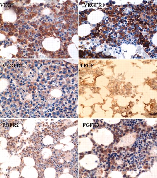
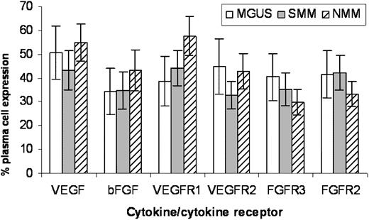
Immunohistochemical studies for angiogenic cytokines and receptors were performed on 57 patients with MGUS (n = 15), SMM (n = 23), and NMM (n = 19). Corresponding MVD values for correlative analysis were obtained from a study of 400 patients with plasma cell disorders.15 Among patients included in the present study, MVD in MGUS was low at 2 (mean, 6; range, 1-19) compared with 6 (mean, 7; range, 2-19) for patients with SMM and 8 (mean, 12; range, 2-37) for patients with NMM. VEGF, bFGF, VEGFR1, VEGFR2, FGFR2, and FGFR3 were expressed by plasma cells in most of the patients (Table 1; Figure 2). When marrow MVD was correlated with cytokine/receptor expression (percentage plasma cells expressing the respective marker) across the entire group, there was a correlation between MVD and expression of VEGF (P = .02), bFGF (P = .008), and VEGFR1 (P = .02). However, the percentages of plasma cells expressing VEGF, bFGF, and their receptors were not significantly different among the 3 groups of patients (P = NS) (Figure 3).
Expression of angiogenic cytokines and their receptors by immunohistochemistry
. | No. patients (%) . | . | . | . | ||
|---|---|---|---|---|---|---|
| Cytokine/cytokine receptor . | MGUS n = 15 . | SMM n = 23 . | NMM n = 19 . | Level of expression . | ||
| VEGF | 7 (47) | 11 (48) | 7 (37) | −/+ | ||
| 8 (53) | 12 (52) | 12 (63) | ++/+++ | |||
| bFGF | 8 (54) | 13 (57) | 8 (42) | −/+ | ||
| 7 (46) | 10 (43) | 11 (58) | ++/+++ | |||
| VEGFR1 | 8 (54) | 10 (44) | 5 (26) | −/+ | ||
| 7 (46) | 13 (56) | 14 (74) | ++/+++ | |||
| VEGFR2 | 7 (47) | 13 (57) | 8 (42) | −/+ | ||
| 8 (53) | 10 (43) | 11 (58) | ++/+++ | |||
| FGFR3 | 7 (46) | 14 (61) | 9 (48) | −/+ | ||
| 8 (54) | 9 (39) | 10 (52) | ++/+++ | |||
| FGFR2 | 7 (47) | 12 (52) | 8 (42) | −/+ | ||
| 8 (54) | 11 (48) | 11 (58) | ++/+++ | |||
Angiogenic cytokine and cytokine receptor expression by immunohistochemistry. Photomicrographs taken during bone marrow biopsy demonstrate positive plasma cell expression of VEGF, bFGF, VEGFR1, VEGFR2, FGFR2, and FGFR3 by immunohistochemistry. Pictures obtained at 100×/0.3 (magnification/aperture).
Angiogenic cytokine and cytokine receptor expression by immunohistochemistry. Photomicrographs taken during bone marrow biopsy demonstrate positive plasma cell expression of VEGF, bFGF, VEGFR1, VEGFR2, FGFR2, and FGFR3 by immunohistochemistry. Pictures obtained at 100×/0.3 (magnification/aperture).
Expression of angiogenic cytokines and cytokine receptors by disease stage. Graph shows no significant correlation between plasma cell expression of angiogenic cytokines and their receptors by immunohistochemistry and disease stage (error bars represent SE).
Expression of angiogenic cytokines and cytokine receptors by disease stage. Graph shows no significant correlation between plasma cell expression of angiogenic cytokines and their receptors by immunohistochemistry and disease stage (error bars represent SE).
Cytokine expression by quantitative RT-PCR techniques
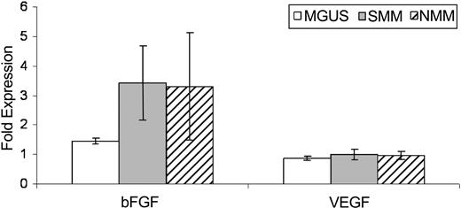
When purified bone marrow plasma cells were examined for cytokine expression by RT-PCR, we found VEGF and bFGF to be expressed by plasma cells from all patients in all 3 groups. We found no significant difference in the level of VEGF mRNA expression between MGUS, SMM, and NMM, with median VEGF/GAPDH ratios of 0.84, 1.12, and 0.80, respectively (P = .74). Similarly, no difference was seen among the 3 groups in the level of bFGF expression, with median bFGF/GAPDH ratios of 1.4, 1.89, and 1.15, respectively (P = .14) (Figure 4).
Quantitative RT-PCR for VEGF and bFGF. Comparison of quantitative RT-PCR results of VEGF and bFGF among the 3 disease stages. Cytokine expression was normalized using the housekeeping gene GAPDH, and results are expressed as cytokine/GAPDH expression ratio. Results represent mean ± SE.
Quantitative RT-PCR for VEGF and bFGF. Comparison of quantitative RT-PCR results of VEGF and bFGF among the 3 disease stages. Cytokine expression was normalized using the housekeeping gene GAPDH, and results are expressed as cytokine/GAPDH expression ratio. Results represent mean ± SE.
Quantitative Western blot analysis of VEGF, bFGF, VEGFR1, and VEGFR2
Western blot analysis for VEGF, bFGF, VEGFR1, and VEGFR2 was initially performed on myeloma cell lines (RPMI 8226, OCI-My 5, U266, JJN3, OPM2, KAS 6/1) and human umbilical vein endothelial cells (HUVECs) for optimization. All cell lines studied showed expression of VEGF, bFGF, VEGFR1, and VEGFR2.
Results of quantitative Western blot analysis on plasma cells and from patients and healthy controls are given on Table 2, and results for VEGF are represented in Figure 5. Essentially no significant differences were observed in expression of these cytokines/receptors between the studied groups. However, the 18-kd cytosolic isoform of bFGF was only present in 2 of 4 MM patient samples and in 1 of 2 SMM patient samples, but it was not present in any of the control samples studied. In contrast, the 24-kd isoforms were detected in all patient and control samples.
Quantitation of VEGF, bFGF, VEGFR1, and VEGFR2 by Western blot analysis
. | Mean ratio . | . | . | . | |||
|---|---|---|---|---|---|---|---|
| Plasma cell group . | VEGF/immunoglobulin* . | bFGF/immunoglobulin† . | VEGFR1/immunoglobulin‡ . | VEGFR2/immunoglobulin§ . | |||
| Normal | 0.6 | 1.9 | NA | NA | |||
| SMM | 0.5 | 2.5 | 1.5 | 2.2 | |||
| MM | 0.5 | 2.4 | 1.4 | 3.1 | |||
Mean ratios represent densitometric readings for cytokine/receptor bands corrected to the corresponding mouse immunoglobulin band derived from the immunoprecipitation antibody. NA indicates not applicable.
Normal, n = 2; SMM, n = 1; MM, n = 2.
Normal, n = 3; SMM, n = 2; MM, n = 4.
SMM, n = 3; MM, n = 5.
SMM, n = 3; MM, n = 5.
Expression of VEGF using Western blot analysis. Representative blots of VEGF expression by plasma cells in normal bone marrow, MM, and SMM after immunoprecipitation. The ARH 77 cell line was used as an additional control. Densitometric readings for cytokine/receptor bands were obtained and corrected to the corresponding mouse immunoglobulin band (shown below the VEGF band) derived from the immunoprecipitation antibody.
Expression of VEGF using Western blot analysis. Representative blots of VEGF expression by plasma cells in normal bone marrow, MM, and SMM after immunoprecipitation. The ARH 77 cell line was used as an additional control. Densitometric readings for cytokine/receptor bands were obtained and corrected to the corresponding mouse immunoglobulin band (shown below the VEGF band) derived from the immunoprecipitation antibody.
Human in vitro angiogenesis assay
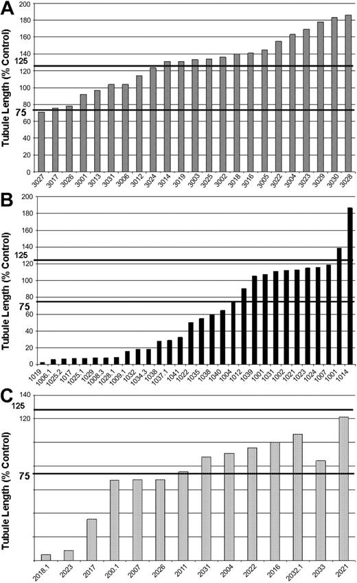
Plasma from patients with myeloma resulted in a significantly higher degree of tubule formation than from patients with SMM or MGUS. Total tubule length as a percentage of NT controls varied significantly among the 3 groups (medians, 41 [range, 3-187] in MGUS, 80 [5-121] in SMM, and 133 [71-186] in NMM, respectively; P < .001). Fourteen (61%) of 23 of NMM samples stimulated angiogenesis, whereas none of the 14 SMM samples and only 2 (7%) of 30 of MGUS samples were stimulatory (P < .001) (Figure 6A-C).
Results of human in vitro angiogenesis assay by disease stage. Bar charts show the degree of tubule formation (total tubule length) with the human in vitro angiogenesis assay expressed as a percentage of NT control. Results demonstrate stimulation of angiogenesis by the bone marrow plasma of patients with newly diagnosed myeloma (A), MGUS (B), and SMM (C) with marked inhibition of angiogenesis by the bone marrow plasma of most patients with MGUS.
Results of human in vitro angiogenesis assay by disease stage. Bar charts show the degree of tubule formation (total tubule length) with the human in vitro angiogenesis assay expressed as a percentage of NT control. Results demonstrate stimulation of angiogenesis by the bone marrow plasma of patients with newly diagnosed myeloma (A), MGUS (B), and SMM (C) with marked inhibition of angiogenesis by the bone marrow plasma of most patients with MGUS.
Interestingly, 19 (63%) of 30 MGUS marrow plasma samples inhibited angiogenesis in this assay compared with 6 (43%) of 14 SMM samples and only 1 (4%) of 23 NMM samples (P < .001) (gFigure 6A-C). In fact, 11 MGUS samples strongly inhibited tubule formation to less than 20% of NT control and resembled wells treated with suramin (negative control wells). Two normal marrow plasma samples also showed evidence of inhibition (26% and 68% of NT control).
Eight MGUS samples with inhibitory activity were heated to 100°C for 10 minutes and were studied again in the in vitro angiogenesis assay. None of the samples lost the ability to inhibit angiogenesis with heating, indicating that this inhibitory activity was heat stable (median inhibition, 11% of NT control baseline [range, 4%-51%] vs 6% of NT control [range, 1%-25%]; Spearman ρ, 0.83; P = .02).
Two of the MGUS samples that were inhibitory (5% and 34% of NT control, respectively) were mixed in a 1:1 ratio with bone marrow plasma known to be noninhibitory in the assay (163% and 115% of NT control, respectively). Mixing was unable to fully overcome the inhibition (35% and 68%, respectively). The mixing study was repeated in the patient with 5% inhibitory activity, and similar results were obtained. Four inhibitory samples (3 MGUS and 1 normal) were tested in the in vitro angiogenesis assay with added VEGF (2 ng/mL, the concentration of VEGF used as positive control). Adding VEGF could not overcome the inhibitory effect in any of the studied cases. Results were 5%, 17%, 29%, and 60% of NT control for no VEGF compared with 10%, 20%, 28%, and 76% of NT control for added VEGF, respectively. To confirm the inability of VEGF to abrogate the inhibitory activity of MGUS bone marrow plasma, we added VEGF at a higher concentration (8 ng/mL, 4 times the concentration of VEGF used as positive control in the assay) to bone marrow plasma from an MGUS patient with known inhibitor. VEGF at this concentration was still unable to overcome the inhibition—7% of NT control (no VEGF) compared with 13% of NT control (with added VEGF).
Four of the MGUS inhibitory samples were fractionated based on molecular weight using size-exclusion chromatography. Using this method, more than 40 fractions were obtained and run on the in vitro angiogenesis assay. The inhibitor was consistently present in the fraction, corresponding to a molecular weight of less than 10 kd. The other fractions were not inhibitory. This size excludes immunoglobulins. In addition, there was no correlation between serum monoclonal protein level and angiogenic ability in this assay, in all samples (ρ, 0.05; P = .7), or inhibitory samples alone (rho, 0.23; P = .3). Among strongly inhibitory MGUS samples (less than 20% of NT control), the serum M spike ranged from 0.5 to 4.1 g/dL.
We studied peripheral blood plasma to determine whether inhibitory activity was restricted to marrow or diluted in the circulation. Of 20 peripheral blood samples studied (5 controls, 7 MGUS, 7 NMM, and 1 SMM), none showed evidence of inhibitory activity (range, 131%-341% of NT control). This includes 2 MGUS patients with known inhibitors in the marrow plasma. Five inhibitory MGUS samples were tested multiple times in the assay, with no significant difference in degree of inhibition.
Discussion
Bone marrow angiogenesis plays an important role in the pathogenesis and progression of MM and in other hematologic malignancies and solid tumors. In myeloma, the degree of bone marrow angiogenesis, as estimated by MVD, has been shown to be an important prognostic factor for survival.7,9,10 In a study of 400 patients, the degree of bone marrow angiogenesis was found to progressively increase across the spectrum of plasma cell proliferative disorders.15 Other studies also have confirmed the correlation between MVD and disease stage.10,26
Studies so far point to the presence of multiple autocrine and paracrine interactions between malignant plasma cells and other residents of the bone marrow microenvironment, especially stromal cells, endothelial cells, and bone cells mediated by various cytokines, growth factors, and adhesion molecules.27 A variety of cytokines (IL-1β, tumor necrosis factor-α [TNF-α], transforming growth factor-β [TGF-β], IL-6, bFGF, VEGF, HGF)16,28-33 have been implicated in this complex cascade of interactions. Of these, VEGF and bFGF are considered key participants, and have been of particular interest in myeloma.19-21,34,35 VEGF activity is mediated primarily through 2 receptors, VEGFR-1 and VEGFR-2.36,37 Similar findings have been described in the context of bFGF. Bone marrow stromal cells express FGF receptors, and, on stimulation by bFGF secreted by myeloma cells, they release IL-6, which in turn stimulates the plasma cells.32
In this study we show the correlation between these 2 cytokines and the MVD, consistent with a prominent role for them in the angiogenic process in myeloma. However, our data show that the expression of VEGF, bFGF, and their receptors is not significantly different between MGUS, SMM, and MM in various assays at the protein and at the mRNA level. This suggests that the up-regulation of VEGF and bFGF by individual plasma cells is probably not causally involved in the induction of angiogenesis in plasma cell disorders, at least not in a major way. The data also suggest that the gradual increase in MVD with disease progression may be related in part to the cumulative angiogenic effect of increasing numbers of plasma cells in the marrow rather than increased VEGF/bFGF expression by individual plasma cells. Based on previous data regarding the paracrine effects of VEGF/bFGF in myeloma, we hypothesize that a positive feedback loop of increasing tumor burden, resulting in increased VEGF and bFGF and vice versa, may amplify the process of angiogenesis, but it does not appear to be the initial trigger. Our study also provides evidence of heterogeneity in terms of angiogenic cytokine expression between myeloma cells from the same patient, with only a subset of neoplastic cells showing expression for each cytokine studied (Figure 3).
In animal models with other tumor types, overexpression of VEGF and bFGF results in rapid tumor growth and enhanced vascularity.38 There is also a reduction in angiogenesis and tumor necrosis after blockade of these cytokines, indicating a central role for VEGF and bFGF in tumor angiogenesis. Our findings in myeloma, though not contradictory, do not support such a primary role for VEGF and bFGF overexpression in the induction of angiogenesis in myeloma; however, as discussed in the previous paragraph, these cytokines are likely involved in sustaining and increasing the observed angiogenic response. One explanation for the apparent discrepancy is that the angiogenic switch is the result of an alteration in the balance between proangiogenic and antiangiogenic stimuli in a given tumor, and the specific mediators involved likely vary across tumor types.39 In fact, as discussed, angiogenesis induction may involve a loss of angiogenesis inhibitory activity.
In a previous study evaluating the angiogenic potential of marrow-derived plasma, Vacca et al,40 using the chorioallantoic membrane (CAM) assay, demonstrated higher angiogenic activity with marrow from active myeloma compared with inactive myeloma or MGUS. They also found a linear correlation between the bone marrow MVD in these patients and the degree of activity seen in the CAM assay for the corresponding marrow. Neutralizing antibodies to bFGF were capable of abrogating this angiogenic activity to some degree.40 In addition, conditioned plasma from patients with active myeloma was able to stimulate HUVECS better than plasma from inactive MM or MGUS.40 A higher number of endothelial cell precursors has been demonstrated in the marrow of patients with active myeloma than in those with treated myeloma or MGUS or with normal marrow,41 again a reflection of the increased angiogenic potential in MM.
In this study we show, using a novel human in vitro angiogenesis assay, that the increased MVD demonstrated in NMM in several studies is matched by a significantly higher angiogenic potential in NMM bone marrow plasma compared with SMM and MGUS bone marrow plasma. Using the human in vitro angiogenesis assay overcomes criticisms regarding other angiogenic assays, such as the CAM assay, in which the receptors and vessels are not derived from humans. It also overcomes the limitations in accurately assessing bone marrow MVD and of using MVD alone as a marker of bone marrow angiogenesis. More important, we show that the difference in angiogenesis between MGUS and NMM partly results from a loss of angiogenesis inhibitory activity in NMM that is normally present in MGUS. This represents a major finding. The presence of an inhibitor rather than lower levels of or no proangiogenic cytokines in MGUS is supported by the fact that the mean tubule lengths of the vessels are below those of the NT control and often approach levels of inhibition seen in the suramin negative control well. In addition, inhibition was not overcome by adding VEGF or by mixing studies with noninhibitory marrow plasma.
Additional experiments suggest that the inhibitory activity seen in MGUS is also present in normal bone marrow plasma. However, as expected, this inhibitory activity is not seen in blood plasma, even among patients with MGUS with strong inhibitory activity in the bone marrow plasma. We think this is because the secreted marrow inhibitor is probably diluted in the blood circulation, thus altering the balance between proangiogenic and antiangiogenic cytokines. Sometimes even bone marrow can be inadvertently diluted with blood, depending on the patient, the person performing the procedure, or the volume aspirated. In fact, in some MGUS patients and healthy controls with no evidence of inhibitory activity in bone marrow plasma, it might be caused by dilution of marrow plasma with blood. Our data suggested that the assay used was reliable and reproducible.
Additional studies show that the inhibitory activity is likely one or more small protein/polypeptide factors with molecular weights of less than 10 kd each. Heat stability, molecular weight, and lack of correlation between inhibitory activity and monoclonal immunoglobulin levels argue against any role by the monoclonal protein or other immunoglobulins in this process. The inhibitor also appeared to have been independent of VEGF because high concentrations of VEGF that would otherwise have shown markedly increased angiogenesis in this assay were completely inhibited by plasma with inhibitory activity.
Our paper has some limitations. The number of patients studied for quantitative Western blot analysis was small given the need for significantly large amounts of plasma cells. In addition, we did not study the secreted levels of VEGF in plasma cell-conditioned media, though earlier studies by us show no difference in VEGF levels in blood plasma using enzyme-linked immunosorbent assay (ELISA; data not shown). Moreover, up-regulating VEGF and other angiogenic cytokines may require contact with stromal cells in the bone marrow microenvironment, and assays performed on sorted, unbound plasma cells may provide misleading answers. The expression of angiogenic cytokines by stromal cells has also not been studied. We are planning additional studies to address these questions.
We maintain our hypothesis that increased angiogenesis is causally involved in MGUS to MM progression. This angiogenic switch probably plays a critical role in the progression of myeloma given our recent findings that the risk for progression of solitary plasmacytoma (a solid tumor equivalent) is elevated in the presence of increased angiogenesis.22 Our study shows that the angiogenic switch may involve loss of a normal angiogenesis inhibitor, tilting the balance between proangiogenic and antiangiogenic stimuli. Overexpression of VEGF and bFGF are likely not involved in the induction of angiogenesis because there is no evidence to suggest overexpression of either cytokine (or their receptors) by individual plasma cells. Rather, a positive feedback loop of increasing tumor burden resulting in increased VEGF and bFGF, and vice versa, probably exists and may account, at least in part, for the increase in angiogenesis seen in myeloma. We are proceeding with several studies, including one on mass spectroscopy to identify the inhibitor(s) in MGUS bone marrow plasma. Further studies, especially microarray studies evaluating gene expression differences between different stages of myeloma, will likely shed more light on this phenomenon. In addition, studies looking at serial bone marrow samples from individual patients at different stages of the disease will shed more light on the biology behind this progression.
Prepublished online as Blood First Edition Paper, May 6, 2004; DOI 10.1182/blood-2003-11-3811.
Supported in part by the Multiple Myeloma Research Foundation and by National Cancer Institute grants CA 93842, CA 85818, CA 100080, CA 107476, and CA62242. S.V.R. is the recipient of a Leukemia and Lymphoma Society Translational Research Award and is supported by the Goldman Philanthropic Partnerships.
S.K. and T.E.W. contributed equally to the study.
The publication costs of this article were defrayed in part by page charge payment. Therefore, and solely to indicate this fact, this article is hereby marked “advertisement” in accordance with 18 U.S.C. section 1734.


This feature is available to Subscribers Only
Sign In or Create an Account Close Modal