Abstract
Understanding iron metabolism has been enhanced by identification of genes for iron deficiency mouse mutants. We characterized the genetics and iron metabolism of the severe anemia mutant hea (hereditary erythroblastic anemia), which is lethal at 5 to 7 days. The hea mutation results in reduced red blood cell number, hematocrit, and hemoglobin. The hea mice also have elevated Zn protoporphyrin and serum iron. Blood smears from hea mice are abnormal with elevated numbers of smudge cells. Aspects of the hea anemia can be transferred by hematopoietic stem cell transplantation. Neonatal hea mice show a similar hematologic phenotype to the flaky skin (fsn) mutant. We mapped the hea gene near the fsn locus on mouse chromosome 17 and show that the mutants are allelic. Both tissue iron overloading and elevated serum iron are also found in hea and fsn neonates. There is a shift from iron overloading to iron deficiency as fsn mice age. The fsn anemia is cured by an iron-supplemented diet, suggesting an iron utilization defect. When this diet is removed there is reversion to anemia with concomitant loss of overloaded iron stores. We speculate that the hea/fsn gene is required for iron uptake into erythropoietic cells and for kidney iron reabsorption.
Introduction
The genes involved in absorbing iron from the gut and transporting it to the bone marrow have been uncovered from studying defects of iron metabolism in human beings, mice, and zebrafish.1-9 Divalent metal transporter-1 (DMT1) is the iron transporter on the apical surface of intestinal epithelial cells that facilitates the transport of iron from the intestinal lumen into enterocytes in cooperation with the ferric reductase encoded by the gene Dcytb1 (duodenal cytochrome b).2,10-12 The transport of iron through the basolateral surface of intestinal enterocytes into the circulation is carried out by a protein called ferroportin (gene symbol Fpn-1), which requires the ceruloplasmin-like ferroxidase hephaestin (gene symbol Heph).3,4 Genes playing essential roles in iron homeostasis have yet to be identified. These include the gene responsible for reducing iron to the ferrous state for its transport by DMT1 in cells other than the intestinal epithelium. A novel gene affecting iron metabolism may be identified by studying 2 additional anemia mouse mutants: hea (hereditary erythroblastic anemia) and fsn (flaky skin).
The hea mouse mutant arose spontaneously on the CFO strain in Japan and is inherited in an autosomal recessive manner.13 Affected homozygous hea/hea mice are identified by their pale color at birth. These mice suffer from severe anemia in the homozygous state (hea/hea) and die at 1 week of age. The hea mice exhibit elevated numbers of nucleated red blood cells (erythroblasts) in the peripheral circulation.
In this paper we report that C57BL/6J–hea mutant is allelic to another anemia mutant, the flaky skin mouse.14 The fsn mouse is an autosomal recessive mutant with severe anemia that is lethal at 3 months of age. The fsn anemia phenotype includes a defect in iron metabolism. It has been shown that significant levels of radioactive iron are detected in the urine of fsn mice within 24 hours after treatment with radioactive iron by mouth.14 Data from this study were consistent with an inability of fsn mice to appropriately use iron. These mice are also affected with an abnormal skin phenotype described as a flaky, crusty skin from which the mutant mouse derives its name.14 In addition, these mice have abnormalities in the immune system.15-17
Here we present a study of the hea and fsn mice, including (1) the mapping of the hea locus to chromosome 17; (2) genetic studies showing that hea is allelic to fsn; and (3) a further hematologic characterization of the anemia mutants including that of iron metabolism. Analysis of tissue iron stores in these anemia mutants indicates that there is significant age-dependent tissue iron loss. The fsn anemia phenotype is cured while on an iron-supplemented diet but reverts to anemia when the diet is removed. There is also a significant loss of tissue iron stores in adult fsn mice after removal of the iron-supplemented diet. We hypothesize that hea and fsn mice have a defect in iron metabolism.
Materials and methods
Mice
The hea mutation, which arose spontaneously on the CFO strain, was moved to the C57BL/6J background by repeated matings of +/hea carriers to C57BL/6J for more than 10 generations to produce a congenic line. In addition, the +/hea carrier mice were repetitively bred to the WB/Re strain to produce another congenic line. The resultant C57BL/6J–+/hea and WB/Re–+/hea congenic lines were mated to each other to produce WBB6F1–hea/hea mice and normal controls for all hematology studies. The hea mice were recognized by their pallor at birth. However, heterozygous +/hea mice could not be distinguished from +/+ littermates and therefore were only identified by mating with a known +/hea carrier to determine whether anemic offspring were born. Therefore, normal littermates to hea/hea mice are classified as having the +/? genotype. The BALB/cJ–+/fsn mice (which originally arose on the A/J strain) were obtained from The Jackson Laboratory (Bar Harbor, ME). BALB/cJ–fsn/fsn mice were produced from the mating of +/fsn carriers and were identified at birth by their pale color.14
Hematology
Peripheral blood samples were obtained in hematocrit tubes from neonatal WBB6F1–hea/hea pups, BALB/cJ fsn/fsn, and normal littermates. Red blood cells were counted with a Beckman Coulter Counter (model Z1; Fullerton, CA). Hematocrit percentage was assessed using an Adams Hematocrit Reader (Becton Dickinson, Parsippany, NJ). The mean cell volumes (MCVs) were calculated from these values. Hemoglobin concentration of the red cells lysed in Drabkin solution (Total Hemoglobin Kit; Sigma Diagnostics, St Louis, MO) was measured in a Perkin-Elmer Lambda 40 spectrophotometer (Norwalk, CT) at a wavelength of 530 nm. Zinc protoporphyrin was measured using a Hematofluorometer Model 206D (Aviv Biomedical, Lakewood, NJ). This instrument measures zinc protoporphyrin's fluorescence and displays results in micrograms per deciliter. Transferrin was analyzed by cellulose acetate electrophoresis by the method of Sassa and Bernstein18 followed by scanning with a Beckman Appraise Densitometer (Beckman Coulter). Blood cell smears were stained with Wright-Giemsa stain (Fisher, Pittsburgh, PA) for light microscopy examination. Smudge cell counts were counted manually per 1000 cells.
The fragility of erythrocyte membranes was examined by measuring lysis in the presence of an NaCl gradient ranging from 0.85% to 0.10%. A sample of 10 μL whole blood was added to 1.99 mL of each solution of the gradient, mixed, and incubated at room temperature for 20 minutes. Cells were then centrifuged at 1000 g (2000 rpm) for 5 minutes. The supernatant was measured at 540 nm in the Perkin-Elmer Lambda 40 spectrophotometer.
Genetic mapping
To determine the chromosomal location of the hea mutation, C57BL/6J–+/hea mice were mated to the MOLD/Rk strain. A backcross of the B6MOLDF1–+/hea progeny to C57BL/6J–+/hea mice was used to generate 100 hea/hea offspring. The hea/hea pups were the only animals analyzed, because their genotype was the only one that could be identified in the offspring of this backcross. The hea/hea mice were scored by the presence of smudge cells on blood smears. High molecular weight DNA was extracted from spleens of the hea/hea backcross mice using the Super Quik Gene DNA extraction kit (Analytical Genetic Testing Center, Denver, CO). These DNA samples were genotyped with primer pairs (Research Genetics, Huntsville, AL) for a series of simple sequence length polymorphisms as previously described.19 The PigF gene (phosphatidylinositol glycan, class F) was mapped in the backcross using a full-length 1-kb PigF cDNA (a gift from Mark Fleming, Harvard Medical School, Boston, MA) as a probe in Southern blot analysis with techniques as previously described.20 The results of the mapping studies were analyzed using the Map Manager (version 2.5) computer program (K. Manly, Roswell Park Institute, Buffalo, NY) to determine the relative chromosomal position of the hea locus.
Transplantation of fetal liver cell populations
Transplantation of fetal liver cells was performed as previously described.21 Timed pregnancies were used to isolate fetal livers from female 16-day gestation fetuses. Fetal livers were dissected, placed in sterile phosphatebuffered saline (PBS), and the cells dissociated by repeated flushing with a 23-gauge needle. After the single cell suspension was washed with PBS and centrifuged, a cell count was made and 2 × 10⁶ cells were injected into the tail vein of female W/Wv recipients with hypoplastic bone marrow.22 Repopulation of the bone marrow was monitored by detecting the switch of the diffuse (recipient) to single (donor) β-globins in recipient mice using cellulose acetate electrophoresis.23 DNA extraction from fetal livers and polymerase chain reaction (PCR) amplification of a unique flanking marker to the hea locus (CA dinucleotide repeat polymorphism found in contig NT039658 at position nt13807192) was performed using the Extract-N-Amp Tissue PCR kit (Sigma Diagnostics). Genotyping of this marker in fetuses was determined by the presence of a unique 200-bp band in hea mice versus a 184 bp band for the normal allele. The sex of the fetuses was determined by PCR amplification of Y chromosome–24 and X chromosome–(DXMit26) specific markers.
Fe metabolism
Serum was isolated from peripheral blood samples of hea, fsn, and normal littermate control animals. Serum iron levels were measured by the Vitros 950 Clinical Chemistry Dry Reagent System (Ortho-Clinical Diagnostics, Rochester, NY). The method involved the release of Fe3+ from transferrin by acidic conditions and reduction of Fe3+ to Fe2+ by ascorbic acid. The concentration of Fe2+ is measured by reading reflection density of an Fe2+ dye-colored complex at 600 nm. Tissue iron from killed hea/hea, fsn/fsn, and normal control mice were analyzed for nonheme iron as previously described.25 Liver and spleen samples were oven dried overnight, weighed, and digested in an acid mixture (3 M hydrochloric acid, 10% trichloroacetic acid) at 65° C for 20 hours. An aliquot of 400 μL of each acid extract was mixed with 1.6 mL bathophenanthroline chromagen reagent (Sigma Diagnostics). The absorbance was measured at a wavelength of 535 nm, and the results were recorded as micrograms of icon per gram of tissue. For the iron overloading study, mice were fed either a standard diet (Purina 5001 Plus butylated hydroxytoluene), which contains 0.02% wt/wt iron, or fed the same diet with 2% wt/wt iron (Harlan Teklad, Madison, WI). The Hemoccult SENSA kit (Beckman Coulter) was used to detect blood (heme) in fresh stool samples from fsn and normal mice.
Chemistry
Kinetic enzyme studies were performed for glucose-6-phosphate dehydrogenase, phosphogluconate dehydrogenase, hexokinase, glucose phosphate isomerase, triose phosphate isomerase, phosphoglucomutase, and pyruvate kinase.26 The presence of heme in urine was detected using urinalysis dipsticks (Multistix 8 SG dipsticks; Bayer, Elkhart, IN).
Histology
Necropsies were done on 5 each of hea, fsn, and normal littermate control mice after carbon dioxide asphyxiation. Tissues collected immediately after death were preserved in 10% buffered neutral formalin and blocked for routine paraffin infiltration. Skin samples were adhered to cardboard supports prior to fixation, and sections were cut at 5 μm and stained with hematoxylin and eosin (HE). Detection of iron in liver tissue samples was performed using Perls stain.
Results
Phenotype of hea mice
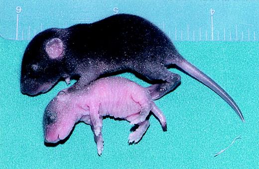
The hea mutant spontaneously arose on the CFO strain and was transferred to the C57BL/6J strain to produce a congenic line. A new phenotype emerged in B6–hea/hea mice (Figure 1), which includes necrosis of the tail similar to the necrotic tail tip seen in the mk (microcytic anemia) mouse iron metabolism mutant. The skin of 7-day-old hea/hea pups showed dried crusty skin similar to that seen for the flaky skin (fsn) mutant, which also suffers from both severe anemia and a psoriasis-like condition. Histologic studies of skin from B6–hea mice showed that there was superficial epidermal necrosis, hyperkeratosis, and basal epidermal hyperplasia (data not shown).
Phenotype of anemic hea pup showing growth retardation and skin abnormalities. A normal pup is pictured above and compared to an anemic hea/hea pup shown below with skin pallor, crusty skin with infoldings, and blackened necrotic tail tip.
Phenotype of anemic hea pup showing growth retardation and skin abnormalities. A normal pup is pictured above and compared to an anemic hea/hea pup shown below with skin pallor, crusty skin with infoldings, and blackened necrotic tail tip.
Hematology
Blood samples obtained from 1-week-old WBB6F1–hea/hea mice and +/? littermates showed marked disturbances in red blood cell parameters (Table 1). Homozygous hea/hea pups showed a significant reduction in red blood cell (RBC) number, hematocrit (Hct), and hemoglobin (Hb). However, the mean cell volume (MCV) was within the normal range compared with normal +/? littermates. Interestingly, in hea mice there was a significant elevation (P < .001) in both zinc protoporphyrin levels and serum iron, suggesting ineffective utilization of iron and deficient hemoglobin production. There was no significant difference in red blood cell osmotic fragility between hea pups and normal littermates (data not shown).
Hematologic data from 1-week-old WBB6F1-hea/hea and +/? normal littermates (n = 6)
Genotype . | RBC count, × 1012/L . | Hct . | MCV, fL . | Hb level, g/L . | Zn protoporphyrin level, μg/dL . | Serum Fe level, μM; n=5 . |
|---|---|---|---|---|---|---|
| hea/hea | 2.73 ± 0.34* | 0.252 ± 0.012† | 97.4 ± 10.0 | 60 ± 3† | 129 ± 5† | 71.958 ± 5.37† |
| +/? | 4.52 ± 0.21 | 0.385 ± 0.013 | 85.7 ± 2.8 | 107 ± 04 | 77 ± 3 | 27.387 ± 1.611 |
Data are mean ± standard error of the mean. RBC indicates red blood cell; Hct, hematocrit; and Hb, hemoglobin.
Mean value for hea/hea is significantly different from +/? littermates by t test at P < .05.
Mean value for hea/hea is significantly different from +/? littermates by t test at P < .001.
Blood smears taken from hea/hea mice were quite striking compared with +/? normal littermates and show marked polychromasia, anisocytosis, poikilocytosis, an elevated number of erythroblasts, naked nuclei, and smudge cells (Figure 2). The smudge cells in hea/hea blood smears appeared to be derived from broken nucleated red blood cells. The presence of many smudge cells was characteristic of the hea phenotype and closely resembled the blood smears of fsn mice, which showed the same disturbances in red blood cell morphology.
Peripheral blood smears from 7-day-old hea and fsn mice show several disturbances in red cell morphology. Marked anisocytosis, polychromasia, poikilocytosis, nucleated red cells, naked nuclei, and smudge cells are observed in hea and fsn pup blood smears as compared with a normal pup littermate. Micrographs were prepared using an Olympus VANOXT AH2 Version microscope and an Olympus camera (both from Olympus, Melville, NY). High dry objective lenses with a numerical aperture of 1.25 and an objective of 60× at a magnification of 600× were used. The images were scanned and labeled with Adobe Photoshop software.
Peripheral blood smears from 7-day-old hea and fsn mice show several disturbances in red cell morphology. Marked anisocytosis, polychromasia, poikilocytosis, nucleated red cells, naked nuclei, and smudge cells are observed in hea and fsn pup blood smears as compared with a normal pup littermate. Micrographs were prepared using an Olympus VANOXT AH2 Version microscope and an Olympus camera (both from Olympus, Melville, NY). High dry objective lenses with a numerical aperture of 1.25 and an objective of 60× at a magnification of 600× were used. The images were scanned and labeled with Adobe Photoshop software.
Similar findings for the blood values in hea mice were also found in fsn mice (Table 2). Adult homozygous fsn/fsn mice showed a reduction in RBC number, Hct, and Hb. The MCV for fsn mice indicated the presence of microcytic red cells (fsn/fsn: 43.3 ± 3.6 fL versus +/?: 53.3 ± 1.9 fL). Zinc protoporphyrin was elevated in fsn mice, as it is in hea mice. However, serum iron levels in fsn adult 6-week-old mice were reduced when compared with normal littermates (fsn/fsn: 25.06 ± 1.074 μM [140 ± 6 μg/dL] versus +/?: 60.86 ± 3.759 μM [340 ± 21 μg/dL]). This was in contrast to 1-week-old hea pups, which exhibited elevated levels of serum iron (Table 1) that were later explained to be an age-related difference. Collectively, the hematologic and skin pathology data suggested a link between the hea and fsn mutant.
Hematologic data from adult BALB/cByJ-fsn/fsn and +/? normal littermates (n = 4)
Genotype . | RBC count, × 1012/L . | Hct . | MCV, fL . | Hb level, g/L . | Zn protoporphyrin level, μg/dL; n = 8 . | Serum Fe level, μM; n = 5 . |
|---|---|---|---|---|---|---|
| fsn/fsn | 6.29 ± 0.50* | 0.268 ± 0.01† | 43.3 ± 3.6* | 44 ± 3† | 114 ± 6† | 25.06 ± 1.074† |
| +/? | 10.00 ± 0.50 | 0.531 ± 0.024 | 53.3 ± 1.9 | 157 ± 5 | 42 ± 1 | 60.86 ± 3.759 |
Data are mean ± standard error of the mean. Abbreviations are explained in a footnote to Table 1.
Mean value for fsn/fsn is significantly different from +/? littermates by t test at P < .05.
Mean value for fsn/fsn is significantly different from +/? littermates by t test at P < .001.
Genetics
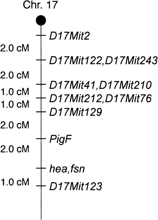
Chromosomal localization of the hea locus was determined by backcross analysis. C57BL/6J–+/hea mice were mated to the MOLD/Rk strain, and the B6MOLDF1–+/hea offspring were backcrossed to C57BL/6J–+/hea carriers. Only anemic hea/hea pups generated by the backcross were used for mapping because the genotype of any other backcross offspring could not be identified. Exactly 100 hea/hea backcross pups were genotyped. Microsatellite genetic markers (dinucleotide repeats) and a PigF EcoRI restriction fragment length polymorphism (RFLP) were scored for the backcross mice. The PigF EcoRI RFLP consisted of 9.2- and 6.3-kb bands in B6–hea mice and 15-, 6.3-, and 5.2-kb bands in MOLD/Rk. The results of the mapping study indicated that the hea locus was located on chromosome 17 with close linkage to D17Mit123 and the PigF gene (Figure 3). This position of the hea locus on chromosome 17 brought it in close proximity to the fsn locus (http://www.informatics.jax.org). The similar location of the 2 loci and the resemblance in hematology data between the 2 anemia mutants led us to predict that hea and fsn were likely the same locus. Therefore, allelic testing was performed to establish the genetic relationship between the hea and fsn mice. C57BL/6J–+/hea mice were mated to BALB/cJ–+/fsn carriers to determine whether anemic offspring could be produced. Two litters with anemic pups were produced from this cross (2 anemic pups and 6 normal pups in each litter). The generation of anemic pups from this cross indicated that hea and fsn are mutations of the same gene. Analysis of blood smears taken from the hea/fsn compound heterozygous mice showed the presence of smudge cells, anisocytosis, poikilocytosis, and polychromasia: abnormalities associated with both hea and fsn anemia mutants. The hea/fsn compound heterozygote mice lived to 2 months of age, which differed from the 1-week and 3-month life span of hea and fsn mice. Hematologic data for red blood cell number, hemoglobin levels, and hematocrit for the hea/fsn mice were similar to those of hea and fsn mice (data not shown).
The hea locus maps to the distal end of mouse chromosome 17 near the fsn locus. Relative linkage of genetic markers with the hea locus on mouse chromosome 17 near the established location for the fsn mutant locus. Relative distance in centimorgans (cM) is shown.
The hea locus maps to the distal end of mouse chromosome 17 near the fsn locus. Relative linkage of genetic markers with the hea locus on mouse chromosome 17 near the established location for the fsn mutant locus. Relative distance in centimorgans (cM) is shown.
Biochemistry and histologic studies
Biochemical studies on blood samples from hea and fsn mice indicated there are no defects in all enzymes tested (data not shown). Osmotic fragility studies indicated that blood cells from both fsn and hea mutants were the same as from normal mice (data not shown). No heme is detected in the urine from hea or fsn mice.
Histologic and pathologic analyses of tissues from hea pups, fsn mice, and normal littermate controls indicated that heart, kidney, lung, gastrointestinal tract, brain, and pancreas were unremarkable in hea and fsn mice and are similar to normal controls (data not shown). However, there was marked extramedullary hematopoiesis in the livers and spleens of hea and fsn mice. Bone marrow was normal in hea and fsn mice as compared with normal controls. The tail lesion seen in hea pups was essentially coagulative necrosis. Perls staining of liver samples from hea mice showed an accumulation of iron in hepatocytes but not in the macrophages of liver or spleen.
Transplantation of fetal liver cell populations
It is difficult to obtain an adequate amount of hea bone marrow to perform experimental transplantation. The alternative method is to use fetal liver cells, which are as potent as hematopoietic stem cells. Approximately 16 weeks after transplantation, changes were noted in the mice that received hea transplants, including significantly elevated protoporphyrin (hea: 145 ± 21 μg/dL versus normal: 54 ± 5 μg/dL) and other aspects of the hea anemia (abnormal blood smears with anisocytosis, polychromasia, and poikilocytosis). Hct, Hb levels, and MCV were reduced for mice that received hea transplants but were not statistically significant from recipients of normal fetal liver transplants (Table 3). The transfer of characteristics of the hea anemia supports the idea that a defect in hematopoietic stem cells contributes to the hea phenotype. The skin phenotype and reduced RBC number seen in hea pups did not appear in mice receiving hea transplants.
Blood data after fetal liver transplantation
. | RBC count, × 1012/L . | Hb level, g/L . | Hct . | Zn protoporphyrin level, μg/dL . | MCV, fL . |
|---|---|---|---|---|---|
| Prior to fetal liver transplantation; n = 7 | 5.9 ± 0.2 | 148 ± 6 | 0.41 ± 0.01 | 56 ± 4 | 70 ± 2 |
| After transplantation: normal; n = 4 | 7.5 ± 0.6 | 163 ± 17 | 0.44 ± 0.03 | 54 ± 5* | 59 ± 6 |
| After transplantation: hea; n = 3 | 7.5 ± 0.3 | 110 ± 12 | 0.34 ± 0.05 | 145 ± 21* | 45 ± 3 |
Data are mean ± standard error of the mean. Abbreviations are explained in a footnote to Table 1.
Mean value for hea/hea posttransplantation values is significantly different from normal transplantation by t test at P < .05; all other values between hea and normal transplantations are not statistically significant.
Iron metabolism
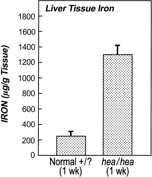
To determine the effect of the hea mutation on iron storage, we analyzed the amount of liver iron stores in 5 hea/hea versus 5 +/? mice. Figure 4 shows striking differences in the amount of iron stored in the liver of hea mice versus normal littermates at 1 week of age (hea/hea: 1302 ± 120 μg/g dry weight tissue versus +/?: 250 ± 68 μg/g dry weight tissue). The liver was iron overloaded in hea mice compared with the normal +/? pups. This was consistent with the finding of elevated serum iron levels in hea mice at the same age.
Liver tissue iron stores in hea mice are overloaded compared with normal littermates. Livers from 1-week-old hea and normal littermates were isolated and nonheme tissue iron was measured. Tissue iron levels in livers from hea mice were 400% higher than in normal littermates. Error bars indicate SEM.
Liver tissue iron stores in hea mice are overloaded compared with normal littermates. Livers from 1-week-old hea and normal littermates were isolated and nonheme tissue iron was measured. Tissue iron levels in livers from hea mice were 400% higher than in normal littermates. Error bars indicate SEM.
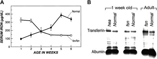
An examination of serum iron and transferrin levels was conducted for fsn mice to determine the similarity between hea and fsn animals. At 1 week of age, fsn mice exhibited elevated serum iron (fsn/fsn: 330 μg/dL versus +/?: 97 μg/dL) compared with normal littermates (Figure 5A). However, serum iron levels for fsn adult mice (6 weeks or older) were decreased (fsn/fsn: 140 μg/dL versus +/?: 340 μg/dL) compared with normal (Table 2). A successive study of serum iron levels from 1 week to 5 weeks of age was conducted for 5 fsn mice and 5 normal littermates, and the results are shown in Figure 5A. As the fsn mice aged, the serum iron levels decreased so that, at 6 weeks of age, the levels were well below normal. Transferrin was found to be slightly elevated in 1-week-old hea and fsn pups compared with normal littermates (n = 3 for each group), as shown in Figure 5B. However, scanning of cellulose acetate strips indicated that fsn adult mice showed about a 3-fold increase in transferrin levels over normal littermates. The increase in transferrin levels in adult fsn mice coincides with the decrease in serum iron levels found as fsn mice age. The elevated transferrin in fsn mice gives further support to a defect in iron metabolism.
Age-dependent loss of serum iron with concomitant elevated transferrin level in fsn mice. (A) Serum iron was measured in fsn and normal mice as they aged from 1-week-old pups to 5-week-old adults. Serum iron decreases as fsn mice age, whereas serum iron levels rise with age in normal mice. (B) Transferrin levels were assessed by electrophoresis of sera samples on cellulose acetate strips, and bands were visualized by staining with Ponceau S. Sera were collected from 1-week-old fsn and hea pups and adult fsn mice with age-matched normal littermate controls. The cellulose acetate strips were scanned and relative intensities noted for transferrin and albumin. Ratios of these 2 proteins were calculated and compared to normal. Transferrin is slightly elevated in hea pups (1.3 times more transferrin compared with normal littermates). Adult fsn mice were determined to have 2.8 times more transferrin than normal littermates (P < .001). Error bars indicate SEM.
Age-dependent loss of serum iron with concomitant elevated transferrin level in fsn mice. (A) Serum iron was measured in fsn and normal mice as they aged from 1-week-old pups to 5-week-old adults. Serum iron decreases as fsn mice age, whereas serum iron levels rise with age in normal mice. (B) Transferrin levels were assessed by electrophoresis of sera samples on cellulose acetate strips, and bands were visualized by staining with Ponceau S. Sera were collected from 1-week-old fsn and hea pups and adult fsn mice with age-matched normal littermate controls. The cellulose acetate strips were scanned and relative intensities noted for transferrin and albumin. Ratios of these 2 proteins were calculated and compared to normal. Transferrin is slightly elevated in hea pups (1.3 times more transferrin compared with normal littermates). Adult fsn mice were determined to have 2.8 times more transferrin than normal littermates (P < .001). Error bars indicate SEM.
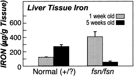
The decrease of serum iron in fsn mice was also concomitant with a loss of tissue iron. Two groups of 5 fsn and 5 normal littermates at 1 week and 5 weeks of age were killed and studied for tissue iron content in liver. The results are shown in Figure 6 and indicate that there was a significant reduction (P < .05) in liver tissue iron storage from 1-week-old pups (402 ± 82 μg/g tissue) to 5-week-old adult mice (156 ± 8 μg/g tissue). Conversely, normal mice showed an increase in liver iron storage between 1 week and 5 weeks of age (115 ± 1 to 275 ± 19 μg/g tissue). Pathological analyses of fsn mice indicated that there were no sites of extensive bleeding that might have accounted for the collective iron loss as the fsn mice age. Fecal occult blood (hemoccult) tests were negative on fresh stool samples from 5 normal mice. Of the 5 fsn mice tested, 2 were negative and 3 were slightly positive and interpreted as a very faint level (300 μg/g hemoglobin according to the kit manufacturer). This was considered insufficient (0.3 μg/g iron) to explain the loss of iron in fsn mice.
Age-dependent loss of liver tissue iron stores in fsn mice. Age-dependent loss of tissue iron stores is seen in fsn mice as iron stores are elevated in 1-week-old pups and are lost from adult fsn mice at 5 weeks of age. Conversely, normal littermates show an elevation of liver tissue iron stores. Error bars indicate SEM.
Age-dependent loss of liver tissue iron stores in fsn mice. Age-dependent loss of tissue iron stores is seen in fsn mice as iron stores are elevated in 1-week-old pups and are lost from adult fsn mice at 5 weeks of age. Conversely, normal littermates show an elevation of liver tissue iron stores. Error bars indicate SEM.
Fe-supplemented diet studies: cure of fsn anemia phenotype
The serum and tissue iron data suggested that the anemia of fsn mice was due, in part, to the loss of iron. To determine the role of iron metabolism on the phenotype of fsn mice, we treated the fsn mutant mice with an iron-supplemented diet. Three pairs of 5-week-old homozygous fsn/fsn mice and normal littermate (+/?) controls were placed on either an iron-rich diet (2% iron by weight) or a normal control diet (0.02% iron) for 2 weeks. The mice were examined for anemia status before and after treatment. Table 4 summarizes the results for RBC number, Hct, and Hb levels. The fsn mice placed on the control diet for 2 weeks continued to show the anemic state. However, the fsn mice placed on the iron-supplemented diet exhibited normal RBC number, Hct, and Hb concentration. Therefore, the anemia of fsn mice was cured by an iron-supplemented diet. Another aspect of the fsn phenotype that improved on the iron-supplemented diet was normal growth. At 7 weeks of age, fsn mice on the Fe-supplemented diet for 3 weeks were the same size as normal mice (fsn/fsn: 21.8 ± 0.6 g versus +/?: 22.7 ± 1.4 g), whereas fsn mice on a regular diet still showed growth retardation (fsn/fsn: 15.8 ± 0.3 g versus +/?: 22.7 ± 1.6 g). Although the skin phenotype of iron-treated fsn mice did not revert to normal, the skin lesions did not worsen as in untreated fsn mice. The life span of fsn mice on the iron-supplemented diet was about the same as in untreated fsn mutants.
Rescue of fsn/fsn anemia with iron-supplemented diet (n = 3)
Genotype . | RBC count, ×1012/L . | Hct . | Hb level, g/L . | |||
|---|---|---|---|---|---|---|
| Prior to iron-supplemented diet | ||||||
| fsn/fsn | 5.87 ± 0.36* | 0.263 ± 0.012† | 43 ± 3† | |||
| Normal: +/? | 9.85 ± 0.68 | 0.512 ± 0.019 | 156 ± 7 | |||
| Two wk on iron-supplemented diet | ||||||
| fsn/fsn | 8.97 ± 0.80 | 0.60 ± 0.003 | 150 ± 2 | |||
| Normal: +/? | 9.03 ± 0.55 | 0.57 ± 0.012 | 176 ± 3 | |||
| Two wk on control diet | ||||||
| fsn/fsn | 6.59 ± 0.03† | 0.34 ± 0.003† | 59 ± 4† | |||
| Normal: +/? | 9.69 ± 0.10 | 0.60 ± 0.015 | 178 ± 6 | |||
Data are mean ± standard error of the mean. Abbreviations are explained in a footnote to Table 1.
Mean value for fsn/fsn is significantly different from +/? littermates by t test at P < .05.
Mean value for fsn/fsn is significantly different from +/? littermates by t test at P < .001.
Iron-supplemented diet studies: fate of iron stores
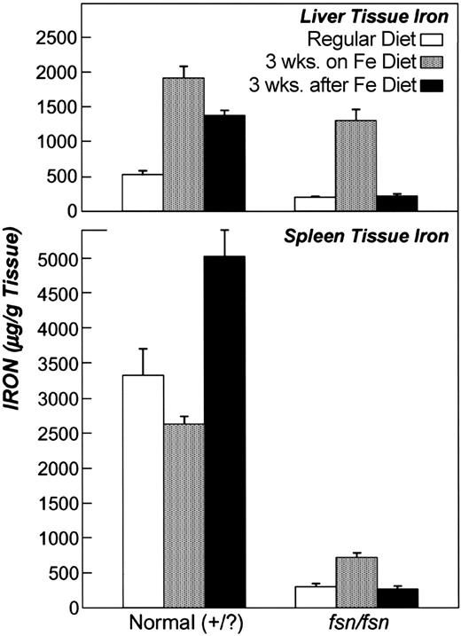
The rescue of the fsn anemia led to questions regarding the state of tissue iron stores in fsn mice treated with the iron diet. A quantitative analysis of tissue iron storage was performed for fsn mice receiving curative treatment by the iron-supplemented diet. Figure 7 shows the collective results of these studies and also includes the tissue iron levels of fsn and normal mice (n = 5 for each) on a standard diet (Figure 7, open bars). In the iron diet study, 5 fsn and normal (+/?) littermates were placed on iron diet for 3 weeks, killed, and tissue iron levels assessed. Figure 7 shows that both fsn and normal (+/?) littermates had iron-overloaded liver tissue when placed on the iron-supplemented diet for 3 weeks (Figure 7, stippled bars). However, tissue iron stores in spleen from normal mice were significantly higher (2622 ± 108 μg/g) than seen in spleen from fsn mice (713 ± 74 μg/g), which reflects the poor iron utilization by fsn mice. In a separate group of mice, the effect of removing the iron-supplemented diet from the iron-overloaded fsn and normal mice was then examined to ascertain the fate of the iron stores. Five fsn and 5 normal littermates were placed on iron diet (2% iron) for 3 weeks and subsequently switched to a control diet (0.02% iron) for an additional 3 weeks after which the animals were killed. The results for liver iron stores are shown in Figure 7 and indicate that fsn mice lost a significantly larger amount (P < .05) of iron stores (Figure 7, solid bars) than did normal mice during the 3 weeks after the iron diet was removed. A striking loss of iron stores was seen in fsn liver (1300 to 203 μg/g) as compared with the less substantial loss of liver iron in normal mice (1909 to 1390 μg/g) after the supplemented diet was removed. Decreased iron stores were also seen in the spleens of fsn mice (713 to 270 μg/g). Conversely, the tissue iron overloading (5023 μg/g) in spleens of normal mice was still present after the diet was removed. The loss of tissue iron overloading in these fsn mice is likely due to the same mechanism that leads to serum iron deficiency in fsn mice with aging.
Loss of tissue iron stores in fsn mice after removal of supplemented iron diet. Tissue iron levels in liver (top) are elevated in fsn and normal adult littermates while on a 2% supplemented iron diet compared with iron stores in mice on a regular diet. Liver tissue iron levels drop significantly in fsn mice after the iron diet is eliminated, whereas normal mice maintain iron overloading. Spleen tissue iron (bottom) in adult fsn mice on a 2% iron-supplemented diet is significantly lower than that found in normal adult littermates. Loss of spleen iron stores is seen for fsn mice when the iron diet is removed, whereas normal mice remain with highly elevated iron in spleen tissue. Error bars indicate SEM.
Loss of tissue iron stores in fsn mice after removal of supplemented iron diet. Tissue iron levels in liver (top) are elevated in fsn and normal adult littermates while on a 2% supplemented iron diet compared with iron stores in mice on a regular diet. Liver tissue iron levels drop significantly in fsn mice after the iron diet is eliminated, whereas normal mice maintain iron overloading. Spleen tissue iron (bottom) in adult fsn mice on a 2% iron-supplemented diet is significantly lower than that found in normal adult littermates. Loss of spleen iron stores is seen for fsn mice when the iron diet is removed, whereas normal mice remain with highly elevated iron in spleen tissue. Error bars indicate SEM.
The hematology of fsn mice was then examined 3 weeks after the iron diet was removed. A summary of the hematologic data for these mice is shown in Table 5. As before, the fsn mice were cured of their anemia when placed on the iron diet for 3 weeks. However, fsn mice returned to the anemia state 3 weeks after removal of the iron diet. Reduction in hemoglobin and hematocrit and an elevation in protoporphyrin levels were observed. The reversal to anemia in fsn mice coincides with the loss of tissue iron.
Alteration in hematology phenotype of fsn/fsn mice after removal of Fe-supplemented diet (n = 5)
Genotype . | Hb level, g/L . | Hct . | Zn protoporphyrin level, μg/dL . | ||
|---|---|---|---|---|---|
| Three weeks on iron-supplemented diet | |||||
| fsn/fsn | 143 ± 6 | 0.516 ± 0.009 | 39 ± 1 | ||
| Normal: +/? | 181 ± 7 | 0.594 ± 0.007 | 30 ± 1 | ||
| Three weeks after iron-supplemented diet terminated | |||||
| fsn/fsn | 86 ± 18* | 0.316 ± 0.04* | 50 ± 7* | ||
| Normal: +/? | 166 ± 7 | 0.514 ± 0.013 | 26 ± 1 | ||
Data are mean ± standard error of the mean.
Mean value for fsn/fsn is significantly different from +/? littermates by t test at P < .05.
Finally, an assessment of smudge cell levels in peripheral blood was examined in fsn mice on and off the iron-supplemented diet. Prior to treatment, fsn mice exhibited an elevated number of smudge cells (96 ± 5 smudge cells per 1000 blood cells). During the iron diet treatment the number of smudge cells in fsn blood dropped to the same level as normal (0 to 1 smudge cells per 1000 blood cells) but increased 3 weeks after the iron diet was removed, to a level of 32 ± 5 smudge cells per 1000 blood cells. Therefore, a change in the number of smudge cells in fsn mice also correlates with the change in phenotype on and off the iron-supplemented diet.
Discussion
The transfer of the hea mutation from the CFO strain to C57BL/6J strain led to the emergence of a psoriasis-like skin phenotype. The skin phenotype of hea mice is almost identical to that of another mouse mutant with both anemia and skin lesions called flaky skin (fsn). The anemia of hea and fsn mice was strikingly similar, and both have large numbers of smudge cells in their peripheral blood smears. The high number of smudge cells, which are likely derived from nucleated red blood cells, reflect the increased erythropoietic activity and the premature release of these cells into the circulation in an attempt to compensate for the severe anemia in these mice. The hea locus was mapped to the most distal end of mouse chromosome 17 near the fsn locus. The similarity in the skin and anemia phenotype led us to predict that these independently derived mutants are likely to be caused by defects in the same gene. An allelic test was performed, and compound heterozygote mice were generated. These mice had the skin lesions and anemia of both hea and fsn mice. Therefore, these mutants result from mutations in the same gene. The phenotype is more severe in hea mice and may be due to strain background differences (hea is on the C57BL/6J strain while fsn is on the BALB/cJ strain) or to the severity of the hea mutation.
We propose that a primary defect in iron metabolism could be the cause of the anemia phenotype seen in hea and fsn mice. This hypothesis is supported by the rescue of the anemia phenotype by supplementation of their diet with 2% iron. The kidney may be partially involved in the generation of the anemia phenotype in fsn and hea mice. We have shown that serum iron and tissue iron stores are lost as fsn mice age. Beamer et al showed that there is substantial loss of urinary iron in fsn mice as detected by using radioactive iron provided by mouth.14 This might explain why fsn mice lose iron stores as they age from neonates to maturity. Iron stores are also lost in adults after the use of an iron supplementation diet has been stopped (Figure 7). Perhaps the loss of iron in fsn mice may be due to lack of reabsorption of iron in the kidney. Iron has been shown to be filtrated and reabsorbed in the kidney in metabolically significant amounts.27 The iron transporter DMT1 has been shown to be localized at the sites where iron is reabsorbed (the loop of Henle and the collecting ducts of the nephron), suggesting that DMT1 provides the molecular mechanism for iron reabsorption.28 The hea and fsn gene may encode a protein that is involved in this pathway and, if defective, may lead to loss of iron in the urine. The fsn mouse also has decreased levels of 2 other divalent metals (Mg2+ and Zn2+) known to be transported by DMT1.14
The skin lesion phenotype in hea and fsn is similar to other mutant mice with defects in metal ion metabolism. The iron deficiency mutant microcytic anemia (mk) shows an abnormal skin phenotype including skin lesions, scaling of all body skin, and ringlike constrictions on the tail.29 The mouse mutant lethal milk (lm) is caused by defective metal ion utilization involving the zinc metal and leads to a skin phenotype including progressive hair loss, dermatitis, and skin lesions.30
A possible explanation for the skin phenotype in fsn mice has been previously proposed based on findings of immunologic defects in these mice.15-17 It has been suggested that an autoimmune disease leads to the skin phenotype in fsn mice. The role of the immune system in the fsn phenotype has been examined by mating fsn mice to scid (severe combined immune deficiency) mice in which there is an absence of T and B cells.31 When double homozygous (fsn/fsn, scid/scid) mice are produced, the skin defects and anemia remain as part of the fsn phenotype. Because scid mice do not have T or B cells, it would seem unlikely that an autoimmune disease could be the underlying reason for the skin defects in fsn mice, because fsn/fsn, scid/scid double homozygotes still show skin lesions.
Bone marrow transplantation (BMT) of fsn cells into scid mice results in the skin phenotype being transferred to recipient mice after 10 weeks of observation.31 A possible explanation for the transfer of skin lesions to BMT recipient mice may come from a common thread between the skin immune defense system and iron metabolism: the monocyte/macrophage system. Psoriasis is accompanied by the infiltration of skin macrophages (dendrocytes), which are derived from the monocyte/macrophage lineage. Kupffer macrophage cells of the liver are required for the iron cycle. Both types of macrophages derive from a common cell: the monocyte. Perhaps both the skin defects and the iron-deficient anemia in hea and fsn mice are due, in part, to a defect in the monoctye/macrophage system.
Our hypothesis is that iron utilization is defective in both hea and fsn mice. It is interesting that fsn mice overcome this problem when given an iron-supplemented diet. We speculate that the additional iron overcomes some threshold by which iron is utilized such that the large amounts of iron can compensate for the defect in iron utilization and rescue the anemia phenotype.
The loss of urinary iron in fsn mice has relevance to treatment of iron overloading in hemochromatosis and transfusion-dependent anemias including beta-thalassemia and sickle cell anemia (in which transfusion prevents stroke). The identification of the hea/fsn gene may lead to new therapy involving the excretion of iron into the urine. Understanding the etiology of the hea and fsn phenotype will require identification of the mutated gene. Positional cloning of the hea/fsn gene should be facilitated by the use of these independently derived mutants.
Prepublished online as Blood First Edition Paper, May 20, 2004; DOI 10.1182/blood-2004-01-0076.
The publication costs of this article were defrayed in part by page charge payment. Therefore, and solely to indicate this fact, this article is hereby marked “advertisement” in accordance with 18 U.S.C. section 1734.
The authors wish to thank Mark Fleming and Nancy Andrews for helpful discussions during the development of this work and Sharrion Russell for administrative assistance.


This feature is available to Subscribers Only
Sign In or Create an Account Close Modal