Abstract
In vitro studies show that BCR/ABL-expressing hematopoietic cells exhibit altered adhesion properties. No in vivo studies show whether the altered adhesion properties affect BCR/ABL leukemo-genesis. Using mice with homozygous inactivation of genes encoding the 2 adhesion molecules P-selectin and intercellular adhesion molecule-1 (ICAM1), we show that the mutant mice develop BCR/ABL-induced chronic myeloid leukemia (CML)-like leukemia at a significantly faster rate than do wild-type (WT) mice. Lack of P-selectin and ICAM1 did not have a significant effect on the development of B-cell acute lymphoblastic leukemia (BALL) induced by BCR/ABL. Using mice deficient for P-selectin or ICAM1 alone, we show that P-selectin plays a major role in the acceleration of CML-like leukemia. Lack of P-selectin resulted in early release of BCR/ABL-expressing myeloid progenitors from bone marrow, appearing to alter the biologic properties of leukemic cells rather than their growth rate by increasing their homing to the lungs, causing fatal lung hemorrhages. These results indicate that adhesion of BCR/ABL-expressing myeloid progenitors to marrow stroma through P-selectin and ICAM1 play an inhibitory role in the development of CML-like disease, suggesting that improvement of adhesion between BCR/ABL-expressing myeloid progenitor cells and bone marrow stroma may be of therapeutic value for human CML. (Blood. 2004;104:2163-2171)
Introduction
Human Philadelphia chromosome-positive (Ph+) leukemias are caused by the chimeric BCR/ABL oncogene resulting from the balanced t(9;22) chromosomal translocation joining the BCR gene on chromosome 22 and the ABL1 gene on chromosome 9. Ph+ leukemias include chronic myeloid leukemia (CML) and B-cell acute lymphoblastic leukemia (B-ALL). CML has a triphasic clinical course: a chronic phase; an accelerated phase; and a terminal phase known as blast crisis, a condition resembling acute leukemia, in which myeloid or lymphoid blasts fail to differentiate. CML is characterized by abnormal accumulation and infiltration of BCR/ABL-expressing hematopoietic cells in peripheral blood, spleen, and liver, and has been shown to have a defect in progenitor-stromal interaction.1,2 The c-Abl protein contains a large noncatalytic C-terminal domain with actin-binding functions, and fusion with BCR activates c-Abl kinase activity and increases its association with the actin cytoskeleton.3,4 This actin-binding function of the BCR/ABL oncoprotein has been found to contribute to the transformation of hematopoietic progenitor cells in Ph+ leukemias by abrogating interleukin-3 (IL-3) dependence and inducing defects in cell adhesion.4 Interestingly, this BCR/ABL-induced adhesion defect does not depend on BCR/ABL kinase activity.5
Interaction between cells and extracellular matrix components through adhesion molecules regulates cell proliferation. However, the effect of cell-stromal interaction on cell proliferation remains controversial, as both stimulation and inhibition of proliferation by this interaction have been described. For example, it has been reported that adhesion of hematopoietic cells to fibronectin stimulates proliferation of normal and BCR/ABL-expressing hematopoietic cells.6-10 In contrast, other studies have reported that fibronectin mediates adhesion of hematopoietic progenitors to bone marrow stroma, and that this cell-stromal interaction inhibits proliferation of progenitors.2,11 Despite these contradictory results, however, the above studies are consistent in showing that cell adhesion regulates proliferation of bone marrow progenitors. Several lines of evidence have shown that BCR/ABL-expressing hematopoietic progenitors and mature neutrophils in bone marrow exhibit altered adhesion properties, suggesting that adhesion of these cells to stromal elements may play a role in the pathogenesis of CML. An in vitro study of marrow hematopoietic progenitors showed that blast colony-forming cells (B1-CFC) isolated from CML patients had a reduced capacity to adhere to steroid-treated stroma,12 explaining the premature release of Ph+ hematopoietic progenitor cells from the marrow of CML patients into the blood.13 In another study, granulocytes isolated from the blood of CML patients were shown to have reduced adhesion properties.14
Previous studies have shown that both P-selectin and ICAM1 are involved in normal leukocyte function and cell adhesion. For example, P-selectin and ICAM1 were shown to participate in the regulation of normal leukocyte function by controlling the rolling phase of the leukocyte adhesion cascade and the migration of neutrophils to loci of inflammation.14-17 Previous studies using P-selectin and ICAM1 knockout mice have shown that transmigration of neutrophils into the peritoneum is completely absent and inflammatory and immune responses are impaired in the absence of these genes.15,17 P-selectin also has been shown to play a significant role in initiating adhesion of primitive human hematopoietic progenitor cells.18 A reduced binding to P-selectin has been observed for Ph+ granulocytes from CML patients.14 ICAM1 has been shown to be important in the cytokine-activated and chemotactic transmigration of neutrophils, myelocytic cell binding to endothelial cells, and differentiation in myeloid cell lines.15,19 ICAM1 also has been shown to be expressed on hematopoietic CD34+ bone marrow cells and to mediate the adhesion of progenitors to the marrow.20 Although both P-selectin and ICAM1 are adhesion molecules, the mechanisms by which they regulate cell adhesion are quite different. P-selectin mainly intereacts with its carbohydrate ligands,21 whereas ICAM1 interacts with β2 integrins.22 However, it is not known whether P-selectin and ICAM1 are involved in the development of Ph+ leukemias. In this study, we used single- and double-gene knockout mice to investigate the roles of P-selectin and ICAM1 in the development of CML and B-ALL in our mouse leukemia model system. We found that P-selectin/ICAM1-deficient mice developed CML significantly faster than did wild-type (WT) mice, and that P-selectin, but not ICAM1, plays a major role in the disease process. The accelerated leukemia development was due to an early release of BCR/ABL-expressing progenitor cells from bone marrow and migration of myeloid leukemic cells into the lungs of the diseased mice. P-selectin/ICAM1 deficiency had no significant effect on survival of mice with BCR/ABL-induced B-ALL.
Materials and methods
Mouse strains
Wild-type C57BL/6J mice and C57BL/6 mice with homozygous null mutations in the gene encoding P-selectin (B6.129S7-Selptm1Bay) or ICAM1 (B6.129S7-Icam1tm1Bay) and in both P-selectin and ICAM1 genes (B6.129S7-Icam1tm1BaySelptm1Bay) were obtained from the Jackson Laboratory (Bar Harbor, ME).
Bone marrow transduction and transplantation
The cDNA for P210 BCR/ABL (b3a2 isoform) in the retroviral vector MSCV-IRES-GFP (murine stem cell virus-internal ribosomal entry site-green fluorescence protein) was used for all experiments. Helper-free retroviral stocks were generated by transient transfection of 293T cells as described previously23 and titered by FACS analysis of the infection efficiency of NIH3T3 cells with the retrovirus. At a 1:4 dilution of the retroviral stock, the percentage of GFP-positive cells is more than 80%. To induce CML-like leukemia, bone marrow cells from donor mice (6-8 weeks old) were transduced with the BCR/ABL retrovirus, followed by transplantation into recipient mice (6-8 weeks old) as previously described.23 Briefly, donor mice were pretreated with 5-fluorouracil (5-FU; 200 mg/kg) through intravenous tail injection. Four days later, bone marrow cells were harvested, prestimulated with IL-3, IL-6, and stem cell factor (SCF), and subjected to 2 rounds of cosedimentation with retroviral stock. The transduced marrow cells were transplanted by lateral tail vein injection into recipient mice of the same genetic background as the donor mice. In a C57BL/6 background, recipients of wild-type BCR-ABL-transduced marrow develop myeloproliferative disease closely resembling the chronic phase of human CML.24 To model B-ALL, we transduced bone marrow from non-5-FU-treated donors without cytokines, and under these conditions all recipients develop fatal acute B-lymphoblastic leukemia (data not shown). Prior to transplantation, all recipient mice received 2 doses of 550-cGy gamma irradiation separated by 3 hours. Recipient mice were evaluated daily for symptoms of B-ALL, including weight and heat loss, splenomegaly, and overall signs of morbidity (decreased level of activity). Hematopoietic tissues and peripheral blood samples were collected from the diseased mice for further pathological and FACS analyses.
Flow cytometry
Hematopoietic cells were collected from peripheral blood, bone marrow, spleen, and pleural cavity of the diseased mice, and red blood cells were lysed with NH4Cl red blood cell (RBC) lysis buffer (pH 7.4). The cells were washed with phosphate buffered saline (PBS), blocked with FC receptor blocking antibody, and stained with B220-PE/CD19-APC for B cells, Mac1-PE/Gr1-APC for monocytes and neutrophils, and Sca1-PE/c-kit-APC for hematopoietic stem cells. After staining, the cells were washed once with PBS and subjected to FACS analysis.
Histology
The lungs and spleens from normal and diseased mice were fixed in Bouin fixative (Fisher Scientific, Pittsburgh, PA) for 24 hours at room temperature, followed by an overnight rinse in water. Ten-μm sections were stained with hematoxylin and eosin (H&E) and observed using a compound microscope (Leica, Heidelberg, Germany). For preparation of cryosections, the lungs were embedded in optimal cutting temperature (OCT) medium (Sakura, Torrance, CA) and frozen at -20°C. Ten-μm frozen sections containing the GFP-expressing or dye-labeled cells were observed using a fluorescence microscope (Leica). All sections were imaged with a 2.5 × PH1 objective or a 20 × PH1 objective (NPLan, Leica; NA, 0.25 and 0.40, respectively). All images were imported into MetaMorph software (Molecular Devices, Downingtown, PA) as a series of tagged image files. All images were then constructed using Adobe Photoshop 6.0 (Adobe, San Jose, CA).
Bone marrow colony formation assay
Bone marrow cells from wild-type and P-selectin-/-/ICAM1-/- mice were transduced with MSCV-P210-IRES-GFP retrovirus, and the transduced cells were assayed for colony formation as described previously.25
Bone marrow cell homing assay
Bone marrow cells isolated from 5-FU-primed wild-type or P-selectin-/- mice were prestimulated with IL-3, IL-6, and SCF, and subjected to 2 rounds of cosedimentation with green fluorescence protein (GFP) retrovirus or labeled with a red fluorescent dye PKH26 (Sigma, St Louis) according to the manufacturer's instruction. Five million of the transduced or 3 million of chemically labeled marrow cells were transplanted by lateral tail vein injection into each recipient mouse of the same genetic background as the donor mouse. At 3 hours after the transplantation, GFP-positive or PKH26-positive cells from bone marrow, peripheral blood, and the spleens of the recipient mice were analyzed by FACS, and the labeled cells in the lung tissue were visualized on 10 μM cryosections.
Statistics
Differences in survival among groups in all experiments were compared using the log-rank test. Differences in white blood cell count, spleen weight, and percentages of GFP-positive B cells, myeloid cells, and marrow progenitor cells in noninjected mice were compared using t test. The percentages of GFP-positive B cells, myeloid cells, and marrow stem cells in BCR/ABL-transduced mice were compared using ANOVA. A P value less than .05 was considered statistically significant.
Results
Lack of P-selectin and ICAM1 accelerates BCR/ABL-induced CML-like disease
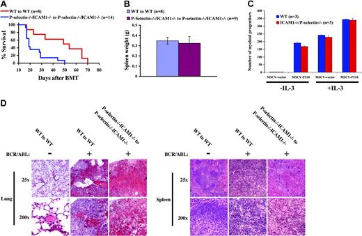
The effects of P-selectin and ICAM1 on BCR/ABL-induced CML-like disease were examined using P-selectin/ICAM1 homozygous double knockout mice in our retroviral bone marrow transduction/transplantation model system.23 P-selectin/ICAM1-deficient mice were used as both donors and recipients, and in the control group wild-type mice were used as both donors and recipients of BCR/ABL-transduced bone marrow cells. We hypothesized that P-selectin and ICAM1 might be required for adhesion of BCR/ABL-expressing progenitors to stromal cells in bone marrow, and that P-selectin and/or ICAM1 deficiency would result in an early release of leukemic progenitor cells from bone marrow, causing accelerated death of the diseased mice. In our previous studies using a bone marrow transplantation model,23,24 5.0 × 10⁵ donor bone marrow cells were injected into each recipient mouse, and the mice developed CML-like disease and died within several weeks. To observe more clearly whether and to what extent the disease was accelerated in the present studies, the cell dose was reduced to 1.0 × 10⁵ donor cells per recipient mouse. We found that P-selectin/ICAM1-deficient mice developed CML-like disease much more rapidly than did wild-type mice (P = .001); 50% of P-selectin/ICAM1-deficient mice died within 3 weeks of bone marrow transplantation, whereas 50% of wild-type mice survived until 7 weeks following transplantation (Figure 1A).
Lack of P-selectin and ICAM1 accelerates the development of CML-like leukemia induced by BCR/ABL (A) Kaplan-Meier survival curve for recipients of marrow transduced by BCR/ABL. Recipient mice received a cell dose of 1 × 10⁵ cells/mouse. There were significant differences in survival between wild-type (WT) recipients of transduced WT marrow and P-selectin/ICAM1-deficient recipients of transduced P-selectin/ICAM1-deficient marrow (P = .001, log-rank test). (B) Mean spleen weight (mean values ± SD) of leukemic mice (measured at the time of morbidity or death). There was no significant difference in spleen weight between the 2 transplant groups (P = .363, t test). (C) Bone marrow colony formation assay. Bone marrow cells from wild-type and P-selectin-/-/ICAM1-/- mice were transduced with MSCV-P210-IRES-GFP or the parental control (MSCV-IRES-GFP) retrovirus, and the transduced cells were assayed for colony formation. The average number of colonies (mean values ± SD) from triplicate platings was determined in the absence or presence of 100 pg/mL murine IL-3. (D) Pathological analysis (hematoxylin/eosin staining) of the lungs and spleens of CML mice at the time of morbidity and death. In the normal control group, nontransduced wild-type bone marrow cells were transplanted into lethally irradiated wild-type recipient mice. In contrast to normal control mice, BCR/ABL-transduced WT to WT and P-selectin-/-/ICAM1-/- to P-selectin-/-/ICAM1-/- transplant groups showed complete infiltration of the lungs with myeloid leukemic cells and severe lung hemorrhages; P-selectin-/-/ICAM1-/- to P-selectin-/-/ICAM1-/- transplant mice showed more severe and larger areas of lung hemorrhages. In contrast to the normal control group, BCR/ABL-transduced WT to WT and P-selectin-/-/ICAM1-/- to P-selectin-/-/ICAM1-/- transplant groups demonstrated complete disruption of follicular architecture of the spleen by infiltrating myeloid leukemic cells.
Lack of P-selectin and ICAM1 accelerates the development of CML-like leukemia induced by BCR/ABL (A) Kaplan-Meier survival curve for recipients of marrow transduced by BCR/ABL. Recipient mice received a cell dose of 1 × 10⁵ cells/mouse. There were significant differences in survival between wild-type (WT) recipients of transduced WT marrow and P-selectin/ICAM1-deficient recipients of transduced P-selectin/ICAM1-deficient marrow (P = .001, log-rank test). (B) Mean spleen weight (mean values ± SD) of leukemic mice (measured at the time of morbidity or death). There was no significant difference in spleen weight between the 2 transplant groups (P = .363, t test). (C) Bone marrow colony formation assay. Bone marrow cells from wild-type and P-selectin-/-/ICAM1-/- mice were transduced with MSCV-P210-IRES-GFP or the parental control (MSCV-IRES-GFP) retrovirus, and the transduced cells were assayed for colony formation. The average number of colonies (mean values ± SD) from triplicate platings was determined in the absence or presence of 100 pg/mL murine IL-3. (D) Pathological analysis (hematoxylin/eosin staining) of the lungs and spleens of CML mice at the time of morbidity and death. In the normal control group, nontransduced wild-type bone marrow cells were transplanted into lethally irradiated wild-type recipient mice. In contrast to normal control mice, BCR/ABL-transduced WT to WT and P-selectin-/-/ICAM1-/- to P-selectin-/-/ICAM1-/- transplant groups showed complete infiltration of the lungs with myeloid leukemic cells and severe lung hemorrhages; P-selectin-/-/ICAM1-/- to P-selectin-/-/ICAM1-/- transplant mice showed more severe and larger areas of lung hemorrhages. In contrast to the normal control group, BCR/ABL-transduced WT to WT and P-selectin-/-/ICAM1-/- to P-selectin-/-/ICAM1-/- transplant groups demonstrated complete disruption of follicular architecture of the spleen by infiltrating myeloid leukemic cells.
All wild-type and P-selectin/ICAM1 double-knockout mice developed the fatal CML-like disease with similar pathological changes, characterized primarily by elevated white blood cell counts in peripheral blood and infiltration of leukemic cells in bone marrow, spleen, liver, and lungs. All mice had extensive lung hemorrhages at the time of death. Although P-selectin/ICAM1-deficient mice developed CML-like disease more rapidly than did wild-type mice, there was no significant difference in spleen weight at death between these 2 groups (P = .363) (Figure 1B). Because the spleen is the main reservoir of myeloid leukemic cells, this observation suggests that the acceleration of CML-like leukemia in the absence of P-selectin/ICAM1 was unlikely to be due to an increased proliferation of leukemic cells. To confirm this, we directly compared the growth of BCR-ABL-expressing wild-type myeloid cells with that of P-selectin-/-/ICAM-/- myeloid cells in an in vitro bone marrow colony assay and found that there were no significant differences in growth between wild-type and P-selectin-/-/ICAM-/- bone marrow myeloid cells (Figure 1C). It is possible that the lack of P-selectin and ICAM1 might cause acceleration of CML-like disease through a non-growth-related mechanism, such as by changing the adhesion properties of leukemic cells in bone marrow and other tissues. Because pulmonary hemorrhage is the major cause of death for the CML mice,23 we tested this possibility by examining the pathological changes in the lungs at death. In contrast to the lungs of normal mice, those of wild-type and P-selectin/ICAM1-deficient CML mice were largely infiltrated with leukemic cells, resulting in destruction of lung architecture and severe hemorrhages (Figure 1D). Relatively, the lungs of P-selectin/ICAM1-deficient CML mice exhibited more diffuse and larger areas of hemorrhage, whereas smaller and patchier areas of lung hemorrhage were observed in wild-type CML mice (Figure 1D). The differences in severity of lung hemorrhage between the 2 groups appeared to correlate with the differences in their survival times (Figure 1A). There was also severe destruction of spleen architecture of wild-type and P-selectin/ICAM1-deficient CML mice (Figure 1D).
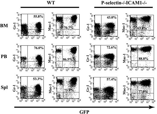
To further characterize P-selectin/ICAM1-deficient CML mice, we analyzed GFP-positive myeloid (Gr-1/Mac-1) cells in bone marrow, peripheral blood, and spleen by FACS analysis at the time of death (Figure 2). Percentages of Gr-1+/Mac-1+ cell in all these tissues were similar in wild-type and P-selectin/ICAM1-deficient CML mice, indicating that mice in both groups developed a similar myeloid leukemia. It is possible that the accelerated CML disease in the absence of P-selectin and ICAM1 may be due to differences in the composition of normal bone marrow progenitors or the efficiency of retroviral transduction in the double-knockout mice. To exclude the first possibility, we analyzed normal bone marrow cells from noninjected wild-type and P-selectin/ICAM1-deficient mice by FACS. There were no significant differences between the 2 groups of mice in the percentages of myeloid progenitors identified by the cell surface markers c-kit, Sca-1, Gr-1, and Mac-1 (Figure 3A). To exclude the second possibility, bone marrow cells transduced with the BCR/ABL retrovirus were cultured in the presence of SCF, IL-3, and IL-6 for 2 days after the 2-round retroviral infection, then the cells were analyzed by FACS for GFP-expressing Gr-1- or Mac-1-positive cells. The percentages of GFP/Gr-1- or GFP/Mac-1-positive myeloid cells were similar for wild-type P-selectin/ICAM1-deficient mice, indicating that there was no significant difference between wild-type and P-selectin/ICAM1-deficient mice in susceptibility to BCR/ABL retroviral transduction (Figure 3B).
FACS analysis shows the development of a similar CML-like leukemia in the presence and absence of P-selectin and ICAM1. WT to WT and P-selectin-/-/ICAM1-/- to P-selectin-/-/ICAM1-/- mice showed similar percentages of GFP+/Gr-1+ and GFP+/Mac-1+ myeloid cells in bone marrow (BM; P = .495), peripheral blood (PB; P = .857), and spleen (Spl; P = .997) (2 × 2 ANOVA).
FACS analysis shows the development of a similar CML-like leukemia in the presence and absence of P-selectin and ICAM1. WT to WT and P-selectin-/-/ICAM1-/- to P-selectin-/-/ICAM1-/- mice showed similar percentages of GFP+/Gr-1+ and GFP+/Mac-1+ myeloid cells in bone marrow (BM; P = .495), peripheral blood (PB; P = .857), and spleen (Spl; P = .997) (2 × 2 ANOVA).
FACS analysis of BCR/ABL retroviral transduction efficiency of wild-type and P-selectin-/-/ICAM1-/- bone marrow cells. (A) Wild-type and P-selectin-/-/ICAM1-/- mice have similar percentages (mean values ± SD) of myeloid (Gr-1- and Mac-1-positive) and lymphoid (B220- and CD19-positive) progenitor cells and hematopoietic stem cells (c-kit- and Sca-1-positive) in bone marrow. One-way ANOVA revealed no statistically significant differences between the 2 groups (P > .05 for all markers). (B) Bone marrow cells from wild-type and P-selectin-/-/ICAM1-/- mice were equally susceptible to transduction by BCR/ABL retrovirus. Bone marrow cells were transduced with BCR/ABL retrovirus, cultured with IL-3, IL-6, and stem cell factor for 2 days, and analyzed by FACS for total GFP-positive cells.
FACS analysis of BCR/ABL retroviral transduction efficiency of wild-type and P-selectin-/-/ICAM1-/- bone marrow cells. (A) Wild-type and P-selectin-/-/ICAM1-/- mice have similar percentages (mean values ± SD) of myeloid (Gr-1- and Mac-1-positive) and lymphoid (B220- and CD19-positive) progenitor cells and hematopoietic stem cells (c-kit- and Sca-1-positive) in bone marrow. One-way ANOVA revealed no statistically significant differences between the 2 groups (P > .05 for all markers). (B) Bone marrow cells from wild-type and P-selectin-/-/ICAM1-/- mice were equally susceptible to transduction by BCR/ABL retrovirus. Bone marrow cells were transduced with BCR/ABL retrovirus, cultured with IL-3, IL-6, and stem cell factor for 2 days, and analyzed by FACS for total GFP-positive cells.
P-selectin deficiency plays a major role in the acceleration of CML-like leukemia
To assess the individual roles of P-selectin and ICAM1 in the acceleration of BCR/ABL-induced CML-like disease, we used mice lacking either P-selectin or ICAM1 and used wild-type and P-selectin/ICAM1-deficient mice as controls. In this experiment, we increased the dose of BCR/ABL-transduced bone marrow cells from 1.0 × 10⁵/mouse (Figure 1) to 2.0 × 10⁵/mouse to allow more rapid development of CML-like disease, facilitating comparison between the experimental groups. There was no significant difference in the rate of disease development between P-selectin-deficient and P-selectin/ICAM1-deficient mice (Figure 4A). ICAM1-deficient mice had a slight but statistically significant acceleration of CML development compared to wild-type mice (P = .001), but developed the disease significantly more slowly than P-selectin-deficient (P = .007) and P-selectin/ICAM1-deficient mice (P = .012). Taken together, these results indicate that P-selectin deficiency is the major cause of the acceleration of CML-like disease, but that ICAM1 also plays a role in acceleration of the disease. Although P-selectin- and ICAM1-deficient mice both developed CML-like disease more rapidly than did wild-type mice (Figure 4A), all mice developed the same disease, as indicated by FACS analysis of Gr-1+/GFP+ in bone marrow, peripheral blood, and spleen (data not shown). There was no significant difference at death among the 4 groups in white blood cell counts (P = .730) (Figure 4B) or spleen weight (P = .078) (Figure 4C), suggesting that acceleration of the disease in the absence of P-selectin or ICAM1, as well as when both P-selectin and ICAM1 were absent, was likely not through promotion of the growth of leukemic cells (Figure 1).
P-selectin deficiency plays a major role in acceleration of CML-like leukemia induced by BCR/ABL (A) Kaplan-Meier survival curve for recipients of marrow transduced by BCR/ABL. Recipient mice received a cell dose of 2 × 10⁵ cells/mouse. Mice lacking P-selectin only and lacking both P-selectin and ICAM1 had a significant acceleration in the development of CML-like leukemia compared to both control (WT) mice and mice lacking ICAM1 only (P = .001). ICAM1-deficient mice also had a slight but statistically significant acceleration of CML development compared to control mice (P = .000). There were no significant differences (mean values ± SD) in white blood cell counts (B) and spleen weights (C) among the 4 transplant groups.
P-selectin deficiency plays a major role in acceleration of CML-like leukemia induced by BCR/ABL (A) Kaplan-Meier survival curve for recipients of marrow transduced by BCR/ABL. Recipient mice received a cell dose of 2 × 10⁵ cells/mouse. Mice lacking P-selectin only and lacking both P-selectin and ICAM1 had a significant acceleration in the development of CML-like leukemia compared to both control (WT) mice and mice lacking ICAM1 only (P = .001). ICAM1-deficient mice also had a slight but statistically significant acceleration of CML development compared to control mice (P = .000). There were no significant differences (mean values ± SD) in white blood cell counts (B) and spleen weights (C) among the 4 transplant groups.
Lack of P-selectin results in early release of BCR/ABL-expressing myeloid progenitors from bone marrow
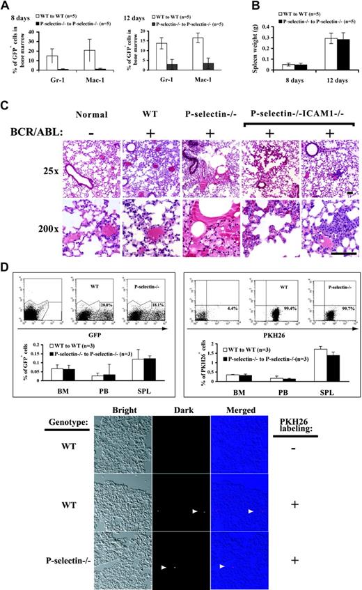
As described in Figures 4B and 4C it is likely that deficiency of both P-selectin and ICAM1 caused the acceleration of CML-like disease through alteration of biologic properties of myeloid leukemic cells, rather than of their growth rate. We hypothesized that P-selectin/ICAM1 deficiency might reduce the adhesion of myeloid leukemic cells to the bone marrow stroma, allowing early release of BCR/ABL-expressing myeloid cells into the blood circulation. Because we determined that P-selectin deficiency played a major role in causing the acceleration of CML-like disease, we investigated whether P-selectin affects the adhesion of myeloid progenitor cells to bone marrow stroma. To do this, we killed wild-type and P-selectin-deficient CML mice at days 8 and 12 after bone marrow transplantation and analyzed by FACS the bone marrow cells that were positive for both GFP and Gr-1 (GFP+/Gr-1+) or Mac-1 (GFP+/Mac-1+) (Figure 5A). The mean percentage of GFP+/Gr-1+ and GFP+/Mac-1+ cells measured on both days was significantly greater in wild-type CML mice (15.0% and 20.8% for day 8, and 13.7% and 16.5% for day 12) than in P-selectin-deficient CML mice (0.93% and 1.15% for day 8, and 2.9% and 3.5% for day 12) (P = .007 and 0.023 for day 8 and P = .039 and .047 for day 12, respectively). These results indicate that in the absence of the adhesion molecule P-selectin, BCR/ABL-expressing myeloid progenitor cells are poorly retained in the bone marrow and leave earlier than wild-type BCR/ABL-expressing progenitor cells.
Lack of P-selectin changes the biologic properties of BCR/ABL-expressing myeloid progenitor cells in bone marrow. (A) Lack of P-selectin results in the early release of BCR/ABL-expressing myeloid progenitor cells from bone marrow. Bone marrow cells from wild-type and P-selectin-/- mice that received transplants of BCR/ABL-transduced bone marrow cells were analyzed by FACS at days 8 and 12 after bone marrow transplantation. At both days 8 and 12, the percentage (mean values ± SD) of GFP+/Gr-1+ and GFP+/Mac-1+ cells was significantly higher in bone marrow cells of wild-type CML mice than in those of P-selectin-/- CML mice (P = .007 and .023 for day 8 and P = .039 and .047 for day 12, respectively). (B) Mean spleen weight (mean values ± SD) of the diseased mice at days 8 and 12 after bone marrow transplantation. At both days, there was no significant difference in spleen weight between wild-type and P-selectin-/-/ICAM1-/-CML mice (P = .833 and 0.784 for days 8 and 12, respectively). (C) Pathological analysis (hematoxylin/eosin staining) of the lungs of CML mice (n = 5 for each transplant group) at day 12 after bone marrow transplantation. In the normal control group, nontransduced wild-type bone marrow cells were transplanted into lethally irradiated wild-type recipient mice. In contrast to normal control mice, BCR/ABL-transduced WT to WT, P-selectin-/- to P-selectin-/-, and P-selectin-/-/ICAM1-/- to P-selectin-/-/ICAM1-/- transplant mice showed significant infiltration of the lungs by myeloid leukemic cells. Lung hemorrhages and leukemic cell masses were observed in P-selectin-/- and P-selectin-/-/ICAM1-/-, but not WT CML mice. Scale bars, 100 μM (short bar at 25 ×) and 50 μM (long bar at 200 ×). (D) Lack of P-selectin does not alter the homing properties of the transplanted donor bone marrow cells to bone marrow of the recipient mice. Donor bone marrow cells from wild-type and P-selectin-/- mice were transduced GFP retrovirus, followed by transplantation into wild-type and P-selectin-/- recipient mice, respectively. Prior to the transplantation, the transduced marrow cells were analyzed by FACS for percentage (mean values ± SD) of GFP+ cells (20.0% and 18.1% for the transduced wild-type and P-selectin-/- bone marrow cells, respectively) (top left panel).At 3 hours after the transplantation, cells were isolated from bone marrow, peripheral blood (PB), and spleens (Spl) of the recipient mice, and analyzed by FACS for GFP+ cells. Similar percentages of GFP+ wild-type and P-selectin-/- cells were detected in these locations (P = .86, .86, and .92 for BM, PB, and Spl, respectively). The transduced wild-type and P-selectin-/- donor bone marrow cells were also labeled with a red fluorescent dye PKH26, followed by transplantation into wild-type and P-selectin-/- recipient mice, respectively. Prior to the transplantation, the percentages (mean values ± SD) of PKH26+ cells were 99.4% and 98.7% for wild-type and P-selectin-/- donor cells, respectively (top right panel).At 3 hours after the transplantation, cells were isolated from bone marrow and spleens of the recipient mice and analyzed by FACS for PKH26+ cells. Similar percentages of PKH26+ wild-type and P-selectin-/- cells were detected in these 2 locations (P = .50, .58 and .07 for BM, peripheral blood, and Spl, respectively). Cryosections of the lungs from the mice receiving the PKH26-labeled bone marrow cells were prepared in optimal cutting temperature (OCT) medium, and the labeled cells (arrow heads) on 10-μM sections were visualized under a fluorescent microscope (bottom panel).
Lack of P-selectin changes the biologic properties of BCR/ABL-expressing myeloid progenitor cells in bone marrow. (A) Lack of P-selectin results in the early release of BCR/ABL-expressing myeloid progenitor cells from bone marrow. Bone marrow cells from wild-type and P-selectin-/- mice that received transplants of BCR/ABL-transduced bone marrow cells were analyzed by FACS at days 8 and 12 after bone marrow transplantation. At both days 8 and 12, the percentage (mean values ± SD) of GFP+/Gr-1+ and GFP+/Mac-1+ cells was significantly higher in bone marrow cells of wild-type CML mice than in those of P-selectin-/- CML mice (P = .007 and .023 for day 8 and P = .039 and .047 for day 12, respectively). (B) Mean spleen weight (mean values ± SD) of the diseased mice at days 8 and 12 after bone marrow transplantation. At both days, there was no significant difference in spleen weight between wild-type and P-selectin-/-/ICAM1-/-CML mice (P = .833 and 0.784 for days 8 and 12, respectively). (C) Pathological analysis (hematoxylin/eosin staining) of the lungs of CML mice (n = 5 for each transplant group) at day 12 after bone marrow transplantation. In the normal control group, nontransduced wild-type bone marrow cells were transplanted into lethally irradiated wild-type recipient mice. In contrast to normal control mice, BCR/ABL-transduced WT to WT, P-selectin-/- to P-selectin-/-, and P-selectin-/-/ICAM1-/- to P-selectin-/-/ICAM1-/- transplant mice showed significant infiltration of the lungs by myeloid leukemic cells. Lung hemorrhages and leukemic cell masses were observed in P-selectin-/- and P-selectin-/-/ICAM1-/-, but not WT CML mice. Scale bars, 100 μM (short bar at 25 ×) and 50 μM (long bar at 200 ×). (D) Lack of P-selectin does not alter the homing properties of the transplanted donor bone marrow cells to bone marrow of the recipient mice. Donor bone marrow cells from wild-type and P-selectin-/- mice were transduced GFP retrovirus, followed by transplantation into wild-type and P-selectin-/- recipient mice, respectively. Prior to the transplantation, the transduced marrow cells were analyzed by FACS for percentage (mean values ± SD) of GFP+ cells (20.0% and 18.1% for the transduced wild-type and P-selectin-/- bone marrow cells, respectively) (top left panel).At 3 hours after the transplantation, cells were isolated from bone marrow, peripheral blood (PB), and spleens (Spl) of the recipient mice, and analyzed by FACS for GFP+ cells. Similar percentages of GFP+ wild-type and P-selectin-/- cells were detected in these locations (P = .86, .86, and .92 for BM, PB, and Spl, respectively). The transduced wild-type and P-selectin-/- donor bone marrow cells were also labeled with a red fluorescent dye PKH26, followed by transplantation into wild-type and P-selectin-/- recipient mice, respectively. Prior to the transplantation, the percentages (mean values ± SD) of PKH26+ cells were 99.4% and 98.7% for wild-type and P-selectin-/- donor cells, respectively (top right panel).At 3 hours after the transplantation, cells were isolated from bone marrow and spleens of the recipient mice and analyzed by FACS for PKH26+ cells. Similar percentages of PKH26+ wild-type and P-selectin-/- cells were detected in these 2 locations (P = .50, .58 and .07 for BM, peripheral blood, and Spl, respectively). Cryosections of the lungs from the mice receiving the PKH26-labeled bone marrow cells were prepared in optimal cutting temperature (OCT) medium, and the labeled cells (arrow heads) on 10-μM sections were visualized under a fluorescent microscope (bottom panel).
The early release of BCR/ABL-expressing myeloid progenitor cells from bone marrow may contribute to the accelerated death of the P-selectin-deficient CML mice. To explore this possibility, we first compared the spleen weights of wild-type and P-selectin-deficient CML mice at days 8 and 12 after bone marrow transplantation (Figure 5B). There was no difference in the spleen weight between the 2 groups (P = .343 for day 8 and P = .126 for day 12), suggesting that the prematurely released marrow myeloid leukemic progenitor cells did not accumulate in the spleen. Because CML mice often die of lung hemorrhages caused by infiltration of mature myeloid leukemic cells into the lungs, we next examined lungs of mice at days 8 and 12 after bone marrow transplantation to observe whether lack of P-selectin alone or of both P-selectin and ICAM1 causes earlier pathological changes in the lungs. We found hemorrhages, more severe infiltration of myeloid leukemic cells, and even myeloid tumor masses in the lungs of P-selectin- and P-selectin/ICAM1-deficient CML mice, but not in those of wild-type CML mice (Figure 5C). This observation indicates that in the absence of these adhesion molecules, more BCR/ABL-expressing cells left the bone marrow and migrated to the lungs, causing lung hemorrhages and death of the animals.
It is possible that lack of P-selectin causes an intrinsic defect in the homing of donor marrow cells to bone marrow of the recipient mice. To test this possibility, we used GFP as a marker to examine the homing of WT and P-selectin-/- bone marrow cells at 3 hours after their transfer into WT and P-selectin-/- recipient mice, respectively. We found that similar amounts of GFP-expressing WT or P-selectin-/- marrow cells were detected by FACS in bone marrow of the recipient mice (Figure 5D, top left panel), indicating that P-selectin-/- marrow cells have no defect in their homing to bone marrow. To confirm this result, we chemically labeled the transduced WT or P-selectin-/- marrow cells with a red fluorescent dye PKH26. At 3 hours after their transfer into the recipient mice, similar amounts of the labeled WT or P-selectin-/- donor marrow cells were detected by FACS in the bone marrow of recipient mice (Figure 5D, top right panel), further indicating that P-selectin-/- marrow cells can home to bone marrow of the recipient mice. To observe whether WT or P-selectin-/- bone marrow cells can home to the lung, we also prepared cryosections of the lung tissue from the mice receiving PKH26-labeled bone marrow cells. In some of the fields under 10 × amplification, we observed 1 or 2 labeled cells residing within the lung tissue for both wild-type and P-selectin-/- mice (Figure 5D, bottom panel). This result suggested that both wild-type and P-selectin-/- bone marrow cells could home to the lung after being transferred into the recipient mice. Taken together, these results indicated that the acceleration of CML in the absence of P-selectin was due to the early release of BCR/ABL-expressing progenitor cells from bone marrow but not due to an inability of the cells to home to bone marrow after the donor cell transplantation.
Lack of P-selectin and ICAM-1 does not affect survival of mice with BCR/ABL-induced B-ALL
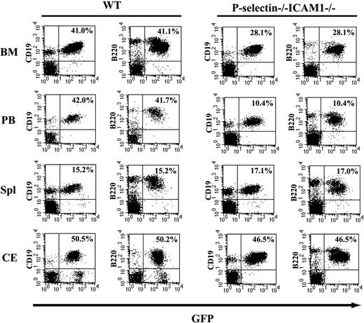
We tested whether lack of P-selectin and ICAM1 also has an effect on survival of the mice with BCR/ABL-induced B-ALL in our retroviral transduction/transplantation system. P-selectin/ICAM1-deficient mice were used as both donors and recipients of BCR/ABL-transduced bone marrow cells, and in the control group wild-type mice were used as both donors and recipients. Survival of P-selectin/ICAM1-deficient mice with B-ALL was similar to that of wild-type B-ALL mice, with 50% of the P-selectin/ICAM1-deficient mice dying of fatal leukemia by 34 days after transplantation, versus 36 days for 50% of wild-type mice (P = .673) (Figure 6A). There was no difference in spleen weight between the 2 groups (P = .474). FACS analysis showed that both P-selectin/ICAM1 and wild-type mice developed B-ALL, as characterized by the infiltration and accumulation of B220+/GFP+ or CD19+/GFP+ B cells in bone marrow, spleen, peripheral blood, and pleural cavity (Figure 7). Interestingly, the mean percentage of B220+/GFP+ and CD19+/GFP+ B cells in peripheral blood was significantly lower in P-selectin/ICAM1-deficient B-ALL mice (10.4 ± 4.0 and 10.4 ± 4.2) than in wild-type B-ALL mice (41.7 ± 6.6 and 42.0 ± 6.5) (P = .002) (Figure 7). This observation suggests that the lack of P-selectin and ICAM1 affected the homing of the leukemic B cells, but that this effect did not alter the survival of the mice (Figure 6A). Because the development of pleural effusion containing leukemic B cells is the major cause of death for B-ALL mice, we compared the percentage of B220+/GFP+ cells in pleural effusions of P-selectin/ICAM1-deficient and wild-type mice with B-ALL and found that there was no significant difference between the 2 groups (P = .260) (Figure 7). This is in agreement with the observation that there was no significant difference in survival (Figure 6A) and spleen weight (Figure 6B) between P-selectin/ICAM1-deficient and wild-type mice with B-ALL. Taken together, these results indicate that the lack of P-selectin and ICAM1 does not affect survival of the mice with BCR/ABL-induced B-ALL.
Lack of P-selectin and ICAM1 does not accelerate the development of BCR/ABL-induced B-ALL. (A) Kaplan-Meier survival curve for recipients of marrow transduced by BCR/ABL. Recipient mice received a cell dose of 1 × 10⁶ cells/mouse. There was no significant difference in survival between wild-type (WT) recipients of transduced WT marrow and P-selectin/ICAM1-deficient recipients of transduced P-selectin/ICAM1-deficient marrow (P = .673). (B) Mean spleen weight (mean values ± SD) of leukemic mice (measured at the time of morbidity or death). There was no significant difference in spleen weight between the 2 transplant groups (P = .474).
Lack of P-selectin and ICAM1 does not accelerate the development of BCR/ABL-induced B-ALL. (A) Kaplan-Meier survival curve for recipients of marrow transduced by BCR/ABL. Recipient mice received a cell dose of 1 × 10⁶ cells/mouse. There was no significant difference in survival between wild-type (WT) recipients of transduced WT marrow and P-selectin/ICAM1-deficient recipients of transduced P-selectin/ICAM1-deficient marrow (P = .673). (B) Mean spleen weight (mean values ± SD) of leukemic mice (measured at the time of morbidity or death). There was no significant difference in spleen weight between the 2 transplant groups (P = .474).
FACS analysis of BCR/ABL-expressing leukemic cells from bone marrow, peripheral blood, spleen, and pleural effusion of mice with B-ALL. Mice in both WT to WT and P-selectin-/-/ICAM1-/- to P-selectin-/-/ICAM1-/- groups showed similar percentages of GFP+/B220+ and GFP+/CD19+ myeloid cells in the spleen (Spl; P = .757 and .752), bone marrow (BM; P = .407 and .412), and pleural effusion (CE; P = .260 and .530), but significantly different percentages of GFP+/B220+ and GFP+/CD19+ myeloid cells in peripheral blood (PB; P = .002 and .006).
FACS analysis of BCR/ABL-expressing leukemic cells from bone marrow, peripheral blood, spleen, and pleural effusion of mice with B-ALL. Mice in both WT to WT and P-selectin-/-/ICAM1-/- to P-selectin-/-/ICAM1-/- groups showed similar percentages of GFP+/B220+ and GFP+/CD19+ myeloid cells in the spleen (Spl; P = .757 and .752), bone marrow (BM; P = .407 and .412), and pleural effusion (CE; P = .260 and .530), but significantly different percentages of GFP+/B220+ and GFP+/CD19+ myeloid cells in peripheral blood (PB; P = .002 and .006).
Discussion
Normal hematopoietic development depends both on the specific interactions of primitive progenitors with cellular and extracellular components of the bone marrow microenvironment and on the trafficking of more developed progenitors to specific sites of differentiation.26 Bone marrow stroma plays critical regulatory and inhibitory role in hematopoietic cell differentiation, proliferation, and migration. Human CML is characterized by abnormal adhesive interactions of hematopoietic progenitor cells with the bone marrow stroma.12 Defective adhesion to stroma could be responsible for the premature release of hematopoietic progenitors from the bone marrow into peripheral circulation, thereby affecting the disease process. Primitive CML progenitors undergo unregulated proliferation even in the presence of normally adherent marrow cells, suggesting that the altered adhesive properties can be attributed to hematopoietic progenitors and not to stromal cells in the marrow.11 Alteration of adhesive interactions between CML hematopoietic progenitors and bone marrow stroma would be expected to affect the disease process. P-selectin and ICAM1 both are critical to the adhesion and migration of neutrophils and likely play important regulatory roles in hematopoietic development.14-16,19 In this study, we show that lack of the adhesion molecules P-selectin and ICAM1 accelerates the development of CML induced by the BCR/ABL oncogene in mice.
Cell culture studies have shown that BCR/ABL-expressing hematopoietic cells exhibit altered adhesion properties, but they disagree on whether these changes in adhesion properties promote or inhibit the proliferation of leukemic cells. On the one hand, BCR/ABL enhances hematopoietic cell adhesion to fibronectin and vascular cell adhesion molecule-1 (VCAM1),27 and adhesion of cells to fibronectin stimulates proliferation of wild-type and BCR/ABL-transfected murine hematopoietic cells.10 These studies suggest that BCR/ABL-transfected cells experience enhanced adhesion to fibronectin and receive growth signals from bone marrow stroma that promote cell proliferation. On the other hand, some studies have shown that direct adhesion of bone marrow progenitor cells to fibronectin can inhibit hematopoietic progenitor proliferation.2,11 Thus, adhesion of BCR/ABL-expressing cells to bone marrow stroma could lead to inhibition of cell proliferation. However, our studies show that lack of the adhesion molecules P-selectin and ICAM1 does not appear to have an effect on proliferation of BCR/ABL-expressing cells. Instead, the lack of these adhesion molecules causes early release of myeloid leukemic progenitors from bone marrow, followed by migration of leukemic cells to the lungs. As a result, leukemic cells cause lung hemorrhages and destruction of lung structures. Thus, our data suggest that the accelerated disease is due not to increased cell proliferation, but to induction of abnormalities in cell adhesion that alter migration and homing of the leukemic cells. We believe that the early-released cells did not accumulate in peripheral blood to increase the white blood cell (WBC) counts in P-selectin-/-/ICAM1-/- mice with CML because they quickly homed to the lungs. This explanation is supported by the fact that the myeloid leukemic cells were found in the lungs 12 days after the bone marrow transplantation. We observed that WBC counts of the diseased mice started to increase significantly at day 12 after the bone marrow transplantation (data not shown).
Although granulocytes from patients with CML exhibit reduced and altered binding to P-selectin,14 there is still no evidence that BCR/ABL can affect the function of ICAM1 that interacts with β2 integrins. We decided to test the role of ICAM1 in CML development because it is expressed on CD34+ bone marrow progenitor cells and plays an important role in mediating the adhesion of progenitors to marrow stroma.20 It is possible that a functional defect of ICAM1 affects pathogenesis of CML. We showed that the lack of ICAM1 alone significantly accelerated the development of CML. We will further study the role of ICAM1 and β2 integrins in CML in the future. Although there is still no evidence for alteration of β2 integrin-mediated cell adhesion in CML, β1 integrins have been shown to be involved in CML pathogenesis. A study by Schofield et al indicates that there is a functional interaction between the β1 integrin very late activation antigen-4 (VLA4) and the IL-3 receptor in regulating cell adhesion and migration.28 Furthermore, it has been reported that CML cells exhibit decreased adhesion to stroma and fibronectin and increased adhesion to several basement membrane components, which is likely to be responsible for the premature release of hematopoietic progenitor cells from the bone marrow into peripheral blood.26 Lack of P-selectin or ICAM1 in either bone marrow progenitors or marrow stroma could contribute to the acceleration of CML development in the mice. We have examined the contribution of donors or recipients that lack P-selectin or ICAM1 in the acceleration of the disease. When the mutant donors and wild-type recipients were used to induce CML in our bone marrow transplantation model system, the development of the disease was significantly accelerated (data not shown), which is similar to the disease acceleration when the mutant mice were used as both donors and recipients. When wild-type donors and the mutant recipients were used to induce CML, the disease acceleration was largely reduced (data not shown). Thus, the absence of ICAM1 is likely to be more important in the progenitor cells than in the bone marrow stroma. In theory, bone marrow environment (including stroma and extracellular matrix) could interact with BCR-ABL-expressing myeloid progenitor cells, and the adhesion molecules P-selectin and ICAM1 may be involved in this process. Our data clearly showed that the lack of P-selectin and ICAM1 caused early release of BCR-ABL-expressing myeloid progenitors from bone marrow and the accumulation of these cells to the lungs of the diseased mice. Our study does not exclude a possibility that other adhesion molecules in marrow environment also may be involved in regulation of adhesion between BCR/ABL-expressing myeloid progenitors and marrow stroma.
The role of P-selectin in adhesion of bone marrow progenitor cells to stroma has been studied indirectly using blocking antibodies. P-selectin glycoprotein ligand-1 (PSGL-1) has been shown to be the sole P-selectin ligand on human hematopoietic progenitor cells.29,30 Blockade of interaction between bone marrow progenitor cells and marrow stroma with anti-PSGL-1 antibody causes about 35% reduction in the homing of the progenitor cells to bone marrow.31 However, PSGL-1 is also the ligand for E-selectin,31 therefore, this homing reduction could be due to the blockade of interaction between PSGL-1 and E-selectin. Studies using mice with P- and E-selectin double-null mutation and mice with E-selectin single-null mutation have suggested that P-selectin may affect marrow progenitor cell homing cooperatively with E-selectin.30,32 Although leukocyte rolling and recruitment to inflammation site are largely reduced in P-selectin-/- mice,16 it has not been shown whether lack of P-selectin alone affects the progenitor cell homing to bone marrow. Our data show that P-selectin-deficient bone marrow cells can home to bone marrow, suggesting that the early release of BCR/ABL-expressing marrow progenitor cells from bone marrow in CML mice are due to the adhesion defects caused by BCR/ABL expression and that these defects are related to P-selectin pathway. It is likely that BCR/ABL could affect multiple adhesion molecules that are functionally linked to P-selectin.
We show that the lower number of B-leukemic cells in the peripheral blood of P-selectin/ICAM-1-deficient mice with B-ALL accompanied with the lower number of B-leukemic cells in the bone marrow. This suggests that the defect in P-selectin/ICAM-1 also reduced adhesion of B-leukemic cells to marrow stroma and the early-released B-leukemic cells did not accumulate in the peripheral blood. We tried to find the homing places for the leukemic cells and found that these cells did not accumulate in the pleural effusion of the mice either, because the similar percentages of B-leukemic cells in the pleural effusion were found in wild-type and P-selectin-/-/ICAM1-/- mice. This is one reason that P-selectin/ICAM-1 defect did not affect the survival of the mice, because the pleural effusion is a major cause of death for mice with B-ALL. The locations of the early-released B-leukemic cells are still unknown, and we will further study this in the future.
The BCR/ABL fusion tyrosine kinase may induce abnormalities in intercellular adhesion by altering the cytoskeletal structure of the cell. Salgi et al found that BCR/ABL colocalized with actin in punctate staining patterns; increased tyrosine phosphorylation of focal adhesion proteins such as paxillin, vinculin, p125FAK, talin, and tensin, and dramatically altered focal adhesion protein-protein interactions.33 Abnormal interactions between these focal adhesion proteins, which are involved in normal integrin signaling, could explain the altered adhesive properties of BCR/ABL-expressing cells.33 Interferon-α and tyrphostin AG957 restore normal adhesive properties to CML hematopoietic progenitors by correcting impaired β1 integrin receptor function.34,35 Tyrphostin AG957 blocks the tyrosine kinase domain of BCR/ABL, which prevents the phosphorylation of focal adhesion proteins including crkl, cbl, paxillin, and PI-3 kinase p85, providing more conclusive evidence that abnormal tyrosine phosphorylation of focal adhesion proteins alters the cytoskeleton and induces abnormal adhesive properties in hematopoietic progenitors.35 β1-integrin-mediated adhesion inhibits proliferation of hematopoietic cells, making defects in β1-integrin-mediated adhesion important to CML development.35
Investigating the effects of adhesion-restoring drugs such as interferons and tyrphostins on mutant mice lacking adhesion function could help dissect the mechanisms through which these drugs restore adhesive properties. This study demonstrates that improvement of adhesion between BCR/ABL-expressing myeloid progenitors and stroma in bone marrow may be of therapeutic value for human CML. It will be interesting to investigate whether the BCR/ABL kinase inhibitor STI571 (Gleevec) can improve adhesion of BCR/ABL-expressing myeloid cells to stroma in bone marrow.
Prepublished online as Blood First Edition Paper, June 22, 2004; DOI 10.1182/blood-2003-09-3033.
S.D.P. and D.S.H. contributed equally to this work.
Supported by the institutional funds from The Jackson Laboratory and grants from the Irving A. Hansen Foundation, The V Foundation for Cancer Research, and Linda Tallen and David Kane Cancer Education and Research Foundation.
An Inside Blood analysis of this article appears in the front of this issue.
The publication costs of this article were defrayed in part by page charge payment. Therefore, and solely to indicate this fact, this article is hereby marked “advertisement” in accordance with 18 U.S.C. section 1734.
The authors thank Leonard Shultz, David Serreze, and Stephen Sampson (The Jackson Laboratory) for the critical reading of the manuscript; Patricia Cherry for the secretarial assistance; and Theodore Duffy for assistance in flow cytometric analysis. D.S. Hong was a summer student of The Jackson Laboratory from Harvard University, Cambridge, MA. S. Li is a Scholar of The V Foundation for Cancer Research.


This feature is available to Subscribers Only
Sign In or Create an Account Close Modal