Comment on Conrotto et al, page 4321
Semaphorins are known to negatively regulate blood vessel formation, for example, by antagonizing endothelial cell integrins. Conrotto and colleagues now demonstrate that semaphorins may also be endowed with proangiogenic properties.
Intense work in recent years has shown that angiogenesis is guided by repellant cues provided by certain members of the semaphorin family of transmembrane, glycosylphosphatidyl-inositol (GPI)–anchored, and secreted proteins. Semaphorins act through plexins, transmembrane molecules thought to be equipped with intrinsic GTPase-activating protein (GAP) activity. In their exciting study published in this issue of Blood, Conrotto and colleagues now present evidence that certain semaphorins provide attracting cues to endothelial cells: exposure of endothelial cells to Sema4D, expressed in secreted or transmembrane forms, leads to endothelial cell migration and sprouting but not mitogenicity, in a manner dependent both on the Sema4D receptor Plexin B1, and the receptor tyrosine kinase Met, but independent of the natural Met ligand, scatter factor/hepatocyte growth factor. It is noteworthy that Sema4D, Plexin B1, and Met share structural motifs in their extracellular domains, including a 500-amino acid residue Sema domain and a cystein-rich stretch of 80 amino acid residues. The work by Controtto et al demonstrates a novel angiogenic pathway leading to formation of vessels apparently indistinguishable from those induced by conventional angiogenic factors such as vascular endothelial growth factor (VEGF).
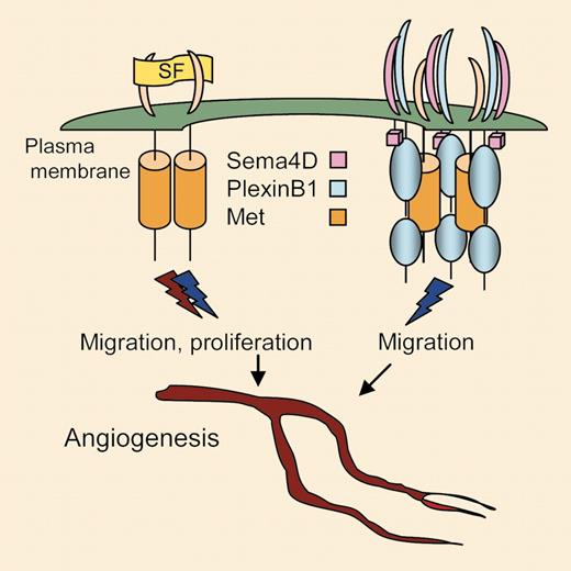
This intriguing new pathway displays several facets of specificity: First, other semaphorins such as Sema3A, Sema4B, or Sema6A do not induce endothelial cell sprouting or chicken embryo angiogenesis. Furthermore, there is no cross-talk between the Sema4D pathway and vascular endothelial growth factor VEGF-induced angiogenesis, or at least, VEGF is not up-regulated by Sema4B and blocking Plexin B1 or Met function does not affect VEGF-induced angiogenesis. Moreover, the Sema4/Plexin B1/Met-induced angiogenesis does not involve Rho/Rho kinases, which have been shown to be critical in VEGF-induced actin reorganization, migration, and angiogenesis.1 Another level of specificity is illustrated by the Met-dependent signals emitted through Sema4B-binding to plexin B1, which lead to actin reorganization and migration but not mitogenicity (see figure). This resembles the contribution of neuropilin-1 to VEGF/VEGFR-2 signaling, which leads to augmented moto- but not mitogenicity.2 Individually, both Plexin B1 (in B lymphocytes) and Met (in endothelial and epithelial cells) are capable of transducing mitogenic responses. The nature of signals transduced by the Sema4D-activated Plexin B1/Met complex leading to cell migration, as well as the extent to which both components, Plexin B1 and Met, directly contribute to the signaling, remain to be explored. The general concept of signaling by receptor tyrosine kinases such as Met is well established and known to involve dimerization of receptor molecules, allowing phosphorylation in trans of positive regulatory tyrosine residues. It is likely that complex formation with Sema4D/Plexin B1 induces formation of Met dimers, but these have different properties than those induced by the natural Met ligand, scatter factor. Possibly, Met dimers induced by Sema4D/Plexin B1 are less stable or the pattern of trans phosphorylation is different, both of consequence for signal transduction. These are exciting and challenging subjects that need to be addressed for us to understand this novel path to blood vessel formation. ▪
Binding of scatter factor (SF) to the extracellular domain of Met expressed in endothelial cells leads to receptor dimerization and signals to migration and proliferation and eventually, angiogenesis. Clustering of Sema4D-bound Plexin B1 induces Met dimerization in the absence of scatter factor and transduction of proangiogenic signals involving cell migration but not proliferation.
Binding of scatter factor (SF) to the extracellular domain of Met expressed in endothelial cells leads to receptor dimerization and signals to migration and proliferation and eventually, angiogenesis. Clustering of Sema4D-bound Plexin B1 induces Met dimerization in the absence of scatter factor and transduction of proangiogenic signals involving cell migration but not proliferation.


This feature is available to Subscribers Only
Sign In or Create an Account Close Modal