Abstract
FLT3 (fms-like tyrosine kinase 3) is constitutively activated in about 30% of patients with acute myeloid leukemia (AML) and represents a disease-specific molecular marker. Although FLT3-LM (length mutation) and TKD (tyrosine kinase domain) mutations have been considered to be mutually exclusive, 1% to 2% of patients carry both mutations. However, the functional and clinical significance of this observation is unclear. We demonstrate that FLT3-ITD-TKD dual mutants induce drug resistance toward PTK inhibitors and cytotoxic agents in in vitro model systems. As molecular mechanisms of resistance, we found that FLT3-ITD-TKD mutants cause hyperactivation of STAT5 (signal transducer and activator of transcription-5), leading to upregulation of Bcl-x(L) and RAD51 and arrest in the G2M phase of the cell cycle. Overexpression of Bcl-x(L) was identified as the critical mediator of drug resistance and recapitulates the PTK inhibitor and daunorubicin-resistant phenotype in FLT3-ITD cells. The combination of rapamycin, a selective mTOR inhibitor, and FLT3 PTK inhibitors restored the drug sensitivity in FLT3 dual mutant–expressing cells. Our data provide the molecular basis for understanding clinical FLT3 PTK inhibitor resistance and point to therapeutical strategies to overcome drug resistance in patients with AML.
Introduction
Activating mutations in the FLT3 (fms-like tyrosine kinase-3) gene play an important role in the pathogenesis of acute myeloid leukemia (AML) and can be found in approximately 25% to 35% of patients with AML. About 30% of AML patients have length mutations in the juxtamembrane domain of FLT3 (FLT3-LM), which consist of internal tandem duplications (ITD) and in some cases also of additional insertions.1-4 Another 7% of patients carry point mutations in the tyrosine kinase domain of FLT3 (FLT3-TKD).5-7 Both kinds of mutations confer interleukin-3 (IL-3)–independent growth to IL-3–dependent hematopoietic cell lines5,8-11 and induce a myeloproliferative syndrome in the mouse bone marrow transplantation model.12
Because of their essential pro-proliferative and antiapoptotic role in AML cells, FLT3 mutants represent a promising molecular target for FLT3 PTK inhibitors. Three compounds with selective FLT3 inhibitory activity are currently evaluated in phase 1/2 clinical trials in patients with AML: PKC412, MLN518, and SU11248.13-15 Initial results have shown that 30% to 50% of patients show a hematological response, but primary and secondary drug resistance represents a major problem. In contrast to the well-characterized mechanisms of drug resistance toward conventional cytotoxic agents, the molecular mechanisms of PTK inhibitor resistance in vivo are poorly understood.
We have previously shown that FLT3-ITD–transformed cells can acquire mutations in the TKD domain (FLT3-ITD-TKD dual mutants) during prolonged exposure to FLT3 PTK inhibitors. These mutations cause resistance by reducing inhibitor affinity to the receptor.16 Similar mechanisms of drug resistance have been described in imatinib-treated patients with chronic myeloid leukemia (CML) that can acquire mutations in the PTK domain of BCR-ABL.17-22 In vitro and in vivo such mutants induce molecular drug resistance by changing the structural conformation and subsequently the access of the inhibitor to the PTK domain. Interestingly, some groups have shown that such mutations in the PTK domain of BCR-ABL can be found in untreated patients with CML and will be selected during inhibitor treatment.19,23,24
Most patients with AML carry either a FLT3-length mutation (FLT3-LM) or a mutation in the tyrosine kinase domain (FLT3-TKD). However, in a subgroup of patients with AML both mutations can be detected. In a study from Thiede et al,7 17 (1.7%) AML patients of a total of 979 had both a FLT3-LM and a TKD mutation, and further analysis of 10 LM+/TKD+ patients demonstrated that 4 of them carried both mutations on the same allele. So far, the biological and clinical relevance of these findings have not been addressed, but 2 studies have shown a worse clinical outcome for patients carrying both mutations compared with patients with a single LM or TKD mutation.25,26 We hypothesized that such dual mutations confer drug resistance not only to PTK inhibitors but also to cytotoxic agents and might be responsible for the poorer clinical prognosis of these patients.
Drug resistance represents the major problem in the treatment of patients with AML with conventional chemotherapy, and several molecular mechanisms have been elucidated. Overexpression of antiapoptotic proteins of the Bcl-2 family (Bcl-2, Bcl-x(L)) causes drug resistance to 122 “standard” chemotherapy agents27 and is associated with a worse clinical outcome in AML patients.28 Another well-characterized mechanism of resistance is overexpression of the ATP-dependent efflux pumps P-gp, BCRP (breast cancer resistance protein), and multidrug resistance protein 1 that reduce intracellular concentrations of cytotoxic agents.29,30
We demonstrate here that cells expressing FLT3-ITD-TKD dual mutants show resistance not only to PTK inhibitors but also to the topoisomerase II inhibitor daunorubicin. Expression of FLT3-ITD-TKD dual mutants in hematopoietic cells induces hyperactivation of signal transducer and activator of transcription-5 (STAT5) and up-regulation of its target RAD51 and the antiapoptotic protein Bcl-x(L) as well as cell-cycle arrest at the G2M checkpoint. Our findings define important mechanisms of drug resistance to FLT3 PTK inhibitors and cytotoxic agents in FLT3 transformed cells. These results will improve therapeutic strategies for a targeted therapy of FLT3-LM-TKD–positive AML.
Materials and methods
Reagents and cell lines
Low-passage murine Ba/F3 cells were obtained from the DSMZ (Deutsche Sammlung von Mikroorganismen und Zellkulturen, Braunschweig, Germany) and were maintained in RPMI-1640 medium with 10% fetal bovine serum (FBS) and 10% WEHI-conditioned medium as a source of murine interleukin-3 (IL-3) when indicated. The PTK-inhibitor SU5614 was obtained from Calbiochem (Calbiochem-Novabiochem, Bad Soden, Germany). Daunorubicin and rapamycin were obtained from Sigma (Taufkirchen, Munich, Germany). The PTK inhibitor PKC412 was kindly provided by Novartis Pharma AG (Basel, Switzerland). Final concentrations of dimethyl sulfoxide in the growth medium did not exceed 0.1%.
Cell proliferation of Ba/F3 cells
Cells were seeded at a density of 4 × 10⁴/mL in the presence or absence of IL-3 and inhibitors or daunorubicin as indicated. Viable cells were counted at 72 hours in a standard hemacytometer after staining with trypan blue. Figures show mean values and standard deviations from 3 independent experiments unless otherwise indicated.
Cell-cycle and apoptosis analysis
Cell-cycle analysis was performed by measuring the DNA content of cell nuclei with propidium iodide (PI) as described previously.31 Assessment of apoptotic cells was carried out by annexin V/7-amino-actinomycin D (7-AAD) staining as recommended by the manufacturer (annexin V-phycoerythrin (PE) apoptosis detection kit, Becton Dickinson, Heidelberg, Germany) using a FacsCalibur flow cytometer (Becton Dickinson, San Jose, CA).
Antibodies
The following antibodies were used: anti–phospho-STAT5-Tyr694, anti-RAD51 (both from New England Biolabs, Frankfurt, Germany), anti-STAT5 (sc-835, Santa Cruz), anti–Bcl-x(L) (sc-8392, Santa Cruz), and anti–β-actin (A-5441, from Sigma).
DNA constructs and vectors
The FLT3-ITD-W51 construct contains a 7 amino acid (AA) duplicated sequence (REYEYDL) inserted between AA601/602 of human FLT3-WT; the FLT3-ITD-NPOS construct contains a 28-AA duplicated sequence (CSSDNEYFYDFREYEYDLKWEFPRENL) inserted between AA611/612 of human FLT3-WT.32 All FLT3 constructs were subcloned in the MSCV-IRES-EYFP/EGFP retroviral expression vector (kindly provided by R. K. Humphries, The Terry Fox Laboratory, Vancouver, University of British Columbia, Canada). The Bcl-x(L) construct was kindly provided by S. J. Korsmeyer (Howard Hughes Medical Institute, Dana-Farber Cancer Institute, Harvard Medical School, Boston, MA).
In vitro mutagenesis
The FLT3-D835Y/N and FLT3-V592A point mutations were introduced into either the FLT3-WT, FLT3-V592A, FLT3-ITD-W51, or the FLT3-ITD-NPOS constructs using the QuikChange Site-Directed Mutagenesis Kit (Stratagene, La Jolla, CA) according to the manufacturer's instructions. The correct sequence of all constructs was confirmed by nucleotide sequencing.
Results
FLT3 PTK inhibitor resistance is restricted to cells expressing dual FLT3-LM-TKD mutations on the same “allele”
The acquisition of distinct point mutations in the TKD domain of PTK—for example, BCR-ABL or FLT3-LM—can cause drug resistance to PTK inhibitors.16-22 To clarify the molecular mechanisms of drug resistance in FLT3-ITD-TKD dual mutant cells, we analyzed whether both mutations have to be present on the same allele to induce PTK inhibitor resistance. For this purpose we generated the following Ba/F3 cell lines: (1) FLT3-ITD-D835Y and FLT3-ITD-D835N (both mutations on the same “allele”) by retroviral transduction with an MIY FLT3-ITD-TKD dual mutant cDNA, (2) FLT3-ITD/D835N and FLT3-ITD/WT (carrying the FLT3 mutations on different “alleles”) by transduction of Ba/F3 MIG+ FLT3-ITD carrying cells (green) with MIY+ (yellow) FLT3-D835N or FLT3-WT cDNA, respectively, and sorting of double-positive (YFP+ and GFP+) cells.
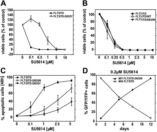
We performed proliferation assays in the presence of different concentrations of the FLT3 PTK inhibitor SU5614 and found that Ba/F3 FLT3-ITD-D835Y (Figure 1A) and FLT3-ITD-D835N (data not shown) dual mutation–expressing cells were partially resistant to the inhibitor. In accordance with these findings, a significantly higher percent of FLT3-ITD cells underwent apoptotic cell death in the presence of the inhibitor compared to FLT3-ITD-D835 dual mutant–expressing cells (Figure 1C). In contrast, our proliferation assay data clearly show that cells transformed with FLT3 constructs located on different “alleles” (FLT3-ITD/D835N and FLT3-ITD/WT) were as sensitive to the inhibitor treatment as FLT3-ITD cells (Figure 1B).
Ba/F3 FLT3-ITD-TKD dual mutants show resistance to the FLT3 PTK inhibitor SU5614. (A) Dose-response curves of the inhibitory activity of SU5614 in Ba/F3 FLT3-ITD (▪) and FLT3-ITD-D835Y cells (○) after 72 hours of incubation. Ba/F3 cells expressing different FLT3 constructs were seeded at a density of 4 × 10⁴ cells/mL in the absence or presence of different concentrations of SU5614. Viable cells were counted after 72 hours by trypan blue exclusion. The growth of cells that were incubated without inhibitor was defined as 100%. Values represent means and standard deviations from 3 independent experiments. (B) Dose-response curves of the inhibitory activity of SU5614 in Ba/F3 FLT3-ITD (▪), FLT3-ITD/WT (○), and FLT3-ITD/D835N cells (▾) after 72 hours of incubation. Values represent means and standard deviations from 3 independent experiments. (C) Ba/F3 cells transduced with the FLT3-ITD (▪), FLT3-ITD-D835N (○), or FLT3-ITD-D835Y constructs (▾) were incubated with different concentrations of SU5614 for 24 hours and were analyzed by flow cytometry after staining with annexin V–PE and 7-AAD. Values represent means and standard deviations from 3 independent experiments. (D) MIG FLT3-ITD (GFP+; ○) and MIY FLT3-ITD-D835N (YFP+)–expressing Ba/F3 cells (▪) were mixed in a ratio of 10:1, and the percentage of GFP+ and YFP+ cells was measured every 3 to 4 days for a time period of 12 days by FACS analysis in the presence of 0.2 μM of SU5614.
Ba/F3 FLT3-ITD-TKD dual mutants show resistance to the FLT3 PTK inhibitor SU5614. (A) Dose-response curves of the inhibitory activity of SU5614 in Ba/F3 FLT3-ITD (▪) and FLT3-ITD-D835Y cells (○) after 72 hours of incubation. Ba/F3 cells expressing different FLT3 constructs were seeded at a density of 4 × 10⁴ cells/mL in the absence or presence of different concentrations of SU5614. Viable cells were counted after 72 hours by trypan blue exclusion. The growth of cells that were incubated without inhibitor was defined as 100%. Values represent means and standard deviations from 3 independent experiments. (B) Dose-response curves of the inhibitory activity of SU5614 in Ba/F3 FLT3-ITD (▪), FLT3-ITD/WT (○), and FLT3-ITD/D835N cells (▾) after 72 hours of incubation. Values represent means and standard deviations from 3 independent experiments. (C) Ba/F3 cells transduced with the FLT3-ITD (▪), FLT3-ITD-D835N (○), or FLT3-ITD-D835Y constructs (▾) were incubated with different concentrations of SU5614 for 24 hours and were analyzed by flow cytometry after staining with annexin V–PE and 7-AAD. Values represent means and standard deviations from 3 independent experiments. (D) MIG FLT3-ITD (GFP+; ○) and MIY FLT3-ITD-D835N (YFP+)–expressing Ba/F3 cells (▪) were mixed in a ratio of 10:1, and the percentage of GFP+ and YFP+ cells was measured every 3 to 4 days for a time period of 12 days by FACS analysis in the presence of 0.2 μM of SU5614.
We also found that FLT3-WT cells in the presence of FLT3 ligand (20 ng/mL) demonstrated a similar sensitivity to SU5614 compared to FLT3-ITD cells (IC50 for both is 0.1 μM, data not shown).
To further analyze the mechanisms of PTK inhibitor resistance, we asked whether structurally different length mutations in the JM region can lead to the differences in the sensitivity to the FLT3 PTK inhibitor SU5614 in the background of the FLT3-D835N construct. For this purpose we generated Ba/F3 cells transformed with FLT3-V592A-D835N and FLT3-NPOS-D835N mutants. The V592A mutation can be found in the AML cell lines Mono Mac1/6 and in AML patient samples and represents the first activating point mutation in the JM region of FLT3.32,33 The FLT3-ITD-NPOS mutant contains a 28-amino acid duplicated sequence inserted between amino acids 611/612 in contrast to the FLT3-ITD-W51 mutant that contains only a 7-amino acid duplicated sequence inserted between amino acids 601/602.12 We found that all these dual mutants show partial resistance to SU5614 although to a different degree (IC50 = 0.3 μM for the V592A-D835N and 1 μM for the NPOS-D835N mutant) (data not shown). Furthermore, we could show that MIY FLT3-ITD-D835Y(N) dual mutant–expressing cells have a competitive growth advantage compared to MIG FLT3-ITD cells in the presence of 0.2 μg/mL of SU5614 (Figure 1D).
Thus, the occurrence of LM and TKD mutations on the same but not on different FLT3 “alleles” induces resistance to the selective FLT3 PTK inhibitor SU5614 that is independent from the kind of TKD and ITD mutation.
FLT3-ITD-TKD dual mutant–expressing Ba/F3 cell lines are partially resistant to the cytotoxic agent daunorubicin
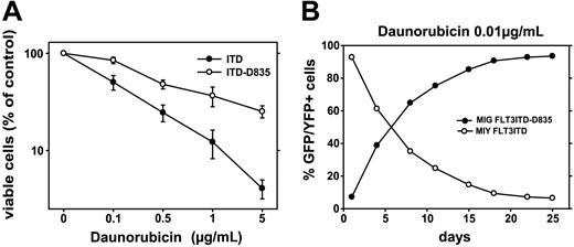
Cellular resistance to cytotoxic drugs represents the most important mechanism for treatment failure and relapse in patients with AML. Many studies have shown that the presence of FLT3-LM mutations and loss of the FLT3 wild-type allele confer a poor prognosis in AML patients.8,34-37 Few data are available on the clinical relevance of FLT3-LM+/TKD+ dual mutations, but preliminary results suggest that these mutations are associated with an even poorer prognosis when compared to patients carrying FLT3-LM alone.25,26 To further characterize the mechanisms leading to treatment failure in AML patients with FLT3 dual mutations, we analyzed the sensitivity of these mutants to the cytotoxic agent daunorubicin in vitro. Our results clearly demonstrate that Ba/F3 FLT3-ITD-D835Y/N cells show a 5-fold higher cellular resistance to daunorubicin than FLT3-ITD expressing Ba/F3 cells in proliferation assays (IC50 = 0.1 and 0.5 μg/mL, respectively) (Figure 2A). These data were confirmed when the percentage of apoptotic cells after daunorubicin incubation was determined by annexin V/7AAD staining (data not shown).
FLT3-ITD-TKD mutants confer resistance to daunorubicin in Ba/F3 cells. (A) Ba/F3 cells transduced with the FLT3-ITD (▪) or FLT3-ITD-D835Y constructs (○) were incubated with different concentrations of daunorubicin for 30 minutes, washed twice with cold PBS, and seeded at a density of 1 × 10⁵/mL. Viable cells were counted after 20 hours by trypan blue exclusion. Values represent means and standard errors from 3 independent experiments. (B) MIG FLT3-ITD (GFP+; ▪) and MIY FLT3-ITD-D835(N/Y) (YFP+)–expressing Ba/F3 cells (○) were mixed in a ratio of 10:1, and the percentage of GFP+ and YFP+ cells was measured every 3 to 4 days for a time period of 25 days by FACS analysis in the presence of 0.1 μg/mL of daunorubicin.
FLT3-ITD-TKD mutants confer resistance to daunorubicin in Ba/F3 cells. (A) Ba/F3 cells transduced with the FLT3-ITD (▪) or FLT3-ITD-D835Y constructs (○) were incubated with different concentrations of daunorubicin for 30 minutes, washed twice with cold PBS, and seeded at a density of 1 × 10⁵/mL. Viable cells were counted after 20 hours by trypan blue exclusion. Values represent means and standard errors from 3 independent experiments. (B) MIG FLT3-ITD (GFP+; ▪) and MIY FLT3-ITD-D835(N/Y) (YFP+)–expressing Ba/F3 cells (○) were mixed in a ratio of 10:1, and the percentage of GFP+ and YFP+ cells was measured every 3 to 4 days for a time period of 25 days by FACS analysis in the presence of 0.1 μg/mL of daunorubicin.
Furthermore, we analyzed whether treatment with daunorubicin might select for cells carrying FLT-ITD dual mutants. For this purpose we mixed Ba/F3 cells expressing the FLT3-ITD-D835Y/N dual construct (YFP+) with cells carrying a FLT3-ITD mutation alone (GFP+) in a ratio of 1:10 in the presence of low concentrations of daunorubicin (0.01 μg/mL). Subsequently, cells were analyzed every 3 to 4 days using flow cytometry for the ratio of GFP/YFP expression. Our results show a significant competitive growth advantage of Ba/F3 FLT3-ITD-D835Y/N cells in the presence of daunorubicin compared to FLT3-ITD cells (Figure 2B).
These data show that FLT3-ITD-TKD dual mutants cause drug resistance to daunorubicin and provide a competitive growth advantage to leukemic subclones expressing these mutants.
FLT3-ITD-TKD dual mutants induce hyperactivation of STAT5 and up-regulation of its downstream targets Bcl-x(L) and RAD51 in Ba/F3 cells
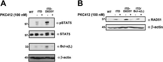
STAT5 is an important downstream target of the constitutively activated FLT3 receptor and is probably responsible for most of its transforming potential in vitro and in vivo.8,9,32 Activation of STAT5 results in altered expression of several genes regulating cell cycle, apoptosis, and proliferation.38 To investigate the mechanisms leading to resistance toward the FLT3 PTK inhibitor SU5614 and cytotoxic agents like daunorubicin by FLT3 dual mutants, we analyzed the activation of STAT5 by Western blot analysis. As shown in Figure 3A, the FLT3-ITD-TKD construct induces a significantly stronger STAT5 activation in Ba/F3 cells compared to the FLT3-ITD construct.
The expression of FLT3-ITD-TKD dual mutants induces hyperactivation of STAT5 and up-regulation of Bcl-x(L) and RAD51. (A) The phosphorylation status of STAT5 in extracts of Ba/F3 FLT3WT, FLT3-ITD, and FLT3-ITD-D835Y/N cells was determined by Western blot analysis using the polyclonal anti-pSTAT5 antibody. The membrane was stripped and reproved with polyclonal anti-STAT5 antibody. Expression of Bcl-x(L) and RAD51 in the same lysates was analyzed by immunoblotting with monoclonal anti–Bcl-x(L) (B) and polyclonal anti-RAD51 (C) antibodies. Identical protein loading in all lanes was confirmed by immunoblotting using an anti–β-actin antibody.
The expression of FLT3-ITD-TKD dual mutants induces hyperactivation of STAT5 and up-regulation of Bcl-x(L) and RAD51. (A) The phosphorylation status of STAT5 in extracts of Ba/F3 FLT3WT, FLT3-ITD, and FLT3-ITD-D835Y/N cells was determined by Western blot analysis using the polyclonal anti-pSTAT5 antibody. The membrane was stripped and reproved with polyclonal anti-STAT5 antibody. Expression of Bcl-x(L) and RAD51 in the same lysates was analyzed by immunoblotting with monoclonal anti–Bcl-x(L) (B) and polyclonal anti-RAD51 (C) antibodies. Identical protein loading in all lanes was confirmed by immunoblotting using an anti–β-actin antibody.
We hypothesized that the increased STAT5 activation and subsequent up-regulation of STAT5 targets like Bcl-x(L) and RAD51 play an essential role in the inhibitor-resistant phenotype of FLT3-LM/TKD dual mutant cells. In Western blot analyses we could clearly show elevated protein levels of Bcl-x(L) and RAD51 in dual mutants compared to FLT3-ITD–expressing cells (Figure 3B-C).
To clarify whether the expression of the FLT3 dual mutant was responsible for the up-regulation of Bcl-x(L) and RAD51, Ba/F3 FLT3-ITD and ITD-D835Y cell lines were incubated with the selective FLT3 PTK inhibitor PKC412, and the expression levels of Bcl-x(L) and RAD51 were determined by Western blot analyses. Figure 4 shows that both STAT5 targets, Bcl-x(L), and RAD51 were strongly down-regulated after FLT3 receptor inhibition.
The FLT3 PTK inhibitor PKC412 down-regulates phosphorylation of STAT5 and expression of Bcl-x(L) and RAD51 in FLT3-ITD and FLT3-ITD-TKD Ba/F3 cells. The expression and phosphorylation of STAT5 in Ba/F3 FLT3-WT, FLT3-ITD, and FLT3-ITD-D835Y–expressing cells treated with 100 nM of PKC412 or untreated cells was determined by Western blot analysis in crude lysates using polyclonal pSTAT5 and anti-STAT5 antibodies as well as monoclonal anti–Bcl-x(L) and anti–β-actin (A), and polyclonal anti-RAD51 and monoclonal anti–β-actin (B) antibodies.
The FLT3 PTK inhibitor PKC412 down-regulates phosphorylation of STAT5 and expression of Bcl-x(L) and RAD51 in FLT3-ITD and FLT3-ITD-TKD Ba/F3 cells. The expression and phosphorylation of STAT5 in Ba/F3 FLT3-WT, FLT3-ITD, and FLT3-ITD-D835Y–expressing cells treated with 100 nM of PKC412 or untreated cells was determined by Western blot analysis in crude lysates using polyclonal pSTAT5 and anti-STAT5 antibodies as well as monoclonal anti–Bcl-x(L) and anti–β-actin (A), and polyclonal anti-RAD51 and monoclonal anti–β-actin (B) antibodies.
These results clearly demonstrate that expression of FLT3-ITD-TKD dual mutants in Ba/F3 cells lead to elevated expression of Bcl-x(L) and RAD51 and point to a crucial role for these STAT5 target genes as mediators of cellular resistance toward FLT3 PTK inhibitors and cytotoxic drugs.
The coexpression of Bcl-x(L) and FLT3-ITD recapitulates the FLT3 PTK inhibitor and daunorubicin-resistant phenotype in Ba/F3 cells
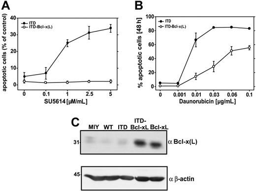
To provide further evidence that the elevated expression of Bcl-x(L) plays a central role for the drug-resistant phenotype in Ba/F3 cells, we generated Ba/F3 cell lines coexpressing FLT3-ITD-W51 and Bcl-x(L). The expression of Bcl-x(L) in these cells was confirmed in Western blot analyses (Figure 5C). We performed proliferation assays for the Ba/F3 FLT3-ITD/Bcl-x(L) cells in the presence of different concentrations of SU5614 (data not shown) and apoptosis assays using annexinV/7AAD staining. Our data clearly show that overexpression of Bcl-x(L) confers resistance of FLT3-ITD Ba/F3 cells toward the selective FLT3 PTK inhibitor SU5614 (Figure 5A). Similar results were obtained in proliferation assays (data not shown) and apoptosis assays (Figure 5B) in the presence of the different concentrations of daunorubicin.
The expression of Bcl-x(L) in FLT3-ITD mutants restores the SU5614- and daunorubicin-resistant phenotype in Ba/F3 cells. Ba/F3 cells transduced with the FLT3-ITD (▪) or FLT3-ITD and Bcl-x(L) constructs (○) were incubated with different concentrations of SU5614 (A) or daunorubicin (B) for 24 hours and were analyzed by flow cytometry after staining with annexin V-PE and 7-AAD. Values represent mean and standard deviation from 3 independent experiments. (C) Bcl-x(L) expression in cells transduced with empty vector, FLT3-WT, FLT3-ITD, FLT3-ITD and Bcl-x(L), or Bcl-x(L) was determined by Western blot analysis. Identical protein loading in all lanes was confirmed by immunoblotting using an anti–β-actin antibody.
The expression of Bcl-x(L) in FLT3-ITD mutants restores the SU5614- and daunorubicin-resistant phenotype in Ba/F3 cells. Ba/F3 cells transduced with the FLT3-ITD (▪) or FLT3-ITD and Bcl-x(L) constructs (○) were incubated with different concentrations of SU5614 (A) or daunorubicin (B) for 24 hours and were analyzed by flow cytometry after staining with annexin V-PE and 7-AAD. Values represent mean and standard deviation from 3 independent experiments. (C) Bcl-x(L) expression in cells transduced with empty vector, FLT3-WT, FLT3-ITD, FLT3-ITD and Bcl-x(L), or Bcl-x(L) was determined by Western blot analysis. Identical protein loading in all lanes was confirmed by immunoblotting using an anti–β-actin antibody.
Expression of FLT3-ITD-TKD dual mutants in Ba/F3 cells leads to G2M cell-cycle arrest after treatment with daunorubicin
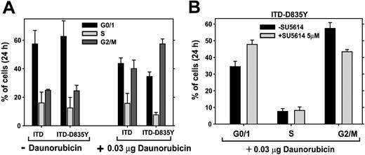
In nontransformed, drug-sensitive cell lines, cytotoxic agents induce apoptosis and accumulation of cells in the G0/G1 phase of the cell cycle. In contrast, transformed cells accumulate in the G2M phase after genotoxic stress that allows prolonged DNA repair and results in cellular resistance toward cytotoxic drugs.39,40 We hypothesized that the same mechanism might play a role in FLT3-ITD-TKD drug-resistant cells. To prove this hypothesis, Ba/F3 FLT3-WT, FLT3-ITD, and FLT3-ITD-TKD cells were exposed for 24 hours to low concentrations of daunorubicin (0.03 μg/mL), and cell-cycle analysis was performed after propidium iodide staining of cell nuclei (Figure 6A). FL-stimulated Ba/F3 FLT3-WT cells demonstrated 95% of hypodiploid cells in the presence of this concentration of daunorubicin (data not shown). Ba/F3 cells transformed with either the ITD or the dual mutant ITD-D835Y constructs accumulate in G2M after exposure to daunorubicin compared to untreated cells. Importantly, a significantly higher percentage of Ba/F3 FLT3-ITD-TKD cells were arrested in the G2M phase after drug treatment compared to FLT3-ITD cells (57.4% in FLT3-ITD-TKD vs 40% in FLT3-ITD cells) (Figure 6A). To prove that FLT3 is responsible for the G2M arrest after DNA damage, we exposed the cells to daunorubicin either in the presence or in the absence of the FLT3 PTK inhibitor SU5614 (5 μM). We found that exposure to SU5614 decreased the percentage of daunorubicin-treated cells in G2M phase from 57.4% to 43% (Figure 6B). Therefore, our results clearly show that FLT3 receptor inhibition allows Ba/F3 FLT3-ITD-D835Y cells to accumulate in G0/G1. Thus, expression of the FLT3 dual mutant in Ba/F3 cells is sufficient to induce G2M cell arrest after exposure to genotoxic stress, thereby contributing to the daunorubicin/PTK inhibitor-resistant phenotype.
Induction of DNA damage-dependent G2M delay in FLT3-ITD-TKD–transformed cells. (A) Ba/F3 FLT3-ITD- and ITD-D835Y–expressing cells (untreated and treated for 24 hours with 0.03 μg/mL of daunorubicin) were analyzed by flow cytometry after staining of DNA with propidium iodide. Filled bars indicate G0/1; light gray bars, S; and dark gray bars, G2/M. (B) Ba/F3 FLT3-ITD and ITD-D835Y cells were incubated with daunorubicin in the absence (▪) or in the presence of 5 μM of SU5614 (▦) for 24 hours, and cell cycle analysis after staining with propidium iodide was performed. Values represent means and standard deviations from 3 independent experiments.
Induction of DNA damage-dependent G2M delay in FLT3-ITD-TKD–transformed cells. (A) Ba/F3 FLT3-ITD- and ITD-D835Y–expressing cells (untreated and treated for 24 hours with 0.03 μg/mL of daunorubicin) were analyzed by flow cytometry after staining of DNA with propidium iodide. Filled bars indicate G0/1; light gray bars, S; and dark gray bars, G2/M. (B) Ba/F3 FLT3-ITD and ITD-D835Y cells were incubated with daunorubicin in the absence (▪) or in the presence of 5 μM of SU5614 (▦) for 24 hours, and cell cycle analysis after staining with propidium iodide was performed. Values represent means and standard deviations from 3 independent experiments.
Rapamycin restores the FLT3 PTK inhibitor sensitivity in FLT3-ITD-D835Y–expressing Ba/F3 cells
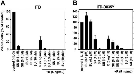
Rapamycin is an inhibitor of the serine threonine kinase mTOR (mammalian target of rapamycin), which positively regulates cell growth and proliferation.41 Recently, Mohi et al42 showed that the combination of rapamycin and protein tyrosine kinase inhibitors can restore the drug sensitivity of imatinib-resistant mutants of BCR-ABL in vitro and in vivo. To overcome the PTK and daunorubicin-resistant phenotype of FLT3 dual mutant–expressing cells, we analyzed the efficacy of such an approach in AML. For this purpose, we performed proliferation assays with either the FLT3 PTK inhibitor SU5614 or rapamycin alone or in combination. As shown in Figure 7, the combination of these compounds completely restored PTK inhibitor sensitivity in FLT3-ITD-TKD cells (Figure 7B) to levels in FLT3-ITD–expressing cells (Figure 7A).
Rapamycin restores the sensitivity of FLT3-ITD-TKD–expressing cells to the FLT3 PTK inhibitor SU5614. Ba/F3 cells expressing FLT3-ITD (A) or FLT3-ITD-TKD (B) constructs were seeded at a density of 4 × 10⁴ cells/mL in the absence or presence of different concentrations of SU5614 without or in combination with 5 ng/mL rapamycin (R). Viable cells were counted after 72 hours by trypan blue exclusion. The growth of cells that were incubated without inhibitor was defined as 100%. Values represent means and standard deviations from 3 independent experiments.
Rapamycin restores the sensitivity of FLT3-ITD-TKD–expressing cells to the FLT3 PTK inhibitor SU5614. Ba/F3 cells expressing FLT3-ITD (A) or FLT3-ITD-TKD (B) constructs were seeded at a density of 4 × 10⁴ cells/mL in the absence or presence of different concentrations of SU5614 without or in combination with 5 ng/mL rapamycin (R). Viable cells were counted after 72 hours by trypan blue exclusion. The growth of cells that were incubated without inhibitor was defined as 100%. Values represent means and standard deviations from 3 independent experiments.
These data demonstrate that the combination of rapamycin and PTK inhibitors shows promising cytotoxic activity not only in FLT-ITD–expressing cells but also in FLT-ITD-TDK dual mutants. Rapamycin might therefore be a promising drug to avoid or overcome PTK inhibitor resistance in patients with AML.
Discussion
Activating mutations in the FLT3 gene represent the most frequent genetic alterations in AML. It has been proposed that mutations in the juxtamembrane domain (FLT3-LM) and tyrosine kinase domain (FLT3-TKD) are mutually exclusive.1,2,5,6 However, 1% to 9% of patients with FLT3-LM also carry a FLT3-TKD mutation.7,43 Moreover, 2 studies have shown that patients carrying both mutations might have a poorer clinical outcome compared to patients with FLT3-LM.25,26
Our data show that expression of FLT3-ITD-TKD dual mutants in hematopoietic cells causes FLT3 PTK inhibitor and cytotoxic drug resistance. As a molecular mechanism of drug resistance, we identified hyperactivation of STAT5 and its downstream targets RAD51 and Bcl-x(L). The essential role of these targets could be demonstrated by coexpression of a FLT3-ITD mutant and Bcl-x(L) that recapitulated the drug-resistant phenotype in Ba/F3 cells. In our model rapamycin, a selective mTOR inhibitor restored drug sensitivity of FLT3 dual mutants to PTK inhibitors, which might represent a powerful compound to avoid or overcome drug resistance but that needs additional confirmation in other experimental models.
Our data clearly show that only the combination of an ITD and a TKD mutation (FLT3-ITD-D835Y/N dual mutations), but not the TKD mutation alone, caused drug resistance to the FLT3 PTK inhibitor SU5614. Furthermore, the combination of different kinds of activating mutations in the JM domain (different length and localization of ITDs and the point mutant V592A) and in the TKD region (D835Y or D835N) resulted in a similar SU5614-resistant phenotype. Therefore, we hypothesize that several different dual mutations in the FLT3 gene are able to change the protein tertiary structure in a way that reduces its affinity for the PTK inhibitor SU5614. In accordance with that is our finding that the FLT3 dual mutants remain sensitive to a structurally unrelated FLT3 PTK inhibitor, PKC412. This finding is of special importance for clinical trials evaluating FLT3 PTK inhibitors in patients with AML. Drug-resistant leukemic subclones that preexist or develop during inhibitor treatment can be efficiently targeted by switching to a structurally different FLT3 PTK inhibitor. The high frequency and rapid development of primary and secondary clinical resistance to FLT3 PTK inhibitors might also justify an upfront combination therapy with PTK inhibitors in AML.13
Although FLT3 dual mutations can be found only in a subgroup of AML patients at diagnosis, subclones of AML blasts expressing these mutations have a significant competitive growth advantage and will probably be selected in vivo during PTK inhibitor treatment. This hypothesis is supported by the clinical observation that a significant number of AML patients show an initial response to PKC412 but become resistant after a few weeks of inhibitor therapy.13
The molecular mechanisms of resistance to another PTK inhibitor, imatinib, have been studied in patients with CML in detail. These studies have shown that distinct preexisting or acquired point mutations in the PTK domain of BCR/ABL lead to alteration of inhibitor binding and therefore to drug resistance.17-22 Our data on FLT3 PTK inhibitors show a similar molecular mechanism of drug resistance. In contrast to patients with CML in chronic phase who show a high rate of cytogenetic responses to imatinib, the high proliferation rate of AML blasts favors the acquisition of new and the selection of preexisting FLT3 mutations, resulting in a PTK inhibitor–resistant phenotype.
Our data demonstrate that FLT3-ITD-TKD dual mutants confer drug resistance to the topoisomerase II inhibitor daunorubicin. In contrast to the PTK inhibitor resistance, this observation cannot be explained by structural alterations in the catalytic pocket and demonstrate that dual mutants have additional functional properties compared to either FLT3-ITD or -TKD mutants alone. Our results clearly show that expression of FLT3-ITD-TKD dual mutants in hematopoietic cells leads to strong activation of STAT5 and therefore to up-regulation of its downstream targets Bcl-x(L) and RAD51, which are known to play a role in resistance to cytotoxic agents. In addition, FLT3-ITD-TKD cells accumulate at the G2M checkpoint after drug treatment, which allows more time for DNA repair.
Presumably, the presence of 2 mutations in the FLT3 gene causes increased kinase activity. This hypothesis is supported by the finding that several FLT3 downstream targets, for example, ERK and STAT5, are strongly activated in FLT3 dual mutant–compared to single mutant–expressing cells. STAT5 regulates the expression of a variety of proteins, among them Bcl-x(L) and RAD51, by direct transcriptional activation. Bcl-x(L) belongs to the family of Bcl-2 proteins and plays an essential role in resistance to apoptotic cell death by preventing membrane hyperpolarization and mitochondrial swelling in response to death stimuli.44 Bcl-x(L) is overexpressed in a high percentage of AML patients and confers a poor prognosis in these patients.45 Our data show that overexpression of Bcl-x(L) in FLT3-ITD–transformed cells confers drug resistance to structurally different PTK inhibitors and cytotoxic drugs. Therefore, overexpression of Bcl-x(L) and Bcl-2 in AML patients will probably result in primary resistance to FLT3-PTK inhibitors, like PKC412.
RAD51 plays a central role in homologous recombinational repair (HRR) of DNA double-strand breaks (DSBs).46 Elevated transcription of RAD51 was shown to be dependent on STAT5 expression in BCR/ABL-transformed cells.47 The role of RAD51 in drug resistance appears to be specific for drugs that induce DSBs, for example, daunorubicin. We found an elevated level of RAD51 in FLT3-ITD-TKD–transformed cells and suppose that among other mechanisms it can protect cells from DNA damage. These results are in accordance with findings from Lundin et al, who observed fewer DSBs and faster HRR of DNA lesions in RAD51-overexpressing cells after etoposide treatment compared to control cells.48 In addition, it was recently demonstrated that RAD51 plays a dual role in both HRR of DSBs and telomere protection from attrition and fusion.49
Eukaryotic cells respond to DNA damage by cell-cycle arrest at the G0/G1 and G2M checkpoints. This transient delay of the cell cycle presumably allows time for DNA repair and induces resistance to cytotoxic drugs in cancer cells.39,40 Our data show that a higher percentage of cells expressing dual FLT3-LM-TKD undergo G2M arrest after treatment with daunorubicin compared to FLT3-ITD cells. The specificity of this mechanism was proven by the use of FLT3-PTK inhibitors that prevent the G2M arrest induced by daunorubicin. In addition to hyperactivation of STAT5 targets, dual FLT3 mutants undergo a G2M arrest after treatment with cytotoxic agents, which probably contributes to the drug-resistant phenotype.
Overcoming cellular drug resistance to cytotoxic agents represents a central goal of cancer therapy. Several approaches are currently evaluated in clinical trials and include Bcl-x(L) antisense strategies, modulators of MDR efflux pumps, or the combination of PTK inhibitors, like imatinib, with cytotoxic agents. Rapamycin is a selective inhibitor of the serine/threonine kinase mTOR, a downstream target of PI3/Akt that positively regulates cell growth and proliferation.50 Our data show that the sensitivity of FLT3-LM/TKD dual mutants to the PTK inhibitor SU5614 can be restored by rapamycin at concentrations that are reached in the plasma when the drug is used as an immunosuppressant after organ transplantation. This interesting finding needs further investigations in in vivo model systems and might represent a promising therapeutic strategy to avoid or overcome PTK inhibitor resistance in AML.
In summary, we have shown that FLT3-LM-TKD dual mutants found in patients with AML induce drug resistance to FLT3 PTK inhibitors and cytotoxic agents. Overexpression of the STAT5 target gene Bcl-x(L) was identified as the critical mediator of drug resistance and was found to mimic the PTK inhibitor and daunorubicin-resistant phenotype in FLT3-ITD cells. The combination of PTK inhibitors with the mTOR inhibitor rapamycin restored drug sensitivity of FLT3-LM-TKD dual mutants. Our data provide the molecular basis for understanding FLT3 PTK inhibitor resistance and show potential strategies to overcome drug resistance in patients with AML.
Prepublished online as Blood First Edition Paper, December 30, 2004; DOI 10.1182/blood-2004-06-2459.
Supported by grants from the Deutsche Forschungsgemeinschaft (DFG Sp556/3-1 and 1-3) and the Deutsche Krebshilfe (10-1997-Sp2).
The publication costs of this article were defrayed in part by page charge payment. Therefore, and solely to indicate this fact, this article is hereby marked “advertisement” in accordance with 18 U.S.C. section 1734.
We thank Susanne Schnittger, Susan King, and Stefan Bohlander for critical reading of the manuscript. We also thank Susanne Schnittger for sharing unpublished results and for critical discussions.


This feature is available to Subscribers Only
Sign In or Create an Account Close Modal