Cross-presentation is a critical process by which antigen is displayed to CD8 T cells to induce tolerance. It is believed that CD8α+ dendritic cells (DCs) are responsible for cross-presentation, suggesting that the CD8α+ DC population is capable of inducing both cross-priming and cross-tolerance to antigen. We found that cross-tolerance against intestinal soluble antigen was abrogated in C57BL/6 mice lacking mesenteric lymph nodes (MLNs) and Peyer patches (PPs), whereas mice lacking PPs alone were capable of developing CD8 T-cell tolerance. CD8α–CD11b+ DCs but not CD8α+ DCs in the MLNs present intestinal antigens to relevant CD8 T cells, while CD8α+ DCs but not CD8α–CD11b+ DCs in the spleen exclusively cross-present intravenous soluble antigen. Thus, CD8α–CD11b+ DCs in the MLNs play a critical role for induction of cross-tolerance to dietary proteins.
Introduction
Soluble and cell-associated antigens (Ags) can be presented to CD8 T cells in a process termed cross-presentation.1 It is believed that this is a major histocompatibility complex (MHC) class I–restricted procedure exclusively mediated by dendritic cells (DCs). Accumulating evidence suggests that cross-presentation provides the immune system with a mechanism by which it prevents autoimmunity and maintains self-tolerance of CD8 T cells against tissue-specific antigens.1,2 For instance, DCs are continuously sampling self-Ag from tissues and cross-presenting them to CD8 T cells in the draining lymph nodes. Under normal conditions, this leads to clonal deletion of the CD8 T cells by a process termed cross-tolerance.2
Mouse DCs can be divided phenotypically by the expression of CD8α, CD11b, CD4, and Gr-1.3 In the past, CD8α+ DCs (mainly CD11b– in spleen) have been referred to as “lymphoid DCs” and CD8α– DCs (mainly CD11b+ in spleen) as “myeloid DCs,” although this terminology is now thought to be inappropriate.4 den Haan et al5 first demonstrated that cell-associated Ag is cross-presented by CD8α+ DCs but not by CD8α– DCs in the spleen. Moreover, a study showed that this subset of DCs is responsible for the induction of cross-tolerance to tissue-associated self-Ag.2 Another study reported that intravenous-soluble Ag is also presented by CD8α+ DCs in the spleen.6 Of note, a recent study demonstrated that following epidermal infection with herpes simples virus, viral Ag is presented by CD8α+ DCs but not by Langerhans cells, implicating cross-presentation in such immunity.7 CD8α+ DCs have also been reported to be responsible for priming cytotoxic T lymphocyte (CTL) immunity against viral infection via subcutaneous, intravenous, or intranasal routes.8 Thus, the current paradigm for cross-presentation is that CD8α+ DCs are responsible for both cross-priming and cross-tolerance to a wide variety of Ags.
Under normal circumstances, the mucosal immune system does not induce a protective response against dietary proteins and mucosal self-Ag.9 In fact, foreign Ags administered by this route have been shown to induce Ag-specific systemic tolerance for CD4 T and CD8 T cells, and B-cell compartments.10-12 Although many studies have investigated the mechanisms of mucosally induced tolerance,9 the component that regulates Ag-induced tolerance remains largely unclear. In particular, the critical anatomy supporting systemic CD8 T-cell tolerance is not well described. Moreover, little is known regarding the type of DCs responsible for tolerance induction to intestinal Ag. In this study, we have characterized the site and the type of DCs responsible for cross-tolerance against intestinal Ag. Our results identify the exclusive and critical role of mesenteric lymph node (MLN)–resident CD8α–CD11b+ DCs in this process.
Materials and methods
Mice
Female C57BL6 mice (The Charles River, Seoul, Korea) were used at the age of 6 to 8 weeks. The OT-I breeding pairs were purchased from The Jackson Laboratory (Bar Harbor, ME), and offspring were checked by polymerase chain reaction (PCR) and by flow cytometry. To generate Peyer patch (PP)–deficient or PP/MLN–double deficient mice, we injected 200 μg lymphotoxin-β receptor-immunoglobulin (LTβR-Ig) or LTβR-Ig plus 200 μg tumor necrosis factor receptor 55-Ig (TNFR55-Ig) into timed pregnant mice, respectively, on day 14 and day 17 of gestation. The offspring were checked for the presence of PP or MLN as described in the Supplemental Figure S1 (see the Supplemental Figures link at the top of the online article at the Blood website). All mice were kept under specific pathogen-free conditions in the Animal Center for Pharmaceutical Research at Seoul National University.
In vivo antigen distribution study
To measure the sites of cross-presentation of ovalbumin (OVA) administered via intravenous or intragastric route, OVA-specific CD8 T cells (> 94% were Vα2+) were isolated from OT-I mice using magnetic beads. Then these cells were labeled with 10 μM CFSE (5,6-carboxyfluorescein diacetate, succinimidyl ester) and then intravenously transferred into their syngenic mice. The next day, mice were given 100 mg intragastric OVA or 5 mg intravenous OVA, respectively. Forty-eight hours later, lymphoid cells from spleen, inguinal lymph nodes (ILNs), MLNs, and PPs of the recipients were analyzed by flow cytometer after staining with anti-Vα2 antibody-phycoerythrin (Ab-PE).
Measurement of cross-tolerance
To induce OVA-specific CD8 T-cell tolerance, mice were given 25 mg intragastric OVA or phosphate-buffered saline (PBS). In some experiments, mice were given 5 mg intravenous OVA. Seven days later, these mice were primed with OVA-loaded syngenic splenocytes. For measuring CTL activity, splenocytes were prepared 7 days after priming and restimulated with OVA peptide-coated EL-4 cells or the OVA-transfectant (E.G7) after mitomycin C (MMC) treatment. Six days later, cells were harvested, and cytolytic activity against E.G7 target cells was measured using a standard Cr-51 release assay. Lysis of EL4 targets was less than 10% in all experiments. Lytic units were calculated by determining the minimum number of effectors required to generate 20% OVA-specific lysis and then dividing this into the total number of effectors generated per responder spleen.
In some experiments, splenocytes were used as responders in enzyme-linked immunospot (ELISPOT) and intracellular cytokine staining assay. Analysis of interferon γ (IFN-γ) production in response to stimulation with 1 μM OVA peptide for 16 hours was performed on MultiScreen-IP high protein–binding 96-well plates (Millipore, Bedford, MA). For intracellular IFN-γ staining, cells were stimulated with 1 μM OVA peptide for 4 hours in the presence of 1 μg/mL GolgiPlug (BD Biosciences, San Jose, CA). Cells were fixed, permeabilized, and stained with antibodies to mouse IFN-γ–PE and CD8–fluorescein isothiocyanate (FITC).
DC isolation from spleen and MLNs
Cells in MLNs or spleens were released by treatment with collagenase D (1 mg/mL; Roche, Indianapolis, IN) and DNase I (50 μg/mL; Sigma, St Louis, MO) for 30 minutes at 37°C, then EDTA (ethylenediaminetetraacetic acid) was added and incubated for 5 minutes. After washing, the cell suspension was loaded on 16% Nycodenz (Sigma) gradients and centrifuged at 170g for 15 minutes. The low-density cells (> 70% CD11c+) were recovered and further sorted using anti-CD11c microbeads following the manufacturer's instructions. CD8α+ DCs were positively selected in the low-density cell (LDC) population by CD8a– microbeads after depletion of CD3+ cells (> 92% are CD11c+). The negatively eluted population was positively sorted with CD11c microbeads and used as CD8α– DCs (< 3% are CD8α+). CD11b+ DCs were isolated from low-density cells by CD11b microbeads (> 89% are CD11c+), and the negatively eluted population was positively sorted with CD11c microbeads and used as CD11b– DCs (< 5% are CD11b+). We also sorted DCs using fluorescence-activated cell sorting (FACS) Vantage after incubation of LDCs with PE-conjugated CD11c Ab and FITC-conjugated CD8α Ab. Gr-1+B220+ cells in low-density cells were sorted and used as plasmacytoid DCs.
Analysis of in vitro division of OVA-specific CD8 T cells by DCs
To obtain OVA-specific CD8 T cells, lymphoid cells from OT-1 mice, previously depleted of CD11c+ cells to prevent the contamination of CD8α+ DCs, were sorted by CD8 microbead and labeled with 10 μM CFSE. These OT-I T cells (2 × 10⁵/well) were cocultured with the sorted DCs (3 × 10⁴/well). Three days later, the cells were labeled with PE-conjugated anti-Vα2 Ab and analyzed by flow cytometer.
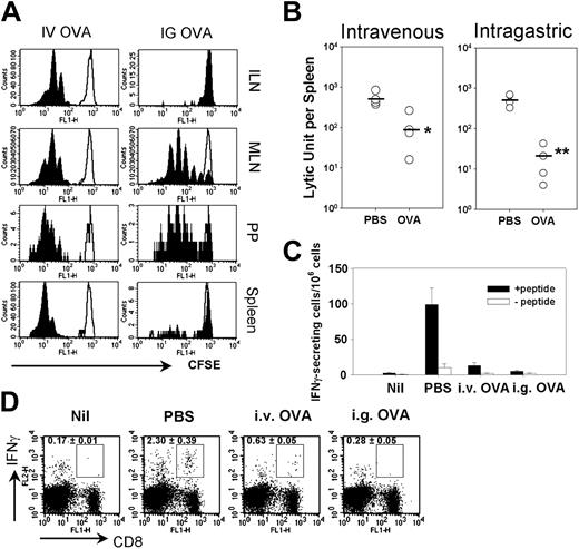
The sites of cross-presentation and induction of cross-tolerance by intravenous and intragastric administration of soluble OVA. (A) CFSE-labeled OT-I T cells were transferred into their syngenic C57BL/6 mice, which then received OVA via intravenous (IV; 5 mg) or intragastric (IG; 25 mg) routes, respectively (filled curve), or PBS as a control (open curve). Forty-eight hours later, lymphoid cells from the indicated secondary lymphoid organs were obtained and analyzed after gating with Vα2+ cells. (B-D) Groups of C57BL/6 mice were given 5 mg intravenous OVA or 25 mg intragastric OVA or PBS. Seven days later, the mice were primed with OVA-loaded splenocytes. (B) Cr-51 release assay was performed at 7 days after priming. The lytic unit was calculated as described in “Materials and methods,” and the mean value is expressed as a bar. (C) ELISPOT assay was performed to assess IFN-γ secretion by antigen-specific cells in response to 1 μM OVA peptide 7 days after priming. Values represent the mean ± SE. (D) Intracellular IFN-γ accumulation was assessed 7 days after priming as described in “Materials and methods.” i.v. indicates intravenous; i.g., intragastric. Means ± SEs from 3 mice per group are shown.
The sites of cross-presentation and induction of cross-tolerance by intravenous and intragastric administration of soluble OVA. (A) CFSE-labeled OT-I T cells were transferred into their syngenic C57BL/6 mice, which then received OVA via intravenous (IV; 5 mg) or intragastric (IG; 25 mg) routes, respectively (filled curve), or PBS as a control (open curve). Forty-eight hours later, lymphoid cells from the indicated secondary lymphoid organs were obtained and analyzed after gating with Vα2+ cells. (B-D) Groups of C57BL/6 mice were given 5 mg intravenous OVA or 25 mg intragastric OVA or PBS. Seven days later, the mice were primed with OVA-loaded splenocytes. (B) Cr-51 release assay was performed at 7 days after priming. The lytic unit was calculated as described in “Materials and methods,” and the mean value is expressed as a bar. (C) ELISPOT assay was performed to assess IFN-γ secretion by antigen-specific cells in response to 1 μM OVA peptide 7 days after priming. Values represent the mean ± SE. (D) Intracellular IFN-γ accumulation was assessed 7 days after priming as described in “Materials and methods.” i.v. indicates intravenous; i.g., intragastric. Means ± SEs from 3 mice per group are shown.
Statistical analysis
Results are expressed as the means ± SEs. Statistical analyses were performed using the Student t test. Each experiment was repeated at least twice.
Results
Sites of cross-presentation and induction of CD8 T-cell tolerance by intravenous and intragastric administration of soluble OVA
To clarify which tissue is involved in the cross-presentation of OVA administered either by the intravenous or intragastric route, CFSE-labeled OVA-specific CD8 T cells from the OT-I T-cell receptor transgenic line (OT-I cells) were adoptively transferred to syngenic C57BL/6 mice that were then given OVA intravenously or intragastrically. Intravenous OVA induced proliferation of OT-I cells in the ILN, MLN, PP, and spleen. Consistent with a previous study,13 intragastric OVA-induced OT-I T-cell proliferation was primarily restricted to the MLNs and PPs but not other lymphoid organs13 (Figure 1A). Since we examined proliferation within 2 days of antigen administration, it is unlikely that proliferating T cells had time to recirculate. Thus, detection of proliferation is indicative of antigen presentation in that organ. When naive B6 mice received soluble OVA via either the intravenous or intragastric route, hyporesponsiveness toward conventional CTL priming was induced (Figure 1B). Consistent with this observation, IFN-γ–producing cells after peptide stimulation were significantly decreased in the spleens of mice given intravenous or intragastric OVA compared with PBS-treated controls (P < .05, Figure 1C-D). This OVA-specific cross-tolerance was observed in oral doses ranging from 10 mg to 100 mg (not shown). Taken together, although both intravenous and intragastric OVA induced OVA-specific systemic CD8 T-cell tolerance, the sites of induction may differ somewhat (although presentation in MLN and PP clearly overlaps).
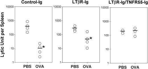
MLN-deficient mice are refractory for the induction of cross-tolerance toward intestinal OVA. We injected 200 μgLTβR-Ig or LTβR-Ig/TNFR55-Ig to timed pregnant mice to generate PP-null or PP/MLN-double deficient mice, respectively (see Supplemental Figure S1). The offspring mice were given 25 mg intragastric OVA or PBS then primed with OVA-loaded splenocytes. Seven days after priming, CTL assay was performed. The mean value of the lytic unit is expressed as a bar. *P < .05 in comparison with PBS-treated group. Data are representative of 2 experiments.
MLN-deficient mice are refractory for the induction of cross-tolerance toward intestinal OVA. We injected 200 μgLTβR-Ig or LTβR-Ig/TNFR55-Ig to timed pregnant mice to generate PP-null or PP/MLN-double deficient mice, respectively (see Supplemental Figure S1). The offspring mice were given 25 mg intragastric OVA or PBS then primed with OVA-loaded splenocytes. Seven days after priming, CTL assay was performed. The mean value of the lytic unit is expressed as a bar. *P < .05 in comparison with PBS-treated group. Data are representative of 2 experiments.
Mesenteric lymph nodes are crucial for cross-tolerance of intestinal antigens
Since proliferation of OT-I cells in response to intragastric OVA was primarily restricted to the MLNs and PPs, we questioned whether either or both of these lymphoid organs were responsible for CD8 cross-tolerance induction to intestinal antigen. To this aim, we injected LTβR-Ig or LTβR-Ig/TNFR55-Ig into timed pregnant B6 mice to generate PP-deficient and PP/MLN-double deficient mice, respectively. As described previously, the offspring of LTβR-Ig–treated mice showed complete lack of PPs and noticeably reduced MLN size compared with wild-type mice.14 No MLNs and PPs were found in the offspring of LTβR-Ig/TNFR55-Ig–treated mice (Supplemental Figure S1). To examine whether these mice could induce CD8 T-cell tolerance to intestinal Ags, both were given intragastric OVA or PBS and then primed intravenously with OVA-loaded splenocytes. In LTβR-Ig–treated mice, we observed significant impairment of cytotoxicity in mice given OVA intragastrically (P < .05), indicating that OVA-specific cross-tolerance could be established in mice lacking PPs (Figure 2). The degree of hyporesponsiveness, however, was slightly less in the PP-deficient mice compared with that of wild type (the lytic units were 63.1 ± 34.2 versus 10.7 ± 4.39, respectively, P = .17). The level of OVA-specific cytotoxicity in PP-null mice was comparable to the control Ig-treated mice, indicating that LTβR-Ig treatment did not affect CTL induction. Most interestingly, however, mice pretreated with LTβR-Ig/TNFR55-Ig lacking PPs and MLNs showed comparable levels of OVA-specific cytotoxicity whether also treated with OVA intragastric or PBS (Figure 2).
These results strongly suggest that MLNs are essential for inducing Ag-specific CD8 T-cell tolerance toward intestinal soluble Ag.
CD8α+ DCs cross-present intravenous OVA in the spleen whereas CD8α– DCs cross-present intragastric OVA in the MLN
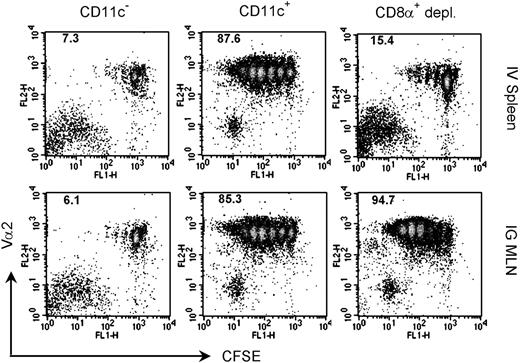
Next, we attempted to identify the cell type responsible for cross-tolerance induction after either intravenous or intragastric OVA administration. We examined antigen-presenting cells (APCs) from the spleen for intravenous OVA and the MLN for intragastric OVA because (1) these organs showed the most vigorous division of OT-I T cells after OVA administration by each route and (2) the MLNs were essential for cross-tolerance of intestinal Ag. To observe the cross-presentation of intestinal OVA more clearly, mice were given 100 mg intragastric OVA. The intragastric OVA was cross-presented in the MLNs at least for 5 days, and the proliferation of OT-I T cells was most vigorous when the T cells were transferred at 14 hours after administration (Supplemental Figure S2). Groups of mice received OVA intravenously or intragastrically, and 14 hours later low-density cells (> 70% CD11c+) were isolated from spleens or MLNs. While depletion of CD11c+ cells abrogated the division of OT-I cells, CD11c+ cells stimulated strong OT-I proliferation by both regimes (Figure 3). This indicates that CD11c+ DCs were exclusively responsible for the cross-presentation of soluble Ag administered via each route.
Consistent with others,6 we found that splenic DCs depleted of CD8α+ cells from mice given intravenous OVA stimulated minimal OT-I cell proliferation, indicating that CD8α+ DCs were primarily responsible for cross-presenting intravenous-soluble Ag (Figure 3). Interestingly, depletion of MLN CD8α+ cells from the MLN population of mice given intragastric OVA showed little reduction in their capacity to stimulate proliferation of OT-I cells (Figure 3). This observation clearly demonstrates that the CD8α– DC subset in the MLNs is involved in cross-presentation of intestinal Ag.
To explore the cross-presentation capacity of CD8α+ DCs and CD8α– DCs directly, we sorted each population using a FACS (Figure 4A). Indeed, CD8α+ DCs from spleen of intravenous OVA-treated mice presented Ag to OT-1 T cells (Figure 4B). Unexpectedly, CD8α+ DCs from the MLNs of mice given intragastric OVA induced little division of OT-I cells. Instead, CD8α– DCs from the same mice induced the proliferation of OT-I cells (Figure 4B-C). These results strongly suggest that intestinal-soluble Ag is mainly cross-presented by CD8α– DCs in the MLNs. Furthermore, when the isolated DCs were incubated with whole OVA, CD8α– DCs in the MLNs more efficiently cross-presented the Ag than CD8α+ DCs, while the reverse was true of the splenic DCs (Figure 4D). However, the failure of CD8α+ MLN DCs to cross-present was not due to a general poor stimulatory nature of these cells, since there was no apparent difference in stimulating OT-I cells when CD8α+ and CD8α– DCs were pulsed with the relevant peptide in vitro (Figure 4E).
Dendritic cells exclusively mediate the cross-presentation of intravenous OVA in the spleen and intragastric OVA in the MLNs. Groups of mice were given 5 mg intravenous OVA or 100 mg intragastric OVA. Fourteen hours later, spleens or MLNs from the respective groups were obtained, and the low-density cells were isolated after collagenase digestion. Low-density cells were further sorted with anti-CD11c microbeads with or without prior depletion with anti-CD8α microbeads. CD11c– cells were prepared from low-density cells after depletion of CD11c+ cells. Each population of cells was cocultured with CFSE-labeled OT-I T cells for 3 days and analyzed by flow cytometry. The percentages of cells that had divided are indicated. Data are representative of 5 experiments.
Dendritic cells exclusively mediate the cross-presentation of intravenous OVA in the spleen and intragastric OVA in the MLNs. Groups of mice were given 5 mg intravenous OVA or 100 mg intragastric OVA. Fourteen hours later, spleens or MLNs from the respective groups were obtained, and the low-density cells were isolated after collagenase digestion. Low-density cells were further sorted with anti-CD11c microbeads with or without prior depletion with anti-CD8α microbeads. CD11c– cells were prepared from low-density cells after depletion of CD11c+ cells. Each population of cells was cocultured with CFSE-labeled OT-I T cells for 3 days and analyzed by flow cytometry. The percentages of cells that had divided are indicated. Data are representative of 5 experiments.
CD8α– DCs but not CD8α+ DCs cross-present intestinal OVA in MLNs. Groups of mice received intravenous (5 mg) or intragastric (100 mg) OVA, and low-density cells were prepared as described in Figure 3. (A) Isolated cells were stained with CD11c and CD8α, and cells in the boxed areas were sorted and used as CD8α+ DCs and CD8α– DCs, respectively. (B-C) CD8α–CD11c+ or CD8α+CD11c+ cells were sorted by FACS and cocultured with CFSE-labeled OT-1 T cells for 3 days. Then they were analyzed by flow cytometer for their division (B) or [3H] methyl thymidine incorporation (C) after an 18-hour pulse. The percentages of cells that had divided are indicated (B). CPM indicates counts per minute (C). (D-E) CD8α–CD11c+ or CD8α+CD11c+ cells were sorted by FACS from the spleens or MLNs of naive C57BL/6 mice then pulsed with 500 μg/mL whole OVA for 6 hours (D) or 1 μM Kb-restricted OVA peptide for 1 hour (E). These cells were cocultured with OT-I T cells for 3 days and pulsed with [3H] methyl thymidine for 18 hours then analyzed for tritium incorporation. (C-E) Values represent the mean ± SE. Data are representative of at least 2 experiments.
CD8α– DCs but not CD8α+ DCs cross-present intestinal OVA in MLNs. Groups of mice received intravenous (5 mg) or intragastric (100 mg) OVA, and low-density cells were prepared as described in Figure 3. (A) Isolated cells were stained with CD11c and CD8α, and cells in the boxed areas were sorted and used as CD8α+ DCs and CD8α– DCs, respectively. (B-C) CD8α–CD11c+ or CD8α+CD11c+ cells were sorted by FACS and cocultured with CFSE-labeled OT-1 T cells for 3 days. Then they were analyzed by flow cytometer for their division (B) or [3H] methyl thymidine incorporation (C) after an 18-hour pulse. The percentages of cells that had divided are indicated (B). CPM indicates counts per minute (C). (D-E) CD8α–CD11c+ or CD8α+CD11c+ cells were sorted by FACS from the spleens or MLNs of naive C57BL/6 mice then pulsed with 500 μg/mL whole OVA for 6 hours (D) or 1 μM Kb-restricted OVA peptide for 1 hour (E). These cells were cocultured with OT-I T cells for 3 days and pulsed with [3H] methyl thymidine for 18 hours then analyzed for tritium incorporation. (C-E) Values represent the mean ± SE. Data are representative of at least 2 experiments.
CD8α–CD11b+ DCs are responsible for cross-presentation of intestinal Ag
DCs can be further divided by surface molecules other than CD8α such as CD11b.12 Previous results indicated that the CD8α–CD11b+ DCs and CD8α–CD11b– DCs reside in almost equal proportion in the MLNs.15 We isolated CD8α– DCs from MLNs after intragastric administration of OVA and further depleted CD11b+ DCs from CD8α– DCs to prepare CD8α–CD11b– DCs. While OT-I cells cocultured with CD8α– DCs showed vigorous dividing, depletion of CD11b+ cells from this population resulted in the loss of OT-I division, indicating that CD8α–CD11b+ DCs in MLNs are responsible for the cross-presentation of intestinal Ag (Figure 5A).
Gr-1+B220+ plasmacytoid DCs (pDCs) were not likely involved in the cross-presentation of intestinal Ag in MLNs for 2 reasons: We observed no division of OT-I cells after coculture with pDCs in MLNs (Figure 5B), and CD11b+ but not CD11b– DCs induced OT-I cell proliferation (pDCs are mainly CD11b–). Overall, we concluded that CD8α–CD11b+ DCs in the MLN is the main APC responsible for cross-presentation of intestinal-soluble Ag, while intravenously administered protein is cross-presented by CD8α+ DCs in the spleen.
Discussion
In the present study, we aimed to clarify the site and the type of antigen-presenting cells involved in cross-tolerance of dietary protein. Our findings demonstrate that (1) MLNs are the site where the inductive event of cross-tolerance to intestinal-soluble Ag occurs and (2) among 4 different DC subsets, CD8α–CD11b+ DCs mainly present intestinal Ag to relevant CD8 T cells. In a previous study the PP was suggested as an inductive site for CD4 T-cell tolerance,16 while another study showed that the MLN is crucial for oral tolerance.17 In terms of cross-tolerance of intestinal Ag, the present study suggests that the MLN is essential for the induction of tolerance. However, we observed that the level of tolerance induced in PP-deficient mice was slightly less compared with wild type. We speculated that this is due to the smaller size of MLNs in the PP-deficient mice, since the LTβR-Ig–treated offspring had reduced MLN size. Thus, we suggest that cross-tolerance of dietary protein is mainly mediated by the CD8α–CD11b+ DCs in MLNs. This is the first characterization of the mucosal lymphoid organ DCs that present dietary Ag to CD8 T cells.
Given that the subset of cross-presenting DCs is different in the spleen and MLNs, DCs sharing the same surface phenotype might not be identical. Consistent with this concept, DCs in liver, thymus, or PP show functionally different properties from splenic DCs.15,18,19 The composition of DC subsets in gut-associated lymphoid tissues (GALTs) is also different from that in the spleen or peripheral lymph nodes.12,15 Therefore, we suggest that the function of DC subsets should be defined with caution and with consideration of their anatomic environment. Our findings suggest that the immune system in GALTs may develop a unique mechanism to cope with intestinal Ag.
CD8α+ DCs are generally involved in the cross-presentation of various forms of Ag. It has been demonstrated that cell-associated Ag is solely cross-presented by CD8α+ lymphoid DCs in the spleen when Ag is injected systemically.5 CD8α+ DCs are also responsible for presenting viral Ag in the spleen or the draining lymph nodes after viral infection via the intravenous, intranasal, or subcutaneous route.8,20 In this case, whether presentation is due to direct infection of cross-presentation has not been resolved. In addition, intravenously injected soluble Ag is cross-presented by CD8α+ DCs in the spleen.6 In contrast, CD8α–CD11b+ DCs appear to be generally involved in CD4 T-cell responses to soluble Ag.6 It has been reported that CD11b+ DCs are also responsible for a protective CD4 T-cell immunity against cutaneous Leishmania.21 Moreover, CD11b+ DCs also present viral Ag to CD4 T cells after infection in the vaginal mucosa.22 Taken together, these results strongly suggest that different DC populations play a critical role for the development of CD4 T- and CD8 T-cell responses against Ag captured by different routes.
CD8α–CD11b+ DCs are the main cross-presenting cells of intestinal OVA. (A) A group of mice was given 100 mg intragastric OVA, and the CD11c+ cells were positively isolated after depletion with anti-CD8α and anti-CD11b microbeads. (B) Gr-1+B220+ cells were isolated from MLN low-density cells of mice treated with 100 mg intragastric OVA using FACS. Sorted cells were cocultured with CFSE-labeled OT-I T cells for 3 days and then analyzed by flow cytometry. The percentages of cells that had undergone division are indicated. Data are representative of 3 experiments.
CD8α–CD11b+ DCs are the main cross-presenting cells of intestinal OVA. (A) A group of mice was given 100 mg intragastric OVA, and the CD11c+ cells were positively isolated after depletion with anti-CD8α and anti-CD11b microbeads. (B) Gr-1+B220+ cells were isolated from MLN low-density cells of mice treated with 100 mg intragastric OVA using FACS. Sorted cells were cocultured with CFSE-labeled OT-I T cells for 3 days and then analyzed by flow cytometry. The percentages of cells that had undergone division are indicated. Data are representative of 3 experiments.
In the present study, we clearly demonstrate that CD8α–CD11b+ DCs rather than CD8α+ DCs cross-present intestinal Ag to CD8 T cells, which is in sharp contrast to the current understanding of cross-presentation against Ag delivered by other routes. Liver sinusoid endothelial cells (LSECs) cross-present intravenously administered soluble Ag and might be involved T-cell tolerance induction.23 In the present study, it is not clear whether intestinal soluble Ag is also cross-presented by LSECs or not. However, it is unlikely these cells might be responsible for the cross-tolerance of intestinal Ag since we showed that the MLN is essential for the tolerance induction.
The actual mechanism responsible for the cross-tolerance against dietary protein by CD8α–CD11b+ DCs remains to be clarified. Since CD8α+ DCs are responsible for both cross-priming and cross-tolerance in lymphoid tissues other than the GALTs,1,2,5,8 we surmise that CD8α–CD11b+ DCs in the GALTs could also be responsible for both cross-priming and cross-tolerance toward intestine-derived proteins. In this case, it is likely that cross-tolerance toward the dietary protein is a default outcome, but providing additional “danger signals” together with a dietary protein would generate cytotoxic T cells. This notion is supported by a report which describes that blocking CD40L prevents CTL induction to oral Ag.24 In addition, we observed that CD40 signals blocked the induction of CD8 T-cell tolerance to intestinal Ag.25
One can ask what role the CD8α+ and CD8α–CD11b– DCs play in the mucosal compartments. According to a recent study by Fleeton et al,26 CD8α+ and CD8a–CD11blo DCs (probably CD8α–CD11b– DCs in the present study) in PPs acquire viral Ag from reovirus-infected intestinal epithelial cells and present the Ag to CD4 T cells. That study suggests that CD8α+ or CD8α–CD11b– DCs might be involved in the CD4 T-cell response against viral infection in the intestine; however, it is not clear whether these types of DCs also cross-present the viral Ag to relevant CD8 T cells and whether the same event occurs in the MLNs. In addition, a recent study by Belz et al27 showed that CD8α–CD11b– DCs as well as CD8α+ DCs present viral Ag in the draining lymph node when mice are infected with virus in the airway. In our study, however, both types of DCs in the MLNs are not primarily involved in the cross-presentation. Therefore, we suggest that the cross-presentation by CD8α–CD11b+ DCs is a novel feature of MLNs rather than a general characteristic of mucosa-associated lymphoid tissue. Thus, within the mucus-associated lymphoid tissues, cross-priming and cross-tolerance may be mediated by different types of DCs.
The gastrointestinal tract is the site where a variety of foreign harmless Ags, such as dietary proteins and commensal bacteria, and self-Ags continuously pass through.12 Studies showed that activated CTLs are found in the intestinal mucosa of patients with inflammatory bowel diseases (IBDs) such as ulcerative colitis and Crohn disease.28,29 In addition, CD8 T cells can cause enterocolitis in response to the intestinal infection with a pathogen expressing relevant Ag.30 Thus, it is likely that cytotoxic CD8 T cells contribute to the pathogenesis of IBDs. Induction of tolerance toward these Ags might be necessary to avoid inflammatory disorders in the gastrointestinal (GI) tract. On the basis of our findings, targeting CD8α–CD11b+ DCs in the MLNs would be beneficial for inducing CD8 T-cell tolerance and for preventing intestinal diseases caused by CTLs. On the other hand, induction of mucosal tolerance is a major obstacle to the development of mucosal vaccine. Designing mucosal vaccine approaches that target CD8α–CD11b+ DCs, and cause their activation, might be effective for enhancing CD8 T-cell–mediated immunity.
Prepublished online as Blood First Edition Paper, March 17, 2005; DOI 10.1182/blood-2004-11-4240.
Supported by a Rheumatism Research Center grant by the Korean Science and Engineering Foundation (R11-2002-098-03002-0).
The online version of the article contains a data supplement.
The publication costs of this article were defrayed in part by page charge payment. Therefore, and solely to indicate this fact, this article is hereby marked “advertisement” in accordance with 18 U.S.C. section 1734.
We thank to Dr William Heath (WEHI, Australia) for critical review and discussion and the entire Kang lab for their help and discussion.


![Figure 4. CD8α– DCs but not CD8α+ DCs cross-present intestinal OVA in MLNs. Groups of mice received intravenous (5 mg) or intragastric (100 mg) OVA, and low-density cells were prepared as described in Figure 3. (A) Isolated cells were stained with CD11c and CD8α, and cells in the boxed areas were sorted and used as CD8α+ DCs and CD8α– DCs, respectively. (B-C) CD8α–CD11c+ or CD8α+CD11c+ cells were sorted by FACS and cocultured with CFSE-labeled OT-1 T cells for 3 days. Then they were analyzed by flow cytometer for their division (B) or [3H] methyl thymidine incorporation (C) after an 18-hour pulse. The percentages of cells that had divided are indicated (B). CPM indicates counts per minute (C). (D-E) CD8α–CD11c+ or CD8α+CD11c+ cells were sorted by FACS from the spleens or MLNs of naive C57BL/6 mice then pulsed with 500 μg/mL whole OVA for 6 hours (D) or 1 μM Kb-restricted OVA peptide for 1 hour (E). These cells were cocultured with OT-I T cells for 3 days and pulsed with [3H] methyl thymidine for 18 hours then analyzed for tritium incorporation. (C-E) Values represent the mean ± SE. Data are representative of at least 2 experiments.](https://ash.silverchair-cdn.com/ash/content_public/journal/blood/106/1/10.1182_blood-2004-11-4240/4/m_zh80130580520004.jpeg?Expires=1765917523&Signature=YaQbntfPxF6Az8RnGBPpzNG5~t-gr0E1LJ1xo0MOA4pzcM20oMhkxiWJyavtD6X0QN4n6PAesdRoFKiw~8KsRGOdaVYbqIq~eMORjIcADRuuOFLA1QWpqXhKG8oDjnES7QKrXfK-Of2vkhfTZaUyRy-Z1QC-wmToS4HeUQebgaiZpfF7GXh7FKkuRqH3hhOjAk4vvcPw3HOP1-O9T~b2wI5bfsNBOegcvyYREPk5mWFeb60p1q~b-KSJEFli5q0ajPnXq0MHY9NNw950YC4uLOzPaHYvR1cjPTbh2WymeGL~fZcFAAS5Qp4uBwNdO0kUwDyiW0JrcDy5eYOWhAQCIw__&Key-Pair-Id=APKAIE5G5CRDK6RD3PGA)

This feature is available to Subscribers Only
Sign In or Create an Account Close Modal