Abstract
Using a new magnetic resonance method that separately estimates the two principal forms of storage iron, ferritin and hemosiderin, in the heart, we examined the relationship between myocardial storage iron fractions and left ventricular function in thalassemia major. In patients with iron overload, the amount of iron in functional and transport pools changes only slightly. Virtually all of the excess is sequestered in storage forms of iron, as ferritin, a diffuse, soluble fraction, and as hemosiderin, an aggregate, insoluble fraction. The two storage forms of iron strongly affect signal intensity in both T2 and T2* weighted images but influence MRI signal decay through different means because of their differences in solubility and in intracellular distribution (
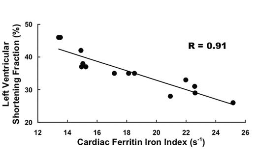
Overall, variation in the ferritin index explained more than 80% of the variation in ventricular function. For comparison, variation in the hemosiderin index, A, accounted for only about 33% of the variation in shortening fraction. Using an empirical calibration to estimate iron concentrations, variation in total (ferritin + hemosiderin) iron accounted for only about 40% of the variation in ventricular function. In patients with thalassemia major, the concentration of ferritin iron may provide a better indicator of the magnitude of the toxic iron pool than the total storage iron concentration. Magnetic resonance determinations of the partition of storage iron between ferritin and hemosiderin may be clinically valuable in evaluating tissue iron toxicity in patients with transfusional iron overload.
Author notes
Corresponding author


This feature is available to Subscribers Only
Sign In or Create an Account Close Modal