High-throughput genomic technology identified an association between a single nucleotide polymorphism (SNP), a proline (P387) rather than the predominant alanine (A387) at position 387 in thrombospondin-4 (TSP-4) and premature myocardial infarction. The inflammatory hypothesis of atherosclerosis invokes a prominent role of leukocytes and cytokines in pathogenesis. As the expression of TSP-4 by vascular cells permits its exposure to circulating leukocytes, the interactions of human neutrophils (polymorphonuclear leukocytes [PMNs]) with both TSP-4 variants were investigated. Phorbol 12-myristate 13-acetate (PMA)–stimulated PMNs adhered and migrated well and equally on the TSP-4 variants. Integrin αMβ2 was identified as the TSP-4 receptor mediating these responses, and the 3 epidermal growth factor (EGF)–like domains of TSP-4 harboring the SNPs interacted with the αMI-domain. Despite the similarity in these responses, the P387 variant induced more robust tyrosine phosphorylation of the stress-related mitogen-activated protein kinases (MAPKs): p38MAPK and c-Jun NH2-terminal kinase (JNK), as well as signal transducer and activator of transcription-1 (STAT1) and heat shock protein 27 (HSP27) than the A387 variant. Additionally, cells adherent to P387 TSP-4 variant released 4-fold more H2O2 and secreted 2-fold more interleukin 8 (IL-8) as compared with the A387. H2O2 release and p38MAPK activation were totally inhibited by blockade of αMβ2. Thus, αMβ2 plays a central role in proinflammatory activities of TSP-4 (P387) and may contribute to the prothrombotic phenotype associated with this variant.
Introduction
Recent high-throughput genotyping has sought to identify single nucleotide polymorphisms (SNP) associated with familial coronary artery disease and myocardial infarction (MI).1-3 One such study detected a highly statistical association between SNPs in 3 of the 5 members of the thrombospondin (TSP) family and MI.1 The disease-associated TSP-4 SNP, a proline at position 387 (P387) rather than the predominant alanine (A387), showed very strong association with MI (adjusted odds ratio, 1.89) and occurred at high frequency (34%) in a white population.1 This association was particularly surprising because no prior data had linked TSP-4 with MI; indeed, very little was known about the function of TSP-4 in vivo. Nevertheless, the association of TSP-4 (P387) with MI has been corroborated in several independent studies4-7 and strongly supports the importance of TSP-4 in cardiovascular pathology.
The thrombospondin family is composed of 5 extracellular matrix glycoproteins. TSP-1 and TSP-2 are the best characterized members of the family and are implicated in diverse responses, including platelet aggregation, inflammatory processes, and regulation of angiogenesis (reviewed in Adams,8 Adams and Lawler,9 and Bornstein et al10 ). TSP-4 mRNA was first identified in heart and skeletal muscles,11 and cellular expression of its mRNA and/or protein has been demonstrated in endothelial and smooth muscle cells from brain and heart.12 Of the limited information on its functions, TSP-4 has been shown to support myoblast adhesion13 and neurite outgrowth14 and to interact with certain matrix proteins.15 We have reported that the TSP-4 variants exert distinct effects on endothelial cells with the proatherogenic TSP-4 variant, (P387), failing to support endothelial cell adhesion and proliferation.12 However, not only endothelium but also leukocytes are crucial to the inflammatory response underlying atherosclerosis. In early stages of the disease, monocytes and neutrophils infiltrate the intima and generate reactive oxygen species (ROS), which provoke oxidative damage to vascular cells.16-18 Indeed, epidemiologic studies indicate that peripheral blood leukocytes, particularly neutrophils, correlate with coronary atherosclerosis and MI.19 In addition, neutrophils are activated in patients with unstable angina and MI and accumulate in eroded atherosclerotic plaques and sites of endothelial cell denudation immediately following bypass grafting.20-22 In view of these findings, we sought to characterize and compare interactions of TSP-4 and its variants with human neutrophils.
Materials and methods
Reagents
Inhibitors of kinases were purchased from Tocris (Ellisville, MO). Antiphosphotyrosine (4G10) and anti-GST (glutathione S transferase) monoclonal antibodies (mAbs) were from Upstate Biotechnology (Lake Placid, NY), PathScan TM Multiplex Western Cocktail II, all anti-p38 mitogen–activated protein kinase (MAPK) Abs were from Cell Signaling Technology (Beverly, MA). Anti-αL mAb was from Dako (Carpinteria, CA). The mAbs to αM (44a), β2 (IB4), and to major histocompatibility complex 1 (MHC-1; W6/32) were from American Type Culture Collection (Rockville, MD). mAbs against other integrins were from Chemicon International (Temecula, CA). Goat anti–human TSP-4 was from Santa Cruz Biotechnology (Santa Cruz, CA). Neutrophil inhibitory factor (NIF) was provided by Corvas International (San Diego, CA). The Cyquant-Cell Proliferation Kit and Amplex Red Hydrogen Peroxide/Peroxidase Assay Kit were from Molecular Probes (Eugene, OR). Quantikine Immunoassay Kits for human interleukin 8 (IL-8), tumor necrosis factor α (TNF-α), and macrophage inflammatory protein 1α (MIP-1α) were from R & D Systems (Minneapolis, MN).
Purification and characterization of TSP-4 variants
Full-length TSP-4 variants, TSP-4 (A387) and TSP-4 (P387), were purified from the supernatants of human embryonic kidney 293 (HEK293) cells stably expressing the proteins as described.12 TSP-4 variant fragments, which harbor residue 387 and consist of 3 type 2 (epidermal growth factor [EGF]–like) repeats (3EGF–TSP-4; amino acid residues 325-491) were expressed as secreted glutathione S transferase (GST)–fusion proteins in HEK293 cells and purified by affinity chromatography on a column of anti-GST.12,23 Purification of all TSP-4 variants was performed in endotoxinfree conditions, and endotoxin levels were undetectable (< 0.1 EU/mL using QCL-1000 Limulus Amebocyte Lysate Assay; Bio-Whittaker, Walkersville, MD). Protein preparations were monitored for purity by sodium dodecyl sulfate–polyacrylamide gel electrophoresis (SDS-PAGE) and Western blotting as described.12 Both variants were properly oligomerized and did not contain aggregates.12 In addition, protein bands detected in SDS-PAGE were analyzed by mass spectrometry and were identified as TSP-4. Representative protein preparations were also subjected to the amino acid analysis, and the relative ratios of amino acids confirmed the purity of the TSP-4 preparations.
Synthetic peptides
The TSP-4 peptides containing the SNPs and the predicted Ca2+-binding sites (TDIDECRNGACWPN and TDIDECRNGPCWPN) were synthesized in the Cleveland Clinic Biotechnology Core. Characterization of the peptides was done by amino acid analysis (W. K. Warren Medical Research, Oklahoma City, OK).
Neutrophil isolation
Granulocytes were isolated from human peripheral blood of healthy volunteers, collected under informed consent, drawn into sterile acid-citratedextrose (1/7 vol 145 mM sodium citrate, pH 4.6, and 2% dextrose) as described.24 Approval for the study was obtained from the Cleveland Clinic Foundation Institutional Review Board in accordance with federal regulations. Informed consent was provided according to the Declaration of Helsinki.
Integrin-expressing cell lines
Cell adhesion
The adhesion of resting or phorbol 12-myristate 13-acetate (PMA)–stimulated (1 nM PMA for 20 minutes at 37°C) polymorphonuclear leukocytes (PMNs) and HEK293 cells to 96-well tissue culture–nontreated plates (Falcon; Becton Dickinson, San Diego, CA) coated with full-length and 3EGF–TSP-4 variants (0-160 nM in phosphate-buffered saline) was performed as described.24,27 In inhibition experiments, cells were pretreated with selected antibodies or other reagents for 20 minutes at 22°C. In all adhesion experiments with 3EGF–TSP-4 variants, control wells were coated with GST at the same molar concentrations as the 3EGF–TSP-4, and the data are presented on subtraction of background adhesion to GST, which did not exceed 15% of cell adhesion to the entire 3EGF–TSP-4. The number of adherent cells was quantified using the Cyquant Cell Proliferation assay kit as described.24,27 Alternatively, cells were processed for H2O2 release, cytokines secretion, or Western blot analysis.
Cell migration
Cell migration was measured as described,24,27 using Costar 24-transwell plates with 3-μm pore polycarbonate filters (Corning, Corning, NY) with full-length TSP-4 (0-350 nM) immobilized on their lower surface. Cells (2 × 10⁵/well) were added to the upper chamber and incubated in a humidified incubator at 37°C and 5% CO2 for 6 hours. For inhibition experiments, the cells were pretreated with the function-blocking mAbs (20 μg/mL) for 20 minutes at 22°C.
H2O2 release
PMA-stimulated PMNs were allowed to adhere to immobilized TSP-4 or the 3EGF–TSP-4 variants (0-300 nM) for up to 4 hours at 37°C in Hanks balanced saline solution containing 1 mM Mg2+, 1 mM Ca2+, and 0.1% bovine serum albumin (BSA). H2O2 was measured in the supernatants of the adherent PMNs using the Amplex Red Hydrogen Peroxide/Peroxidase Assay Kit.
PMN signaling
Lysates of adherent PMNs were prepared,28 and equal amounts of protein were analyzed by Western blotting using selected antibodies and developed with enhanced chemiluminescence (Pierce Chemicals, Rockford, IL). Tyrosine phosphorylation of proteins in Western blots was quantified by laser scanning densitometry using Photoshop (Adobe Systems, San Jose, CA) and the National Institutes of Health (NIH) Image software (Research Services Branch, National Institutes of Health, Bethesda, MD).
Cytokine secretion
Human IL-8, TNF-α, and MIP-1α secreted by PMNs adherent to the full-length or 3EGF–TSP-4 variants (100 nM) were measured in supernatants by enzyme-linked immunosorbent assay (ELISA) using Quantikine Immunoassay Kits (R & D Systems).
Immunostaining
PMA-stimulated human PMNs were seeded onto Lab-Tek Cell Culture second-generation (CC2) Chamber Slides (Nalge Nunc International, Rochester, NY) previously coated with full-length or 3EGF–TSP-4 variants (160 nM, 2 hours, 37°C) at 5 × 10⁵ cells/well in Dulbecco minimal essential medium (DMEM) F-12 media and incubated for 45 minutes (for αM staining) or 30 minutes (for phospho-p38 MAPK staining) at 37°C, 5% CO2. Nonadherent cells were removed by washing, and adherent cells were fixed with 4% paraformaldehyde for 20 minutes at 22°C and stained with biotinylated mAb to αM subunit (clone 44a) or anti–phospho-p38 MAPK for 30 minutes at 22°C followed by incubation with Alexa 488–conjugated avidin (1:500) or Alexa 488–conjugated goat anti–rabbit immunoglobulin G (IgG) for 30 minutes at 22°C (Molecular Probes). The slides were mounted using Vectashield mounting medium (Vector Laboratories, Burlingame, CA) and observed by fluorescence microscopy. All images were analyzed using a Leica DMR microscope equipped with 5 ×/0.12 NA, 10 ×/0.4 NA, 20 ×/0.5 NA, or 40 ×/0.7 NA objective lenses with numeric apertures ranging from 0.12 to 0.7 (Leica Microsystems, Wetzlar, Germany). Images were photographed with a Qimaging Retiga ExiFas camera (Qimaging, Burnaby, BC, Canada) using Image Pro 5.1 software (Media Cybernetics, Silver Spring, MD). All images were processed with Adobe Photoshop 5.0 (Adobe Systems, San Jose, CA).
Binding of TSP-4 variants to αMI-domain
αMI-domain was expressed in Escherichia coli as GST fusion protein,25,26 and the GST tag was removed by treatment of glutathione Sepharose-bound GST-αMI-domain with factor Xa (50 U) for 4 hours at 22°C. Microtiter wells (Corning Costar, Cambridge, MA) were coated with GST-free αMI-domain at 50 μg/mL for 20 hours at 4°C and blocked with 3% BSA for 1 hour at 37°C. 3EGF–TSP-4 variants containing the GST tag (0-1.5 μM) were added to the wells in the presence of 1 mM MgCl2/1 mM CaCl2 and incubated for 4 hours at 37°C. Plates were washed with Tris (tris(hydroxymethyl)aminomethane)–buffered saline (TBS)/1 mM MgCl2/1 mM CaCl2, and bound 3EGF–TSP-4 variants were detected by an ELISA using anti-GST mAb (Upstate Biotechnology), alkaline phosphatase (AP)–conjugated secondary antibody and para-nitrophenylphosphate (Pierce Chemicals) as the substrate, and absorbance at 405 nm (A405) was measured. When the interaction of full-length TSP-4 variants with αMI-domain was examined, the bound TSP-4 was detected with goat anti–TSP-4 (Santa Cruz Biotechnology). In binding isotherm studies, input concentrations of the GST–3EGF–TSP-4 variants required for half-maximal binding to immobilized αMI-domain were estimated using the Sigma Plot software (SPSS, Chicago, IL) in which the data were fitted to the one-site binding equation.
Statistical analyses
The data are expressed as means ± SEMs. To determine the significance of differences between 2 groups, Student t tests were performed using the SPSS; P value less than .05 was considered significant.
Results
PMN adhesion and migration to the TSP-4 variants
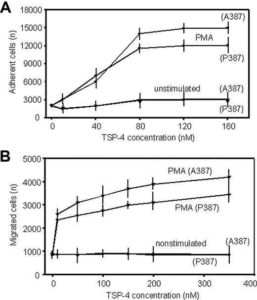
We evaluated the capacity of the TSP-4 variants to support the adhesion and migration of isolated human PMNs. Both TSP-4 variants failed to support adhesion of nonstimulated PMNs (Figure 1A). However, when the PMNs were stimulated with PMA, the cells adhered in a dose-dependent manner. The extent of adhesion of the stimulated PMNs at the various concentrations of the TSP-4 variants was not significantly different. Similar results were obtained in cell migration assays. PMA-stimulated PMNs, but not resting cells, migrated toward both TSP-4 variants; no difference was observed in the extent of PMN migration induced by the 2 TSP-4 variants (Figure 1B).
PMN adhesion and migration to the TSP-4 variants. (A) The 96-well plates were coated with increasing concentrations of TSP-4 variants for 16 hours at 4°C. Resting or PMA-stimulated (1 nM) PMNs were allowed to adhere for 30 minutes at 37°C. Adherent cells were detected using the Cyquant Cell Proliferation kit. (B) The lower filter surface of Boyden chambers was coated with increasing concentrations of TSP-4 variants. Resting or PMA-stimulated PMNs were added to the upper chamber and incubated for 6 hours at 37°C and 5% CO2. The migrated cells were counted using the DNA-based Cyquant Cell Proliferation kit. The data are means ± SEMs of triple measurements from 3 blood donors.
PMN adhesion and migration to the TSP-4 variants. (A) The 96-well plates were coated with increasing concentrations of TSP-4 variants for 16 hours at 4°C. Resting or PMA-stimulated (1 nM) PMNs were allowed to adhere for 30 minutes at 37°C. Adherent cells were detected using the Cyquant Cell Proliferation kit. (B) The lower filter surface of Boyden chambers was coated with increasing concentrations of TSP-4 variants. Resting or PMA-stimulated PMNs were added to the upper chamber and incubated for 6 hours at 37°C and 5% CO2. The migrated cells were counted using the DNA-based Cyquant Cell Proliferation kit. The data are means ± SEMs of triple measurements from 3 blood donors.
Identity of the TSP-4 receptors on stimulated PMNs
In view of the role of integrins in recognition of other TSP family members and their importance in PMN adhesion to numerous substrates, we investigated the role of these receptors in TSP-4 recognition. Function-blocking mAbs against various integrin β subunits were tested as inhibitors of adhesion of PMA-stimulated PMN adhesion to the TSP-4 variants (Figure 2A). The various mAbs tested had very similar effects on adhesion to both TSP-4 variants. mAb IB4, an anti-β2 blocking mAb, completely inhibited adhesion. Additionally, a blocking mAb to the β3 subunit (only αVβ3 is expressed by PMNs), reduced PMN adhesion to both variants by approximately 50%. Blocking mAbs to β1 and β5 subunits and a control mAb to unrelated surface antigen, MHC-1, had no effect.
Similar experiments were performed in cell migration assays (Figure 2B). When PMA-stimulated PMNs were preincubated with the mAb to the β2 integrin subunit (IB4), their migration to both TSP-4 variants was completely blocked. In contrast to the adhesive response, a blocking mAb to the β1 integrin subunit attenuated PMN migration, but the mAb to αVβ3 had no effect. mAbs to β5 integrins or the control mAb had no effect on PMN migration.
Known PMN functions dependent on the β2 integrins are mediated primarily by αMβ2 (macrophage antigen 1 [Mac-1]), αLβ2 (lymphocyte function antigen 1 [LFA-1]), and αXβ2 (p150, 95). We sought to identify which of these β2 integrins is responsible for TSP-4 recognition. Function-blocking mAbs to the αM (clone 44a) and to β2 subunit (clone IB4) completely suppressed PMN adhesion to both TSP-4 variants, whereas blocking mAbs against αL and αX failed to reduce PMN binding (Figure 2C, left) NIF, a high-affinity αMβ2-specific ligand, blocks recognition of many αMβ2 ligands, and it attenuated PMN adhesion to both TSP-4 variants in a dose-dependent manner (Figure 2C, right). Inhibition by NIF was complete at a concentration of approximately 10 nM. Complete inhibition by mAb 44a and NIF also was observed in cell migration assays (data not shown).
To verify the role of αMβ2 in recognition of TSP-4, we assessed the ability of HEK293 cell lines expressing β2 integrins to adhere to both TSP-4 variants. HEK293 cells expressing αMβ2 adhered well to both TSP-4 variants, whereas control (vector-transfected) cells or cells expressing αLβ2 exhibited negligible adhesion (Figure 2D). The interaction of most protein ligands of αMβ2 depends on a region of approximately 200 amino acids, the αMI-domain, in the αM subunit. We have previously described a chimeric receptor in which the αLI-domain in αLβ2 was replaced with the αMI-domain.26 HEK293 cells expressing this chimeric receptor acquired the capacity to adhere to the TSP-4 variants as efficiently as the αMβ2 cells (Figure 2D), indicating that the αMI-domain is a major recognition site for TSP-4. Indeed, recombinant-purified αMI-domain bound both TSP-4 variants in a dose-dependent, saturable manner (Figure 2E). Using the Sigma Plot software, half-maximal binding of TSP-4 (A387) and TSP-4 (P387) to αMI-domain occurred at input TSP-4 concentrations of 320 ± 46.4 and 102 ± 19.3 nM (means ± SEMs; n = 5), respectively. Taken together, these results indicate that TSP-4 is an adhesive substrate of αMβ2 and recognized by the αMI-domain and that the TSP-4 variants are equivalent in their support of PMN adhesion.
Identity of TSP-4 receptors on stimulated PMNs. (A) PMA-stimulated PMNs were pretreated with function-blocking mAbs to various integrin β subunits (20 μg/mL) for 20 minutes at room temperature (RT) and seeded onto polyvinylopyrrolidone (PVP)– or TSP-4–coated plates (80 nM). Alternatively, the pretreated cells were added to the upper chamber of Boyden chambers (B). Adhesion and migration assays were performed as described in Figure 1. (C) PMNs were pretreated with increasing concentrations of NIF (right) or with blocking mAbs (left) to all members of the β2 integrin family for 20 minutes at RT, and their binding to TSP-4 variants was estimated as described in Figure 1. (D) The adhesion assays of HEK293 cells expressing αMβ2, αLβ2, chimeric αLβ2-containing the I-domain of αM (αM-I-αLβ2) and control mock cells to TSP-4–coated plates (80 nM) were performed as described in Figure 1. Cell adhesion to PVP was subtracted as a background. The data are expressed as means ± SEMs of quadruplet wells from 3 blood donors. (E) Microtiter wells were coated with the αMI-domain (5 μg/well) for 20 hours at 4°C and coated afterward with 3% BSA for 1 hour at 37°C. TSP-4 variants (0-1.5 μM) were incubated for 4 hours at 37°C in the presence of magnesium and calcium. After washing, bound TSP-4 variants were detected by an ELISA using anti–TSP-4 Ab. The data are means ± SEMs (n = 5).
Identity of TSP-4 receptors on stimulated PMNs. (A) PMA-stimulated PMNs were pretreated with function-blocking mAbs to various integrin β subunits (20 μg/mL) for 20 minutes at room temperature (RT) and seeded onto polyvinylopyrrolidone (PVP)– or TSP-4–coated plates (80 nM). Alternatively, the pretreated cells were added to the upper chamber of Boyden chambers (B). Adhesion and migration assays were performed as described in Figure 1. (C) PMNs were pretreated with increasing concentrations of NIF (right) or with blocking mAbs (left) to all members of the β2 integrin family for 20 minutes at RT, and their binding to TSP-4 variants was estimated as described in Figure 1. (D) The adhesion assays of HEK293 cells expressing αMβ2, αLβ2, chimeric αLβ2-containing the I-domain of αM (αM-I-αLβ2) and control mock cells to TSP-4–coated plates (80 nM) were performed as described in Figure 1. Cell adhesion to PVP was subtracted as a background. The data are expressed as means ± SEMs of quadruplet wells from 3 blood donors. (E) Microtiter wells were coated with the αMI-domain (5 μg/well) for 20 hours at 4°C and coated afterward with 3% BSA for 1 hour at 37°C. TSP-4 variants (0-1.5 μM) were incubated for 4 hours at 37°C in the presence of magnesium and calcium. After washing, bound TSP-4 variants were detected by an ELISA using anti–TSP-4 Ab. The data are means ± SEMs (n = 5).
Differential PMN cell spreading on the TSP-4 variants
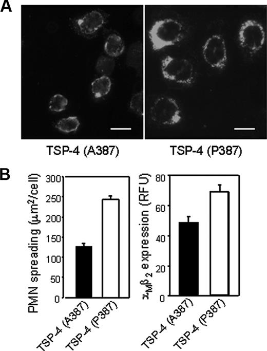
Although PMNs adhered equally to the TSP-4 variants, cells adherent to the pathogenic TSP-4 (P387) variant were significantly more spread and showed higher expression and more clusters of the αMβ2 integrin than did cells adherent to TSP-4 (A387). The PMNs adherent to TSP-4 (P387) were 2-fold larger (243 ± 8.9 μm2/cell) (Figure 3B, left) and 25% brighter (69 ± 4.7 RFU [relative fluorescent unit] staining with mAb 44a) (Figure 3B, right) as compared with those adherent to TSP-4 (A387) (127 ± 7.45 μm2/cell and 56 ± 4.3 RFU, respectively). Thus, despite the similarities in αMβ2-mediated adhesion to the TSP-4 variants, on engagement, PMNs responded differently to the 2 substrates.
TSP-4 (P387) induces proinflammatory responses in human neutrophils
The inflammatory hypothesis of atherosclerosis emphasizes the key roles of leukocyte products in pathogenesis.29 Because oxidative stress is a hallmark of inflammation, we measured the release of H2O2, as a representative ROS, from PMNs adherent to the TSP-4 variants. When stimulated with PMA, the 2 TSP-4 variants induced H2O2 release in a dose-dependent manner (Figure 4). However, the extent of H2O2 production was not the same; TSP-4 (P387) induced approximately 4-fold more H2O2 release than TSP-4 (A387). The concentrations of TSP-4 (P387) and TSP-4 (A387) giving a half-maximal response (EC50) was 4.7 ± 0.4 nM and 21.3 ± 1.8 nM, respectively, in 4 experiments performed with PMNs isolated from 4 blood donors, indicating that the P387 variant is a more potent enhancer of the oxidative burst than the A387 variant. An oxidative burst was not observed in nonstimulated PMNs in the presence of either TSP-4 variants, likely because of a lack of adhesion. The differences in oxidative burst triggered by the TSP-4 variants were observed at low PMA concentrations (1-4 nM). When higher concentrations of PMA were added, PMNs released more H2O2; the maximal oxidative burst (17 nM H2O2/105 cells) at 40 nM PMA was similar on both TSP-4 variants. Next, we examined the production of cytokines from PMNs on exposure to the TSP-4 variants. Secretion of IL-8, TNFα, and MIP-1 was measured. Of these, IL-8 secretion was differentially regulated by the 2 TSP-4 variants, a 2.5-fold increase in its secretion from PMNs adherent to the TSP-4 (P387) variant as compared with TSP-4 (A387) was observed (Table 1). Thus, the TSP-4 (P387) variant enhances proinflammatory functions of human neutrophils.
IL-8 secretion from PMNs adherent to TSP-4 variants
. | IL-8 secretion (pg/mL) . | . | |
|---|---|---|---|
| TSP-4 variant . | 4 h . | 24 h . | |
| PVP | 80 ± 14 | 150 ± 25 | |
| TSP-4 (A387) | 70 ± 10 | 200 ± 35 | |
| TSP-4 (P387) | 145 ± 12 | 500 ± 50 | |
| 3EGF–TSP-4 (A387) | 66 ± 8 | 190 ± 24 | |
| 3EGF–TSP-4 (P387) | 157 ± 12 | 485 ± 30 | |
. | IL-8 secretion (pg/mL) . | . | |
|---|---|---|---|
| TSP-4 variant . | 4 h . | 24 h . | |
| PVP | 80 ± 14 | 150 ± 25 | |
| TSP-4 (A387) | 70 ± 10 | 200 ± 35 | |
| TSP-4 (P387) | 145 ± 12 | 500 ± 50 | |
| 3EGF–TSP-4 (A387) | 66 ± 8 | 190 ± 24 | |
| 3EGF–TSP-4 (P387) | 157 ± 12 | 485 ± 30 | |
PMA-stimulated PMNs were allowed to adhere to the full-length or 3EGF–TSP variants (100 nM) for up to 24 hours. IL-8 levels were measured by ELISA in cell supernatants collected after 4 and 24 hours.
The TSP-4 (P387) variant enhances PMN spreading, αMβ2 surface expression, and clustering. (A) Microscope cover slides were coated with TSP-4 variants for 2 hours at 37°C and coated afterward with 0.5% PVP. PMNs were stimulated with 1 nM PMA and allowed to adhere to the slides for 45 minutes at 37°C. Adherent cells were fixed and stained with biotinylated mAb to αM integrin subunit, followed by incubation with fluorescein isothiocyanate (FITC)–conjugated avidin. Cells were analyzed using a fluorescence microscope (magnification, 1000 ×; × 40/0.7 NA objective). Bar size, 10 μm. (B) Quantification of cell size (area) (left) and brightness intensity (right) was performed using Image Pro-Plus software. A total of 100 cells from 4 blood donors were analyzed, and the data represent mean ± SEM.
The TSP-4 (P387) variant enhances PMN spreading, αMβ2 surface expression, and clustering. (A) Microscope cover slides were coated with TSP-4 variants for 2 hours at 37°C and coated afterward with 0.5% PVP. PMNs were stimulated with 1 nM PMA and allowed to adhere to the slides for 45 minutes at 37°C. Adherent cells were fixed and stained with biotinylated mAb to αM integrin subunit, followed by incubation with fluorescein isothiocyanate (FITC)–conjugated avidin. Cells were analyzed using a fluorescence microscope (magnification, 1000 ×; × 40/0.7 NA objective). Bar size, 10 μm. (B) Quantification of cell size (area) (left) and brightness intensity (right) was performed using Image Pro-Plus software. A total of 100 cells from 4 blood donors were analyzed, and the data represent mean ± SEM.
P387 TSP-4 enhances release of hydrogen peroxide from PMNs. PMNs were untreated or stimulated with 1 nM PMA for 20 minutes at 37°C and allowed to adhere to TSP-4–coated plates for 4 hours at 37°C. Hydrogen peroxide levels were measured in conditioned media using an Amplex Red Assay Kit. The data are means ± SEMs with neutrophils from 4 blood donors with quadruplet measurements made in each assay.
P387 TSP-4 enhances release of hydrogen peroxide from PMNs. PMNs were untreated or stimulated with 1 nM PMA for 20 minutes at 37°C and allowed to adhere to TSP-4–coated plates for 4 hours at 37°C. Hydrogen peroxide levels were measured in conditioned media using an Amplex Red Assay Kit. The data are means ± SEMs with neutrophils from 4 blood donors with quadruplet measurements made in each assay.
Tyrosine phosphorylation in PMNs adherent to the TSP-4 variants
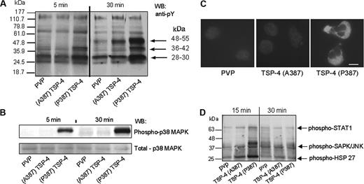
Because the differences in the functional responses of PMN to the TSP-4 variants were αMβ2 mediated, we compared a downstream signaling event, tyrosine phosphorylation, from integrin engagement. PMNs were allowed to adhere to the 2 TSP-4 variants or to PVP. Equivalent amounts of proteins from cell lysates were subjected to SDS-PAGE and Western blotting with an antiphosphotyrosine mAb (Figure 5A). Phosphorylation of the 48- to 55-kDa proteins was observed in the lysates obtained from cells adherent to the TSP-4 variants at 30 minutes but not to PVP. More importantly, phosphorylation of these proteins was much more robust in PMNs adherent to TSP-4 (P387) than to TSP-4 (A387). Additionally, only TSP-4 (P387), but not TSP-4 (A387) or control PVP, induced prominent phosphorylation of proteins at 36 to 42 kDa. TSP-4 (P387) also triggered more intense phosphorylation of the 28- to 30-kDa proteins as compared with TSP-4 (A387) and PVP. These distinctions in tyrosine phosphorylation were observed with the TSP-4 variants at early time points (5-30 minutes). By 60 minutes, substantial dephosphorylation of the proteins had occurred on all substrates (not shown).
Antibodies to specific phosphoproteins were used to identify the target proteins that were differentially affected by the TSP-4 variants. Adhesion of PMNs to either TSP-4 variant did not induce phosphorylation of Akt (protein kinase B) or p53. Among the 3 MAPK pathways, p38MAPK and stress-activated protein kinase (SAPK)/JNK were activated by TSP-4 (P387), but p44/42MAPK (extracellular signal–regulated kinase 1 and 2 [ERK 1/2]) was not phosphorylated. p38MAPK accounted for 1 of the 36- to 42-kDa phosphoproteins (Figure 5B). This kinase was activated exclusively by TSP-4 (P387) and not by TSP-4 (A387) at 5 and 30 minutes. The lower panel of the figure indicates equal loading of total p38MAPK in each sample. These data were confirmed by immunostaining of adherent neutrophils with a mAb to phosphorylated p38MAPK (Figure 5C). PMNs adherent to TSP-4 (P387) were positive, whereas cells adherent to TSP-4 (A387) or PVP were not. HSP27 and STAT1 were more extensively phosphorylated on PMN adhesion to TSP-4 (P387) (Figure 5D). These latter proteins were phosphorylated at the 15- but not at the 5-minute time point, which suggests that their activation occurs downstream of the early activation of p38MAPK.30 In addition, SAPKs and JNK were more robustly phosphorylated on 15 minutes of adhesion to TSP-4 (P387) as compared with TSP-4 (A387) (Figure 5D). Thus, the proatherogenic variant TSP-4 (P387) induces activation of signaling pathways that respond to environmental stresses and inflammatory cytokines (reviewed in Kyriakis and Avruch31 and Dong et al32 ).
TSP-4 (P387)–induced H2O2 release depends on p38MAPK and αMβ2 engagement
PMNs were pretreated with various inhibitors of key kinases: protein kinase A (KT5720), p38MAPK (SB202190), mitogen-activated protein kinase kinase 1 (MEK-1) (PD 98059), phosphatidylinositol 3 (PI3) kinase (Wortmannin, LY294002), and then activation of p38MAPK and H2O2 release were measured on adhesion to TSP-4 (P387). When PMNs were pretreated with inhibitor of MAPKK (PD98059), which is upstream of extracellular-regulated kinases 1/2, a very modest effect on p38MAPK phosphorylation and H2O2 production was observed (Figure 6A-B). In contrast, a p38MAPK inhibitor (SB202190) blocked its activation (Figure 6A) and completely suppressed H2O2 release (Figure 6B), indicating that this kinase is critical for TSP-4 (P387)–dependent H2O2 generation. PKA inhibition also abrogated these 2 events, consistent with data indicating that PKA activation occurs upstream of p38MAPK.33
Next, we sought to determine which of the 2 integrins, αMβ2 or αVβ3, implicated in TSP-4 (P387) adhesion (see Figure 2A) was critical for observed responses. Function-blocking mAbs to αM and β2 subunits completely suppressed p38MAPK phosphorylation, whereas mAbs inhibiting TSP-4 interaction with αVβ3 failed to reduce phosphorylation of p38MAPK (Figure 6C). In addition, RGD peptides, which block ligand interaction with αVβ3, did not inhibit p38MAPK activation. The lower panel of Figure 6C shows equal loading of total p38MAPK despite quantitative differences in adhesion of untreated and mAb-treated cells. In addition, function-blocking mAbs to αMβ2 and NIF significantly attenuated (by 80%-90%) H2O2 production, whereas blocking mAbs to αVβ3, β5, and β1 integrins did not (Figure 6D). These results indicate that, although PMNs adhere to TSP-4 (P387) via αMβ2 and partially αVβ3, it is integrin αMβ2 that mediates TSP-4 (P387)–dependent p38MAPK activation and H2O2 production.
The αMβ2 recognition site resides within 3 type 2 (EGF-like) repeats of TSP-4
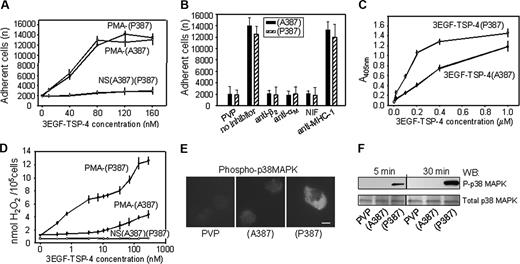
Thrombospondins consist of multiple domains, which fold independently, and TSP fragments have been used extensively to identify various TSP receptors.8-10,15 Thus, we tested the differential functions of TSP-4 variant fragments approximately equal to 50 kDa (residues 325-491), which harbor the 387 SNP and consist of 3 type 2 (EGF-like) repeats23 for recognition by αMβ2. Both 3EGF–TSP-4 variant fragments, like the full-length TSP-4 variants, supported robust and equal adhesion of stimulated PMNs (Figure 7A), which was completely suppressed by function-blocking mAbs to αM (clone 44a), to β2 (IB4) integrin subunit, and by NIF (Figure 7B). These results indicate that αMβ2 recognizes the 3 EGF-like domains of TSP-4. To corroborate these data, we analyzed the direct binding of both 3EGF–TSP-4 variants to purified αMI-domain. As shown in Figure 7C, both TSP-4 variant fragments bound saturably to the αMI-domain, and half-maximal binding of 3EGF–TSP-4 (A387) and 3EGF–TSP-4 (P387) occurred at 300 ± 40 and 130 ± 16 nM concentrations, respectively (means ± SEMs, n = 4). This difference in apparent affinity was statistically significant (Student t test, P < .01). In addition, the 3EGF–TSP-4 (P387) variant induced more robust release of H2O2 (EC50 value, ≈ 4.1 ± 0.4 nM, similar to full-length protein; Figure 7D), secretion of IL-8 (Table 1), and phosphorylation of p38MAPK (Figure 7E) than the 3EGF–TSP-4 (A387) variant. Thus, the αMβ2 binding site resides within the 3 EGF-like domains of TSP-4, and the observed distinct PMN responses to the SNP variants are also attributed to this region. However, small TSP-4 peptides containing the SNPs (TDIDECRNGACWPN and TDIDECRNGPCWPN) did not support adhesion of stimulated PMNs, did not inhibit PMN adhesion to the full-length TSP-4 variants or to the 3EGF TSP-4 fragments, and did not reduce the oxidative burst triggered by the (P387) variant (data not shown).
TSP-4 variants activate different protein tyrosine phosphorylation pathways in adherent PMNs. (A) PMNs were stimulated with PMA (1 nM) and seeded onto plates coated with TSP-4 variants (80 nM) and control PVP and incubated for up to 2 hours at 37°C. After washing, adherent cells were lysed. Equal amounts of total protein in cell lysates were analyzed by Western blot with antiphosphotyrosine mAb. (B) PMNs were treated as described in panel A. Cell lysates were analyzed by Western blot with Abs to phospho-p38 MAPK and total p38 MAPK. (C) Adhesion assay was performed as described in Figure 3A with one exception: cells were incubated for 30 minutes at 37°C. Adherent cells were stained with Ab to phosphorylated p38 MAPK, followed by incubation with FITC-conjugated goat anti–rabbit IgG Ab. Cells were analyzed as described in “Materials and methods.” Bar size, 10 μm. Images captured using a 40 ×/0.7 NA objective lens. (D) The cells were treated as described in panel A, and PMN lysates were analyzed by Western blot using PathScan Multiplex Western cocktail III containing Abs to phosphorylated forms of signal transducer and activator of transcription-1 (STAT1), c-Jun NH2-terminal kinase (JNK), S6 ribosomal protein, and heat shock protein 27 (HSP27).
TSP-4 variants activate different protein tyrosine phosphorylation pathways in adherent PMNs. (A) PMNs were stimulated with PMA (1 nM) and seeded onto plates coated with TSP-4 variants (80 nM) and control PVP and incubated for up to 2 hours at 37°C. After washing, adherent cells were lysed. Equal amounts of total protein in cell lysates were analyzed by Western blot with antiphosphotyrosine mAb. (B) PMNs were treated as described in panel A. Cell lysates were analyzed by Western blot with Abs to phospho-p38 MAPK and total p38 MAPK. (C) Adhesion assay was performed as described in Figure 3A with one exception: cells were incubated for 30 minutes at 37°C. Adherent cells were stained with Ab to phosphorylated p38 MAPK, followed by incubation with FITC-conjugated goat anti–rabbit IgG Ab. Cells were analyzed as described in “Materials and methods.” Bar size, 10 μm. Images captured using a 40 ×/0.7 NA objective lens. (D) The cells were treated as described in panel A, and PMN lysates were analyzed by Western blot using PathScan Multiplex Western cocktail III containing Abs to phosphorylated forms of signal transducer and activator of transcription-1 (STAT1), c-Jun NH2-terminal kinase (JNK), S6 ribosomal protein, and heat shock protein 27 (HSP27).
TSP-4 (P387)–mediated H2O2 release from PMNs depends on activation of p38 MAPK downstream of engagement of αMβ2. PMA-stimulated PMNs were preincubated with specific inhibitors of p38 MAPK (SB202190), ERK 1/2 (PD98059), and protein kinase A (PKA; KT5720) for 30 minutes at RT. Subsequently, the cells were seeded onto (P387) TSP-4–coated plates. Adherent cells were (A) lysed and analyzed on Western blots with Abs to phospho- and total p38 MAPK or (B) measured for H2O2 release as described. (C-D) PMA-stimulated PMNs were pretreated with the (Fab)2 fragments of function-blocking mAbs to various integrins (20 μg/mL), the αMβ2 ligand NIF (10 nM) or RGD peptide (50 μM) for 20 minutes at 37°C. The cells were allowed to adhere to (P387) TSP-4 and p38 MAPK phosphorylation (C), and H2O2 levels (D) were assayed as described. The results are means ± SEMs of quadruplet measurements from 3 blood donors.
TSP-4 (P387)–mediated H2O2 release from PMNs depends on activation of p38 MAPK downstream of engagement of αMβ2. PMA-stimulated PMNs were preincubated with specific inhibitors of p38 MAPK (SB202190), ERK 1/2 (PD98059), and protein kinase A (PKA; KT5720) for 30 minutes at RT. Subsequently, the cells were seeded onto (P387) TSP-4–coated plates. Adherent cells were (A) lysed and analyzed on Western blots with Abs to phospho- and total p38 MAPK or (B) measured for H2O2 release as described. (C-D) PMA-stimulated PMNs were pretreated with the (Fab)2 fragments of function-blocking mAbs to various integrins (20 μg/mL), the αMβ2 ligand NIF (10 nM) or RGD peptide (50 μM) for 20 minutes at 37°C. The cells were allowed to adhere to (P387) TSP-4 and p38 MAPK phosphorylation (C), and H2O2 levels (D) were assayed as described. The results are means ± SEMs of quadruplet measurements from 3 blood donors.
Integrin αMβ2 interacts with a TSP-4 fragment composed of 3 type 2 (EGF-like) domains. (A) The 96-well plates were coated with the TSP-4 variant fragments or control GST (0-160 nM) for 16 hours at 4 °C, and resting or PMA-stimulated PMNs were allowed to adhere for 30 minutes at 37°C. (B) PMA-stimulated PMNs were pretreated with function-blocking mAbs to αMβ2, control mAb to MHC-1 (20 μg/mL), or NIF (10 nM) for 20 minutes at RT, and adhesion was assessed as described in “Materials and methods.” The data are means ± SEMs of quadruplets from 2 blood donors, and background adhesion to GST has been subtracted. (C) The 96-well plates were coated with recombinant-purified αMI-domain (50 μg/mL) for 20 hours at 4 °C and coated afterward with 3% BSA. 3EGF–TSP-4–GST fusion proteins (0-1.5 μM) were added and incubated for 4 hours at 37°C. After washings, the bound proteins were detected with anti-GST mAb by ELISA as described in “Materials and methods.” The results are means ± SEMs (n = 4). (D) Resting or PMA-stimulated (1 nM) PMNs were allowed to adhere to 3EGF–TSP-4–coated plates or control GST for 4 hours at 37°C. Hydrogen peroxide levels were measured as described in Figure 4. The data are means ± SEMs of quadruplet measurements from 3 blood donors. (Panels E and F) PMA-stimulated PMNs (1 nM) were allowed to adhere to the 3EGF–TSP-4 variants (80 nM) and control PVP for up to 30 minutes at 37°C. (E) Cells adherent for 30 minutes were stained with Ab to phosphorylated p38 MAPK and FITC-conjugated secondary Ab. Bar size, 10 μM. (F) Alternatively, equal amounts of total protein in lysates of adherent cells were analyzed by Western blot with Abs to phosphor-p38 MAPK and to p38 MAPK. Images were captured with a 40 ×/0.7 NA objective lens. Results are representative of experiments performed on PMNs isolated from 2 blood donors.
Integrin αMβ2 interacts with a TSP-4 fragment composed of 3 type 2 (EGF-like) domains. (A) The 96-well plates were coated with the TSP-4 variant fragments or control GST (0-160 nM) for 16 hours at 4 °C, and resting or PMA-stimulated PMNs were allowed to adhere for 30 minutes at 37°C. (B) PMA-stimulated PMNs were pretreated with function-blocking mAbs to αMβ2, control mAb to MHC-1 (20 μg/mL), or NIF (10 nM) for 20 minutes at RT, and adhesion was assessed as described in “Materials and methods.” The data are means ± SEMs of quadruplets from 2 blood donors, and background adhesion to GST has been subtracted. (C) The 96-well plates were coated with recombinant-purified αMI-domain (50 μg/mL) for 20 hours at 4 °C and coated afterward with 3% BSA. 3EGF–TSP-4–GST fusion proteins (0-1.5 μM) were added and incubated for 4 hours at 37°C. After washings, the bound proteins were detected with anti-GST mAb by ELISA as described in “Materials and methods.” The results are means ± SEMs (n = 4). (D) Resting or PMA-stimulated (1 nM) PMNs were allowed to adhere to 3EGF–TSP-4–coated plates or control GST for 4 hours at 37°C. Hydrogen peroxide levels were measured as described in Figure 4. The data are means ± SEMs of quadruplet measurements from 3 blood donors. (Panels E and F) PMA-stimulated PMNs (1 nM) were allowed to adhere to the 3EGF–TSP-4 variants (80 nM) and control PVP for up to 30 minutes at 37°C. (E) Cells adherent for 30 minutes were stained with Ab to phosphorylated p38 MAPK and FITC-conjugated secondary Ab. Bar size, 10 μM. (F) Alternatively, equal amounts of total protein in lysates of adherent cells were analyzed by Western blot with Abs to phosphor-p38 MAPK and to p38 MAPK. Images were captured with a 40 ×/0.7 NA objective lens. Results are representative of experiments performed on PMNs isolated from 2 blood donors.
Discussion
In the present study, we demonstrate a previously unrecognized interaction between human leukocytes and TSP-4 and show that the MI risk variant, TSP-4 (P387), and the predominant form, TSP-4 (A387), induce distinct biologic responses in these cells. We also show that αMβ2 is a receptor for TSP-4, the first identified receptor for this TSP family member. Although both TSP-4 variants supported PMN adhesion and migration by engaging αMβ2, the disease-associated variant induced more robust prothrombotic and proinflammatory responses as evidenced by greater release of H2O2 and IL-8. These differences are the consequence of activation of distinct signaling pathways by the variants.
Our identification of αMβ2 as a TSP-4 receptor is based on several lines of evidence: (1) function-blocking mAbs directed to either αM or β2 subunit completely inhibited PMN adhesion to TSP-4; (2) a high-affinity and specific ligand of αMβ2, NIF, also blocked PMN adhesion to TSP-434,35 ; (3) HEK293 cells expressing αMβ2, but not αLβ2 or vector-transfected control cells, responded to TSP-4; (4) a chimeric receptor in which the αMI-domain was placed into the αLβ2 backbone acquired the capacity to recognize TSP-4; and (5) TSP-4 directly interacted with recombinant αMI-domain. This latter observation, together with the specificities of NIF34,35 and mAb 44a36 for the αMI-domain, indicates that this region of the αM subunit plays a crucial role in TSP-4 recognition. Thus, TSP-4 appears to represent an additional ligand of this promiscuous receptor and its I-domain, and its recognition supports both adhesion and migration of PMNs. Although PMN adhesion to TSP-4 was partially mediated by αVβ3, the observed kinase activation and biologic responses (H2O2 production) depended on engagement of αMβ2, legitimizing its classification as a genuine TSP-4 receptor. To date, interactions of TSP-1 with human PMNs have been reported, but the involvement of αMβ2 in recognition remains uncertain. Nathan et al37 demonstrated that PMNs isolated from patients with leukocyte adhesion deficiency (LAD), who lack β2 integrins, as well as normal PMNs treated with blocking mAbs to the integrin, exhibited diminished adherence to TSP-1, indicating involvement of the β2 integrins in TSP-1 binding, whereas Suchard et al38 and Suchard and Boxer39 reported that PMNs from other patients with LAD still adhered to TSP-1. The latest study identified 2 TSP-1 regions involved in PMN recognition: F16-G33 in its heparin binding domain and A784-N823 within its type-3 repeats.40 Our data with the EGF-3 fragments implicate the type-2 repeats of TSP-4 in recognition by αMβ2. Hence, there is not a common site that mediates recognition of both TSP-1 and TSP-4 by αMβ2.
We observed that PMNs increased H2O2 production by 4-fold in response to TSP-4 (P387). Consistent with the observations made with TSP-1 by Nathan,41,42 ROS production was critically dependent on PMN adhesion and only occurred to a very modest extent with cells exposed to ligand in suspension. Our results also are in agreement with other studies indicating that production of ROS by macrophages is not only dependent on αMβ243 but also on activation of p38MAPK.44-46 Although exogenous oxidants can promote p38MAPK activation,47 the time course of events indicated that p38MAPK phosphorylation (5-30 minutes) preceded H2O2 production (first detected at 3 hours, data not shown). Of the inflammatory cytokines measured, TSP-4 P387 enhanced production of IL-8. It is likely that IL-8 secretion also is β2 integrin dependent in view of the data demonstrating that αMβ2-dependent PMN adhesion to fibrinogen resulted in IL-8 production by de novo synthesis and that PMNs isolated from β2-deficient mice are unable to produce MIP-2, a homologue of human IL-8.48,49
Analyses of the tyrosine protein phosphorylation patterns on Western blots of lysates of PMNs adherent to TSP-4 provided direct evidence that the variants induce distinct cellular responses. The TSP-4 P387 variant preferentially activated several kinases. Among these, p38MAPK was selectively activated by engagement of TSP-4 (P387) by αMβ2; its phosphorylation was inhibited by mAbs to αMβ2. Subsequently, active p38 MAPK mediated ROS release by adherent PMNs. In addition, we may speculate that p38 MAPK supported enhanced IL-8 secretion in view of the findings that this kinase promotes stabilization of IL-8 mRNA.50 Activation of p38 MAPK by (P387) TSP-4 is particularly important, because p38 MAPK is a highly proinflammatory kinase, being critically involved in cell migration, regulation of apoptosis and survival,51 oxidative burst,52 and production of IL-6 and TNFα in PMNs and other leukocytes.31,32,53 Thus, p38 MAPK might be an attractive therapeutic target for protection against cardiovascular disease in individuals carrying the P387 variant.54 TSP-4 P387 induced activation of other MAPK family members, heat shock protein (HSP) and c-Jun NH2-terminal kinase (JNK). Both are activated by a variety of environmental stresses and regulate cell apoptosis, production of inflammatory cytokines such as TNFα, IL-12, and IL-1 (reviewed in Kyriakis and Avruch,31 Dong et al,32 and Rane et al55 ). Thus, TSP-4 (P387), in contrast to the TSP-4 A387 variant, preferentially activates stress-related signaling events in human PMNs, contributing to augmented inflammation.
Interestingly, the TSP-4 (P387) variant significantly enhanced PMN spreading as well as αMβ2 expression and clustering. These differences in PMN spreading are consistent with increased p38MAPK activation, which controls spreading of macrophages56 and would more likely lead to distinct PMN responses because H2O2 production depends on PMN spreading.57
The mechanism by which TSP-4 variants induce distinct effects on PMNs is a particularly important issue for future investigations. A387P polymorphism in TSP-4 resides in the third type-2 repeat (EGF-like domain). We did observe that a recombinant fragment of TSP-4 of approximately 50 kDa (residues 325-491) composed of 3 type 2 (EGF-like) repeats23 (3EGF–TSP-4) (Figure 7) retained their differential effects on cell signaling, IL-8, and ROS production. However, short TSP-4 peptides containing the SNPs did not support PMN adhesion and did not inhibit any of tested PMN responses induced by the full-length or shorter TSP-4 variant fragments. Either the short peptide cannot present the αMβ2 recognition site in an appropriate conformation, or the SNP substitutions exert broader conformational changes, which influence the αMβ2 recognition site residing elsewhere within the 3 EGF-like repeats. From homology to other EGF-related domains, it is predicted that the P387 substitution would lead to formation of a unique Ca2+ binding site that would not be present in the A387 variant or with most other substitutions at this position.58,23 Alterations in Ca2+ binding, either through local or global conformational changes, is know to exert profound effects on the structure and function of other TSP family members.59,60 Such postulated conformational differences within EGF-like domains could account for the higher apparent affinity of the P387 variant for the αMI-domain as compared with the A387 variant, which, in turn, could explain the differences in the functional responses elicited by the 2 variants even though both interact with αMβ2. Thus, type-2 repeats in TSPs are important integrin binding sites because they are recognized not only by αMβ2 (in TSP-4) but also by the β1 integrins (in TSP-1).61
Because TSP-4 (P387) promotes endothelial cell detachment,12 PMNs would migrate and adhere to it and produce ROS and IL-8, which are extremely important in the genesis of cardiovascular diseases such as atherosclerosis, thrombosis, and heart failure (reviewed in Husemann et al43 and Cai and Harrison62 ). Thus, TSP-4 P387 creates a microenvironment rich in proatherogenic stimuli and ripe for lesion development.
Prepublished online as Blood First Edition Paper, August 11, 2005; DOI 10.1182/blood-2005-03-1292.
Supported by grants from the American Heart Association (0335088N) (E.P.) and the National Institutes of Health (P50 HL-077107 and R01 HL 66197 [E.F.P.] and K01DK62128 [O.I.S.]).
The publication costs of this article were defrayed in part by page charge payment. Therefore, and solely to indicate this fact, this article is hereby marked “advertisement” in accordance with 18 U.S.C. section 1734.
We thank Dr J. Lawler for the gift of TSP-4 cDNA and Dr L. Zhang for generation of the chimeric αMI-αLβ2 receptor.


This feature is available to Subscribers Only
Sign In or Create an Account Close Modal