Abstract
We developed a novel in vivo multiple myeloma (MM) model by engrafting the interleukin 6 (IL-6)-dependent human MM cell line INA-6 into severe combined immunodeficiency (SCID) mice previously given implants of a human fetal bone chip (SCID-hu mice). INA-6 cells require either exogenous human IL-6 (huIL-6) or bone marrow stromal cells (BMSCs) to proliferate in vitro. In this model, we monitored the in vivo growth of INA-6 cells stably transduced with a green fluorescent protein (GFP) gene (INA-6GFP+ cells). INA-6 MM cells engrafted in SCID-hu mice but not in SCID mice that had not been given implants of human fetal bone. The level of soluble human IL-6 receptor (shuIL-6R) in murine serum and fluorescence imaging of host animals were sensitive indicators of tumor growth. Dexamethasone as well as experimental drugs, such as Atiprimod and B-B4-DM1, were used to confirm the utility of the model for evaluation of anti-MM agents. We report that this model is highly reproducible and allows for evaluation of investigational drugs targeting IL-6-dependent MM cells in the human bone marrow (huBM) milieu. (Blood. 2005;106:713-716)
Introduction
Murine models based on implantation of a human bone marrow (huBM) microenvironment1-6 recapitulate the in vivo pathophysiology of human multiple myeloma (MM).7,8 They have significant advantages over conventional subcutaneous or disseminated human MM xenograft models for the preclinical evaluation of novel agents targeting MM cells and the huBM milieu.9-12 In severe combined immunodeficiency (SCID) mice previously given transplants with a human fetal bone chip (SCID-hu) mice injected with patient MM cells in the huBM allows their expansion and the development of measurable manifestations of MM, including bone lesions and production of human paraprotein.8 Although this model remains the most biologically relevant system for the study of MM pathophysiology, there are limitations in using such in vivo systems for the preclinical investigation of novel agents, particularly in combination therapy, because (1) a limited number of MM cells can be harvested from an individual patient, thus limiting the number of mice that can be injected with cells from a single patient; (2) there is significant patient-to-patient variability in engraftment of disease (2-19 weeks),8 therefore limiting the possibility to group animals; and (3) only a fraction of engrafted specimens produce measurable or comparable paraprotein or osteolytic lesions. In the past, huBM implanted in SCID-hu mice has had major effects on the dissemination pattern not only of primary patient MM cells but also on that of MM cell lines, because human tumor cells preferentially migrate and grow in huBM within SCID-hu mice.7 The majority of available MM cell lines do not require exogenous interleukin 6 (IL-6) for their proliferation in culture, and when engrafted into SCID mice some of these cell lines form tumors in the absence of huBM.13-16 In contrast, primary patient MM cells harvested from huBM are typically IL-6 dependent and depend on huBM for their ability to engraft in mice.8
Here, we describe a novel in vivo SCID-hu model that uses the MM cell line INA-6, which is dependent either on exogenous human IL-6 (huIL-6) or human bone marrow stromal cells (BMSCs) for its growth and survival.17 The huIL-6/BMSC dependence of INA-6 cells is similar to that of primary MM cells.17 Consistent with these requirements, INA-6 cells do not produce tumors in SCID mice when injected subcutaneously or intravenously.17 Here we report that INA-6 cells engraft when injected into human fetal bone chips in SCID-hu mice. This model provides a predictable and reproducible in vivo system for huBM-dependent MM cell growth, which allows for preclinical evaluation of drug targeting the MM cell in the huBM microenvironment.
Study design
Cells and reagents
The establishment and in vitro culture of the huIL-6-dependent MM cell line INA-6 in the presence of 2.5 ng/mL huIL-6 has been previously described.17 INA-6 cells were transduced with the gene for green fluorescent protein (INA-6GFP+) using a lentiviral vector, as previously described.18 Human BMSCs were isolated from patient BM aspirates obtained under protocol approved by the Dana-Farber Cancer Institute review board as previously described.19 Patients provided informed consent according to the Declaration of Helsinki. Atiprimod (N-N-diethyl-8,8-dipropyl-2-azaspiro[4,5] decane-2-propanamine) is a compound that inhibits growth and induces apoptosis of MM cells in vitro.20 A conjugate of murine anti-CD138 monoclonal antibody (mAb) B-B4 and the cytotoxic maytansinoid drug DM1 is a potent anti-MM agent in vitro and in xenograft models in mice.21
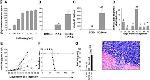
In vitro and in vivo growth of IL-6-dependent INA-6 MM cells. (A) In vitro growth of INA-6 cells in the absence or presence of increasing doses of exogenous huIL-6. (B) In vitro coculture of INA-6 cells and BMSCs in the absence of exogenous IL-6. *P < .05. (C) Levels of shuIL-6R in sera of mice injected with INA-6 cells into human bone implant (SCID-hu, n = 45) or subcutaneously (SCID, n = 8). (D) Clustering of a cohort of 45 SCID-hu mice on the day of first detection of serum shuIL-6R. (E) The kinetics of appearance and evolution of shuIL-6R in SCID-hu mice (n = 16) following engraftment of INA-6 cells (2.5 × 10⁶ cells) injected directly into human bone implant. Each symbol represents a different SCID-hu mouse. (F) Overall survival of SCID-hu (•; n = 16) and SCID mice (○; n = 8) after INA-6 injection (2.5 × 10⁶ cells/mouse). (G) Levels of huIL-6 in sera of SCID or SCID-hu mice given injections of INA-6 cells either subcutaneously or directly into the human bone implant, respectively. Sera were obtained from tail vein in SCID mice (n = 4), or from both tail vein and fetal bone aspirate in SCID-hu mice (n = 6). (H) H&E staining of a human fetal bone section implanted in a SCID-hu mouse and engrafted with INA-6 cells (original magnification ×100). Error bars in panels A-C and G indicate standard error.
In vitro and in vivo growth of IL-6-dependent INA-6 MM cells. (A) In vitro growth of INA-6 cells in the absence or presence of increasing doses of exogenous huIL-6. (B) In vitro coculture of INA-6 cells and BMSCs in the absence of exogenous IL-6. *P < .05. (C) Levels of shuIL-6R in sera of mice injected with INA-6 cells into human bone implant (SCID-hu, n = 45) or subcutaneously (SCID, n = 8). (D) Clustering of a cohort of 45 SCID-hu mice on the day of first detection of serum shuIL-6R. (E) The kinetics of appearance and evolution of shuIL-6R in SCID-hu mice (n = 16) following engraftment of INA-6 cells (2.5 × 10⁶ cells) injected directly into human bone implant. Each symbol represents a different SCID-hu mouse. (F) Overall survival of SCID-hu (•; n = 16) and SCID mice (○; n = 8) after INA-6 injection (2.5 × 10⁶ cells/mouse). (G) Levels of huIL-6 in sera of SCID or SCID-hu mice given injections of INA-6 cells either subcutaneously or directly into the human bone implant, respectively. Sera were obtained from tail vein in SCID mice (n = 4), or from both tail vein and fetal bone aspirate in SCID-hu mice (n = 6). (H) H&E staining of a human fetal bone section implanted in a SCID-hu mouse and engrafted with INA-6 cells (original magnification ×100). Error bars in panels A-C and G indicate standard error.
SCID-hu INA-6 mouse model and in vivo treatments
Six- to 8-week-old male CB-17 SCID mice (Taconic Farms, Germantown, NY) were given subcutaneous implants of human fetal long bone grafts into SCID mice (SCID-hu), as previously described.1-7,22 Four weeks following bone implantation, 2.5 × 10⁶ INA-6 MM cells in 50 μL phosphate-buffered saline (PBS) was injected directly into the human bone implant. Because INA-6 cells release soluble human IL-6 receptor (shuIL-6R), we used this marker to monitor tumor growth and burden of disease in SCID-hu mice. Mouse sera were serially monitored for shuIL-6R levels by enzyme-linked immunosorbent assay (ELISA; R&D Systems, Minneapolis, MN). Mice developed detectable serum shuIL-6R levels approximately 4 weeks following INA-6 cell injection, and then were treated with dexamethasone (1 mg/kg) intraperitoneally for 6 consecutive days or Atiprimod (20 mg/kg) intraperitoneally for a total of 6 alternate days or B-B4-DM1 (150 μg/kg) intravenously for 2 consecutive days. Following treatments, blood samples were collected and sera were analyzed for shuIL-6R levels.
Real-time fluorescence imaging
SCID-hu mice were directly injected in human fetal bone implants with 2.5 × 10⁶ INA-6GFP+ cells and monitored using the Illumatool Bright Light System LT-9900 (Lightools Research, Encinitas, CA), following a cutaneous shave of the tumor area. The images were captured with a Sony DSC-P5 digital camera and analyzed using Image-Pro Discovery software (Media-Cybernetics, Silver Spring, MD).
Histopathologic analysis
Human fetal bone implants were fixed in Bouin solution, embedded in paraffin, sectioned, and stained with hematoxylin and eosin (H&E) for histopathologic examination as previously described.7 Bone implants injected with INA-6GFP+ cells were frozen, sectioned, fixed in methanol/acetone, and serially examined by fluorescence microscopy (Leica DM IRB, Leica Microsystems, Allendale, NJ). The microscope used a Nikon Plan Apo 60×/1.40 numerical aperture oil objective lens. Images were captured with a Nikon Eclipse E800 SPOT camera (Diagnostic Instruments, Sterling Heights, MI) and processed using Adobe Photoshop version 5.5 (Adobe Systems, San Jose, CA).
Statistical analysis
Statistical significance of differences was determined using Student t test. Differences were considered significant when P was below .05.
Results and discussion
The INA-6 cell line17 requires the presence of either exogenous huIL-6 or BMSCs for proliferation in vitro growth, as measured by incorporation of [3H]-thymidine (Figure 1A-B). We tested whether INA-6 cells would engraft in SCID-hu mice given implants of human fetal bone. A cohort of 45 SCID-hu mice were injected directly into the bone implant with INA-6 cells (2.5 × 10⁶/mouse). Because INA-6 cells release shuIL-6R, mice were then monitored serially for serum shuIL-6R levels as an indicator of MM growth. As shown in Figure 1C, INA-6 cells engrafted in SCID-hu mice (n = 45), as indicated by detection of shuIL-6R. The mean value of shuIL-6R at first detection was 4.5 ± 0.9 ng/mL. In contrast, human shuIL-6R was not detected in the serum of control SCID mice without human bone chips (n = 8), injected subcutaneously with INA-6 cells (Figure 1C). As shown in Figure 1D, the onset of elevated shuIL-6R in serum was within a narrow range of 33 to 46 days from cell injection. In all SCID-hu mice, the progressive increase of shuIL-6R has been observed following the initial detection (Figure 1E). The median survival of SCID-hu mice given injections of INA-6 cells was 69 days (Figure 1F). In contrast, no deaths were observed in SCID mice without bone chips up to 200 days of observation following INA-6 cells injection (Figure 1F). Importantly, we found increased serum levels of huIL-6 only in SCID-hu mice engrafted with INA-6 cells with higher concentrations of huIL-6 in fetal bone chip aspirates versus tail vein blood (Figure 1G). Histologic examination of human fetal bone chips following detection of rising levels of shuIL-6R in mouse serum showed a diffuse BM infiltration by MM cells (Figure 1H).
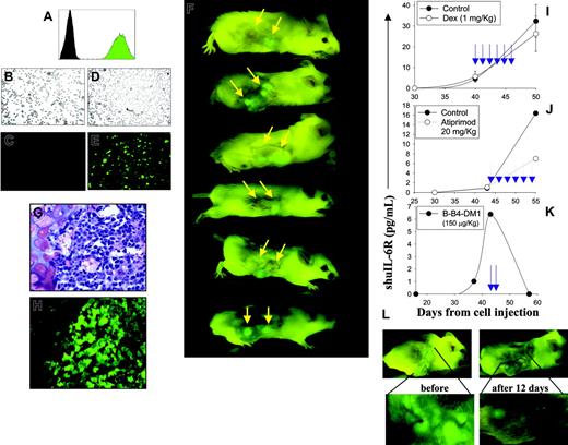
RTFI of SCID-hu mice with MMGFP+ lesions within human fetal bone. (A) Flow cytometric analysis indicates an approximate 2-log difference in mean fluorescence intensity of INA-6GFP+ cells (green peak) versus the parental INA-6 cells (black peak). (B-E) Microscopic analysis of INA-6GFP+ cells transduced with the GFP construct. Panels B and C refer to GFP- cells; panels D and E refer to GFP+ cells analyzed by either contrast-phase (B,D) or fluorescence microscopy (C,E) (original magnification × 100). (F) Representative imaging of 6 different SCID-hu mice following engraftment of INA-6GFP+ cells (2.5 × 10⁶) into the human fetal bone (indicated by yellow arrows). (G-H) Serial sections of a human fetal bone engrafted with INA-6GFP+ cells. H&E staining (G) and fluorescence microscopy (H; original magnification ×200). (I-K) Dexamethasone, Atiprimod, and B-B4-DM1 treatments of SCID-hu mice engrafted with INA-6 cells; levels of shuIL-6R in SCID-hu mice sera were analyzed. The blue arrows indicate the day of treatment. (L) Images of a representative SCID-hu mouse bearing INA-6GFP+ lesions in human fetal bone before and after 2 consecutive days of treatment with B-B4-DM1 (150 μg DM1/kg intravenously). Fetal bone image enlargements are shown.
RTFI of SCID-hu mice with MMGFP+ lesions within human fetal bone. (A) Flow cytometric analysis indicates an approximate 2-log difference in mean fluorescence intensity of INA-6GFP+ cells (green peak) versus the parental INA-6 cells (black peak). (B-E) Microscopic analysis of INA-6GFP+ cells transduced with the GFP construct. Panels B and C refer to GFP- cells; panels D and E refer to GFP+ cells analyzed by either contrast-phase (B,D) or fluorescence microscopy (C,E) (original magnification × 100). (F) Representative imaging of 6 different SCID-hu mice following engraftment of INA-6GFP+ cells (2.5 × 10⁶) into the human fetal bone (indicated by yellow arrows). (G-H) Serial sections of a human fetal bone engrafted with INA-6GFP+ cells. H&E staining (G) and fluorescence microscopy (H; original magnification ×200). (I-K) Dexamethasone, Atiprimod, and B-B4-DM1 treatments of SCID-hu mice engrafted with INA-6 cells; levels of shuIL-6R in SCID-hu mice sera were analyzed. The blue arrows indicate the day of treatment. (L) Images of a representative SCID-hu mouse bearing INA-6GFP+ lesions in human fetal bone before and after 2 consecutive days of treatment with B-B4-DM1 (150 μg DM1/kg intravenously). Fetal bone image enlargements are shown.
We next stably transduced INA-6 cells with a lentiviral vector encoding GFP (INA-6GFP+ cells) (Figure 2A-E) and used these cells for external serial real-time fluorescence imaging (RTFI) of SCID-hu mice. Four weeks following the inoculation of INA-6GFP+ cells in the human bone implants, RTFI of SCID-hu mice demonstrated engraftment of fluorescent MM cells in the implanted human bones (Figure 2F). This was further confirmed by serial histologic analysis of bone implants (Figure 2G-H).
We then evaluated the utility of this model in testing conventional and investigational anti-MM agents. As shown in Figure 2I, treatment of a SCID-hu mouse with dexamethasone did not significantly inhibit the increase in serum shuIL-6R levels, confirming the protective interaction between BMSCs and MM cells as observed in vitro. Treatment with Atiprimod, a novel agent with antiproliferative and apoptotic activity against MM,20 produced a significant inhibition of shuIL-6R accumulation in MM-bearing mice (Figure 2J). Treatment with B-B4-DM1, a novel conjugate of the anti-CD138 mAb with the highly cytotoxic maytansinoid DM1,21 induced tumor regression, as detected by a decrease in both serum shuIL-6R levels and imaging of MMGFP+ lesions (Figure 2K-L). Taken together these results demonstrate that the SCID-hu INA-6GFP+ model can be used to evaluate anti-MM activity of investigational agents in the huBM milieu.
Although the SCID-hu mouse model with primary patient MM cells remains a biologically relevant in vivo system for the study of MM pathophysiology, the inability to produce more than 2 to 4 mice from each patient sample made statistically significant comparisons of data points very difficult. The need has been to have a SCID-hu model that can be used to produce a large number of mice with a uniform cell population so that various treatments can be studied and compared in a consistent fashion. The present model is the first model that uses a uniform cell line that requires the human microenvironment for its in vivo growth in SCID mice and hence recapitulates the disease more accurately and reflects therapeutic effects more akin to what may be expected in patients. In our model, tumor growth may be monitored by measurement of serum shuIL-6R levels, whereas detection of human κ chain, which is produced in very small amounts when cells are cultured in vitro,17 is associated only with the presence of a high burden of disease and does not represent a sensitive marker of early tumor growth. Bone destruction is generally associated with extramedullary disease around the human bone chip and characterizes progression of disease (data not shown). The noninvasive external visualization of bone disease by detection of GFP+ cells allows a detailed, easy, and quick analysis for total number and size of tumors. Thus, this model overcomes the limitations of primary cell SCID-hu model for its reproducibility and predictability and therefore is an important in vivo system for the preclinical evaluation of new agents in MM.
Prepublished online as Blood First Edition Paper, April 7, 2005; DOI 10.1182/blood-2005-01-0373.
Supported by Multiple Myeloma Research Foundation Awards (M.A.S., N.C.M., and K.C.A.); Department of Veterans Affairs merit review grant and the Leukemia and Lymphoma Society Scholar in Translational Research Award (N.C.M.); National Institutes of Health (NIH) grants P50-100707 and PO1-78378 (K.C.A. and N.C.M.); as well as NIH grant RO1-50947 and the Doris Duke Distinguished Clinical Research Scientist Award (K.C.A.). G.S.J is employed by Callisto Pharmaceuticals Inc, whose product (Atiprimod) was used in this study. R.F. and V.S.G. are employed by ImmunoGen Inc, whose potential product (B-B4-DM1) was used in this study, which is presently only under preclinical investigation.
An Inside Blood analysis of this article appears in the front of this issue.
The publication costs of this article were defrayed in part by page charge payment. Therefore, and solely to indicate this fact, this article is hereby marked “advertisement” in accordance with 18 U.S.C. section 1734.


This feature is available to Subscribers Only
Sign In or Create an Account Close Modal