Abstract
Autoantibodies to proteinase 3 (PR3) are involved in the pathogenesis of autoimmune-mediated vasculitis in Wegener granulomatosis (WG). To address the question how the autoantigen PR3 becomes a target of adaptive immunity, we investigated the effect of PR3 on immature dendritic cells (iDCs) in patients with WG, healthy blood donors, and patients with Crohn disease (CD), another granulomatous disease. PR3 induces phenotypic and functional maturation of a fraction of blood monocyte-derived iDCs. PR3-treated DCs express high levels of CD83, a DC-restricted marker of maturation, CD80 and CD86, and HLA-DR. Furthermore, the DCs become fully competent antigen-presenting cells and can induce stimulation of PR3-specific CD4+ T cells, which produce IFN-γ. PR3-maturated DCs derived from WG patients induce a higher IFN-γ response of PR3-specific CD4+ T cells compared with patients with CD and healthy controls. The maturation of DCs mediated through PR3 was inhibited by a serine protease inhibitor, by antibodies directed against the protease-activated receptor-2 (PAR-2), and by inhibition of phospholipase C, suggesting that the interactions of PR3 with PAR-2 are involved in the induction of DC maturation. Wegener autoantigen interacts with a “gateway” receptor (PAR-2) on iDCs in vitro triggering their maturation and licenses them for a T helper 1 (Th1)–type response potentially favoring granuloma formation in WG.
Introduction
One of the key questions with respect to the pathophysiology of human autoimmune diseases is how autoreactivity to particular autoantigens is initiated and maintained and whether the autoantigen itself could have a role in it. Evidence from animal models suggests that dendritic cells (DCs) play a crucial role in initiating and maintaining immune responses not only to foreign antigen but also to self-antigen in autoimmune disease.1 Dendritic cells recognize antigen through distinct pattern-recognition receptors. Data from recently published animal models show that immune responses mediated by such pattern-recognition receptors are important in the transition from autoreactivity to a self-antigen to autoimmune disease.2,3 Moreover, autoantigen presentation has to be sustained in organized lymphoid tissue and facilitated by inflammatory responses.1,3,4 Involvement of such “gateway” receptors could explain why only particular self-antigens are presented efficiently enough to become a target of an autoimmune response and there is not a random response to self-antigens.
Wegener granulomatosis (WG) is a life-threatening autoimmune disease of yet-unknown etiology characterized by granulomatous lesions, an autoimmune small-vessel vasculitis, expansion of T helper 1 (Th1)–type cells, and a highly specific antineutrophil cytoplasmic autoantibody targeting “Wegener autoantigen” proteinase 3 (PR3-ANCA).5-7 Apart from their diagnostic value and correlation with disease activity, PR3-ANCAs play a direct pathogenic role in inducing systemic vasculitis by interacting with neutrophil granulocytes as in vitro and in vivo studies suggest.8 The autoantigen PR3 is abundantly displayed in granulomatous lesions in WG. PR3 expression in such lesions seems to exceed that seen in other granulomatous diseases.9 Moreover, there is preliminary evidence of maturation of autoreactive B cells in early granulomatous lesions in WG as suggested by ANCA-encoding VH genes.10 Therefore, granulomatous lesions themselves could represent a (tertiary) lymphoidlike tissue in which autoantigen is displayed under inflammatory conditions.
Given the strong association and high specificity of PR3-ANCA, the question needs to be addressed by which mechanism PR3 initiates an autoimmune response in WG. As the repertoire of human autoantigens is surprisingly limited, there have to be certain structural, biochemical, or immunologic properties that make the autoantigen immunogenic in predisposed subjects.11 A number of observations suggest that the abundance of an antigen in apoptotic debris can cause an immune response.12 As PR3 can be mobilized upon apoptosis independent from degranulation, expression of PR3 on the surface of apoptotic blebs and ectosomes may render PR3 an antigenic target.13 Protease-activated receptors (PARs) provide a system that detects tissue injury and triggers a set of cellular responses that contribute to various responses including inflammation.14 Since PAR expression has been reported on DCs15,16 and PR3 to activate oral epithelial and nonepithelial cells via the PAR-2,17-19 we wondered whether this receptor could play a role in initiating an adaptive autoimmune response to PR3.
We hypothesized that PR3 may interact and activate antigen-presenting cells (APCs) such as DCs that might initiate an adaptive immune response through induction of PR3-specific CD4+ Th1-type T cells. To test this hypothesis, we first investigated whether PR3 induces maturation of human DCs in healthy blood donors and patients with WG. To see if any effect seen in patients with WG is specific, patients with Crohn disease (CD) were selected as disease controls for another granulomatous inflammatory disorder. We found that Wegener autoantigen induced phenotypical and functional maturation of a fraction of blood monocyte-derived DCs. Moreover, PR3-activated DCs were capable of stimulating autoreactive Th1-type PR3-specific CD4+ T cells. Notably, we showed that PR3-maturated DCs derived from WG patients induce a statistically significant higher response of PR3-specific CD4+ T cells compared with CD patients and healthy donors. Finally, we provide evidence that PR3 induces differentiation of DCs through the PAR-2 pathway.
Patients, materials, and methods
Patient population
Peripheral blood samples were obtained from patients with a well-defined clinical diagnosis of WG (n = 10) and healthy donors (n = 16). The diagnosis of WG was established according to international standards by applying the 1990 classification criteria of the American College of Rheumatology20 and the definitions of the 1992 Chapel Hill Consensus Conference.21 The disease activity in vasculitis patients was documented by using the Birmingham Vasculitis Activity Score (BVAS) at the time the serum samples were collected22 (Table 1). Six ANCA-negative patients with Crohn disease (CD) and active disease served as disease controls for another granulomatous inflammatory disorder.
Demographic data of patients with Wegener granulomatosis
Patient no. . | Age, y/ sex . | PR3-ANCA, U/mL . | Organ involvement . | BVAS . |
|---|---|---|---|---|
| 1 | 53/F | 128 | Ear, nose, and throat; lung; kidney; skin; arthralgias/arthritides; constitutional symptoms; peripheral nervous system | 24 |
| 2 | 73/M | 90 | Ear, nose, and throat; lung; kidney; arthralgias/arthritides; constitutional symptoms; peripheral nervous system | 9 |
| 3 | 51/M | 55 | Ear, nose, and throat; lung; eye | 9 |
| 4 | 62/M | 26 | Ear, nose, and throat; lung; peripheral nervous system; constitutional symptoms | 6 |
| 5 | 63/F | 34 | Ear, nose, and throat; lung; peripheral nervous system; constitutional symptoms; heart; eye; arthralgias/arthritides | 0 |
| 6 | 78/M | 0 | Ear, nose, and throat; lung; constitutional symptoms | 13 |
| 7 | 45/M | 16 | Ear, nose, and throat; lung; kidney; eye; skin; Gl system | 10 |
| 8 | 39/M | 12 | Lung; arthralgias/arthritides; heart; peripheral nervous system; constitutional symptoms | 13 |
| 9 | 63/F | 0 | Ear, nose, and throat; kidney; eye; constitutional symptoms | 0 |
| 10 | 37/M | 0 | Ear, nose, and throat; lung; arthralgias/arthritides; skin; constitutional symptoms | 13 |
ANCA indicates antineutrophil cytoplasmic antibody; BVAS, Birmingham vasculitis activity score; F, female; and M, male.
This study was carried out according to the 1997 Declaration of Helsinki of the World Medical Association. The design of the work has been approved by the ethics committee of the University of Schleswig-Holstein, Campus Lübeck, and each patient gave informed consent prior to participation in the study
Reagents
Anti–PAR-2 mouse monoclonal antibodies (SAM11) were obtained from Santa Cruz Biotechnology (Santa Cruz, CA). Recombinant human tumor necrosis factor-α (rhTNF-α) and propidium iodide (PI) were purchased from BD Biosciences (Heidelberg, Germany); the PAR2 agonist (PAR-2AP, SLIGKV-NH2) and the control peptide (VKGILS-NH2), from Bachem (Veil am Rhein, Germany). Recombinant human granulocyte-macrophage colony-stimulating factor (rhGM-CSF) and human interleukin-4 (rhIL-4) were obtained from R&D System (Wiesbaden, Germany). Human PR3 was obtained from Athens Research & Technology (Athens, GA) and from Wieslab (Lund, Sweden), human leukocyte elastase (HLE) was from Boettger (Berlin, Germany), and human cathepsin G (CG) was obtained from Calbiochem (Beeston, United Kingdom). The purity of the 3 enzymes (PR3, HLE, and CG) was more than 95% by sodium dodecyl sulfate–polyacrylamide gel electrophoresis (SDS-PAGE) and was further confirmed by Western blotting using our anti-PR3 monoclonal antibody WGM2, anti-HLE, and anti-CG (DAKO, Hamburg, Germany) MoAbs, as described earlier.23 Trypsin was purchased from Calbiochem. The recombinant mutated PR3 (PR3S176A: serine was substituted) was a gift from Dr Reiners and Dr Hansen, University of Köln, Germany.24
IL-4 and IFN-γ secretion assay kits were obtained from Miltenyi Biotec (Bergish Gladbach, Germany). Human T-cell enrichment cocktail was from CellSystems (London, United Kingdom).
Dendritic cell culture
Immature DCs (iDCs) were prepared from peripheral blood mononuclear cells (PBMCs), according to the technique of Sallusto et al,25 with some modifications. Briefly, PBMCs were incubated for 75 minutes at 37°C in wells of a 6-well plate, after which nonadherent cells were removed by gently swirling with warm RPMI-1640. DCs were generated from the adherent cells cultured in the presence of 1000 U/mL rhGM-CSF and 1000 U/mL rhIL-4 for 6 days. RPMI-1640 supplemented with 10% heat-inactivated fetal calf serum (FCS), 2 mM glutamine, 100 U/mL penicillin, and 100 μg/mL streptomycin was used for cell culture. On day 6, nonadherent iDCs were collected and transferred to new 6-well plates. Immature DCs were stimulated with PR3 (10 μg/mL), HLE (10 μg/mL), CG (10 μg/mL), and rhTNF-α (50 ng/mL) on the day of transfer and harvested on day 8. A dose-response curve has been established for each protease. RPMI-1640 supplemented with 5% human serum was used for the cell culture.
Analysis of PR3-specific T-cell response
T cells were sorted from PBMCs by using human T-cell enrichment cocktail kit (RosetteSep; CellSystem, St Katharinen, Germany). T cells were plated at 106 cells/well in flat-bottom 6-well plates in RPMI-1640 with 5% human serum. Heat-inactivated PR3 as Ag (10 μg/mL) was added to DC culture medium on day 3.26 The following maturation-inducing agents were used in their respective optimized conditions on day 6 for 2 days: TNF-α (50 ng/mL), enzymatically active PR3 (10 μg/mL), and HLE (10 μg/mL) or TNF-α + PR3 were added to the supplemented culture medium. Autologous DCs prepared as described in “Dendritic cell culture” were added at 10:1 T/DC ratio. DCs and T cells were cocultured at 37°C for 24 hours. T cells stimulated with staphylococcal enterotoxin B (SEB, 1 μg/mL) were used as a positive control. To detect the PR3-specific T-cell frequency, we used a new technique based on the IFN-γ/IL-4 secretion capture assay.27,28 Measurement of IFN-γ/IL-4 secretion by PR3-specific cells was performed according to the manufacturer's protocol. Cells were immediately analyzed by flow cytometry (FACScalibur; Becton Dickinson, San Jose, CA).
Flow cytometry
Cell surface molecule expression of stimulated cells was measured as previously described.7 In brief, stimulated cells were washed with ice-cold PBS and stained with FITC- and PE-labeled antibodies or designed isotype controls for 30 minutes at 4°C. Stained cells were washed, resuspended in PBS/0.1% BSA/0.1% NaN3, and analyzed by a FACScalibur, followed by analysis using CellQuest software (Becton Dickinson). The median fluorescence intensity (MFI) and an MFI ratio of control and PAR-2 antibody MFI values were recorded for each sample. All antibodies were obtained from Becton Dickinson.
Measurement of enzymatic activity and inhibition assays
The enzymatic activity of proteases (PR3, HLE, and CG) was determined by photometrically measuring the hydrolysis of the substrate MeO-Suc-Ala-Ala-Pro-Val-pNA (Sigma, St Louis, MO) for PR3 and HLE and Suc-Ala-Ala-Pro-Phe-pNA (Sigma) for CG, as described earlier.23 To inhibit the enzymatic activity of PR3, the enzyme was preincubated with different concentrations of the serine protease inhibitor phenylmethylsulphonyl fluoride (PMSF) for 30 minutes at 37°C before use.
Immature DCs were pretreated with or without cytochalasin B (CCB, 30 nM) for 30 minutes or cycloheximide (CHX, 1 μg/mL) for 6 hours at 37°C. Then, the cells were stimulated with or without PR3 (10 μg/mL) for 24 hours in the presence or absence of CCB or CHX at 37°C.
To block the cleavage of human PAR-2 peptide by exposure of its tethered ligand with PR3, iDCs were incubated with an anti–PAR-2 Ab (against amino acid residues 37-50 of PAR-2) at 10 μg/mL alone or with PR3 (10 μg/mL) for 48 hours.
To investigate whether PAR-2 activating peptide (SLIGKV-NH2) has any effect on DC maturation, iDCs were cultured with 100 μM synthetic PAR-2 agonist peptides (PAR-2APs) or a control peptide (100 μM) for 48 hours and then analyzed for surface expression of HLA-DR, CD83, and costimulatory molecules by flow cytometry.
Immature DCs were treated with 400 nM U73122 or the control compound U73343 (mock inhibitor) for 1 hour before the exposure to PAR-2AP (100 μM) or PR3 (10 μg/mL) for 24 hours.
Analysis of peptide cleavage by serine proteases
A peptide corresponding to a region spanning the cleavage site of the PAR-2, residues 32 to 45 (32SSKGRSLIGKVDGT45), was synthesized on a 433A Peptide Synthesizer dedicated to Fmoc synthetic chemistry, according to the manufacturer's instructions (Applied Biosystems, Weiterstadt, Germany). PAR-2 peptide (0.5 mg) was digested at 37°C with 10 μg each protease in 200 μL PBS for 30 minutes, 90 minutes, or overnight. Reverse-phase chromatography was performed on an ÄktaPurifier10 with Unicorn Software (GE Healthcare, Uppsala, Sweden). Digested or untreated PAR-2 peptide was separated on a Jupiter C18 4-μ Proteo 90Å column (Phenomenex, Aschaffenburg, Germany).
Cleavage products were analyzed by N-terminal sequencing and mass spectrometry. Fragments of the PAR-2 peptide purified by high-performance liquid chromatography (HPLC) were sequenced by Edman degradation on a Procise protein sequencer with online PTH analyzer (Applied Biosystems) as previously described.29 Mass spectra of peptide fragments were recorded as previously described.30 Briefly, peptide fragments obtained from HPLC were measured by matrix-assisted laser desorption ionization time-of-flight (MALDI-TOF, Reflex III; Bruker Daltonik, Bremen, Germany) in the positive ion mode. Tandem mass spectrometry (MS/MS) analysis was performed by infrared-multiphoton dissociation (IRMPD) on a 7 Tesla ESI-FT-ICR mass spectrometer (APEX II; Bruker Daltonics, Billerica, MA) using doubly protonated precursor ions.
Data analysis
All experiments in this study were performed at least 3 times to confirm the reproducibility of the results. In most experiments, values are represented as mean ± standard deviation (SD) of triplicate assays. The statistical significance of differences between the 2 means was evaluated by the ANOVA test and subsequent posthoc analysis using the Bonferroni test. Values of P below .05 were considered significant.
Results
PR3 up-regulates membrane expression of CD83, HLA-DR, and costimulatory molecules on a fraction of DCs
In this study, we investigated whether PR3 could initiate immunity by inducing human DC maturation. We first examined the effects of PR3, HLE, and CG on the surface maturation phenotype of iDCs.
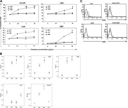
Immature dendritic cells are characterized by low expression of HLA-DR and costimulatory molecules (CD40, CD80, and CD86). The DC-restricted, maturation-associated marker CD83 is not expressed. Immature DCs were further cultured with PR3 and HLE for 48 hours, after which the DCs were collected, stained for markers indicative of maturation, and analyzed by flow cytometry. A dose-response curve was established for each protease (PR3, HLE, and CG; Figure 1A).
Figure 1B shows that PR3 induced the up-regulation of HLA-DR (mean ± standard error of the mean [SEM] MFI ratio for iDCs 19.5 ± 2.0 vs for mature DCs [mDCs] 93.2 ± 5.1, P = .001) and of the costimulatory molecules CD80 (iDCs 4.9 ± 0.6 vs mDCs 20.6 ± 1.6, P = .022) and CD86 (iDCs 2.1 ± 0.3 vs mDCs 6.6 ± 0.6, P = .031) on the surface of DCs. Furthermore, PR3 induced the expression of CD83 (iDCs 1.2 ± 0.3 vs mDCs 4.1 ± 0.5, P = .001), while expression of CD14 was reduced (iDCs 5.2 ± 0.9 vs mDCs 1.0 ± 0.5, P = .001). The induction of maturation by PR3 was equivalent to that seen with TNF-α (the positive control; Figure 1B) and was irreversible, as DCs did not revert to an immature phenotype in the culture. However, DCs did not exhibit a mature phenotype after exposure to HLE or CG (Figure 1A-B).
Blocking protease activity of PR3 inhibits DC maturation
To address whether the ability of PR3 to induce maturation of DCs is due to its protease activity, we examined the effect of a serine protease inhibitor and the effect of a recombinant mutated enzymatically inactive PR3. Incubating PR3 (5 nM) with the potent serine protease inhibitor PMSF (1 mM) for 30 minutes at 37°C abolished the serine protease activity. Accordingly, this condition was used to test the effect of intact versus inactivated serine protease on DC maturation. Figure 1C shows the effect of PMSF and PR3S176A on PR3-induced CD83 expression. The expression of CD83 on DCs stimulated with PMSF-inactivated PR3 and mutated inactive PR3 was significantly lower than that from DCs stimulated with enzymatically active PR3 (P = .021), suggesting that serine protease activity of PR3 is required to induce DC maturation.
DCs exposed to PR3 acquire T-cell stimulatory capacity
To examine whether PR3-treated DCs were able to prime T cells, we measured the cytokine secretion of CD4+ T cells from WG and CD patients and healthy donors by an IFN-γ/IL-4 capture assay. This approach allows the detection of IFN-γ/IL-4 on the surface of cytokine-secreting cells by using a cell surface affinity matrix.24 Purified CD4+ T cells were stimulated by PR3 in the presence of autologous DCs. We then analyzed the pattern of IFN-γ/IL-4 production by CD4+ T lymphocytes. Of interest, both in WG and controls (CD patients and healthy controls) following either PR3- or TNF-α–induced DC activation, T cells differentiated only into IFN-γ–producing cells (Figure 2), not IL-4–producing cells. We did not detect IL-4–producing T cells in any experiment (data not shown). Compared with the nonstimulated control samples and T cells alone, a higher proportion of IFN-γ+ CD4+ cells was detectable in the DC samples treated with PR3 in WG patients as well as in controls. When comparing IFN-γ production between WG and CD patients or healthy controls, we found a significantly higher percentage of IFN-γ–secreting T cells in the WG group, with all maturation stimuli except HLE (ie, for PR3-maturated DCs: healthy controls, 0.16% ± 0.01%; CD, 0.22% ± 0.08%; WG, 0.73% ± 0.13% [P = .02]). Simultaneous stimulation with TNF-α and PR3 was not more potent than PR3 alone in inducing T-cell activation (Figure 2). The percentage of IFN-γ–producing T cells in the group of HLE-maturated DCs was similar to the negative control (iDC) in both patients and healthy controls (Figure 2).
A fraction of monocyte-derived DCs exhibits a mature phenotype after exposure to PR3 but not HLE. Immature DCs cultured for 6 days in IL-4 and GM-CSF were stimulated with PR3, HLE, CG, and TNF-α. After 48 hours, the DCs were stained for CD14, CD80, CD83, CD86, and HLA-DR and analyzed by flow cytometry. Isotype-matched antibodies served as control in all experiments. (A) Dose-response curves for PR3, HLE, and CG; data are presented as mean percentage of positive cells. (B) Expression of CD14, CD80, CD83, CD86, and HLA-DR. The concentration of proteases used (10 μg/mL) is equivalent to the top of the dose-response curve. Data are presented as mean MFI ratios ± SEM. *P < .05; **P < .03; ***P < .001 (Bonferroni test). The experiments pertaining to panels A-B were performed separately. (C) To understand whether the enzymatically active PR3 was required for these effects on DCs, the cells were incubated for 24 hours with enzymatically active PR3, enzymatically inactive mutated PR3, and PMSF and PMSF-pretreated PR3. The expression of CD83 on the cell surface was analyzed by flow cytometry. The histograms show fluorescence values on gated cells. The bold lines represent isotype Abs control. Results are representative of 1 of 3 independent experiments performed in duplicate.
A fraction of monocyte-derived DCs exhibits a mature phenotype after exposure to PR3 but not HLE. Immature DCs cultured for 6 days in IL-4 and GM-CSF were stimulated with PR3, HLE, CG, and TNF-α. After 48 hours, the DCs were stained for CD14, CD80, CD83, CD86, and HLA-DR and analyzed by flow cytometry. Isotype-matched antibodies served as control in all experiments. (A) Dose-response curves for PR3, HLE, and CG; data are presented as mean percentage of positive cells. (B) Expression of CD14, CD80, CD83, CD86, and HLA-DR. The concentration of proteases used (10 μg/mL) is equivalent to the top of the dose-response curve. Data are presented as mean MFI ratios ± SEM. *P < .05; **P < .03; ***P < .001 (Bonferroni test). The experiments pertaining to panels A-B were performed separately. (C) To understand whether the enzymatically active PR3 was required for these effects on DCs, the cells were incubated for 24 hours with enzymatically active PR3, enzymatically inactive mutated PR3, and PMSF and PMSF-pretreated PR3. The expression of CD83 on the cell surface was analyzed by flow cytometry. The histograms show fluorescence values on gated cells. The bold lines represent isotype Abs control. Results are representative of 1 of 3 independent experiments performed in duplicate.
PR3 induces expression of PAR-2 on mDCs
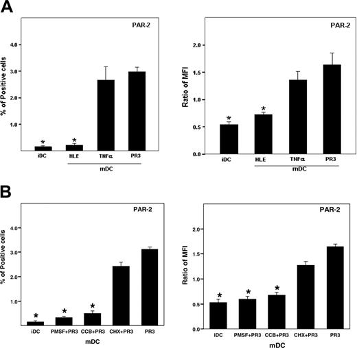
We next extended the investigation to the underlying mechanism by which PR3 activates DCs. We first asked whether PR3 induces expression of PAR-2 on DCs. By flow cytometric analyses, no expression of PAR-2 was detected on the surface of iDCs. The up-regulation by PR3 was detected at 30 minutes of incubation, and reached a plateau at 1 hour. Exposure of the cells to PR3 for up to 24 hours did not further up-regulate the surface expression of PAR-2. The expression of PAR-2 on mDCs was augmented significantly by PR3 treatment (P = .031) but not by HLE (Figure 3A).
DCs gain T-cell stimulatory capacity after exposure to PR3. Mature DCs obtained by TNF-α, PR3, and TNF-α+ PR3 exposure (from day 4 to day 8) were pulsed with PR3 on day 3 of their culture, and were plated at day 6 with autologous CD4+ T cells for 24 hours. Subsequently, the cells were subjected to the IFN-γ/IL-4 capture assay. The cells are labeled as described in “Patients, materials, and methods,” and then analyzed by flow cytometry. In order to confirm that the effect observed for PR3 is specific for this serine protease, similar experiments using HLE were also carried out. Data are plotted as the mean percentage of IFN-γ–producing cells ± SEM (healthy controls [HC], n = 5; WG, n = 10; CD, n = 6). *P < .05.
DCs gain T-cell stimulatory capacity after exposure to PR3. Mature DCs obtained by TNF-α, PR3, and TNF-α+ PR3 exposure (from day 4 to day 8) were pulsed with PR3 on day 3 of their culture, and were plated at day 6 with autologous CD4+ T cells for 24 hours. Subsequently, the cells were subjected to the IFN-γ/IL-4 capture assay. The cells are labeled as described in “Patients, materials, and methods,” and then analyzed by flow cytometry. In order to confirm that the effect observed for PR3 is specific for this serine protease, similar experiments using HLE were also carried out. Data are plotted as the mean percentage of IFN-γ–producing cells ± SEM (healthy controls [HC], n = 5; WG, n = 10; CD, n = 6). *P < .05.
We observed that the effect of PR3 on the expression of PAR-2 was significantly inhibited by sulfonyl fluoride-type serine protease inhibitors such as PMSF. These results indicated that PR3 induced the expression of PAR-2 on mDCs, and the enzymatic activity of PR3 was critical to the induction (Figure 3B). Furthermore, the PR3-induced PAR-2 expression was inhibited by CCB, an inhibitor of actin polymerization, but not by CHX, a protein synthesis inhibitor (Figure 3B). These results indicate that PAR-2 is an inducible receptor from intracellular storage and does not require de novo synthesis.
Inhibitory effect of anti–PAR-2 Ab on DC maturation induced by PR3
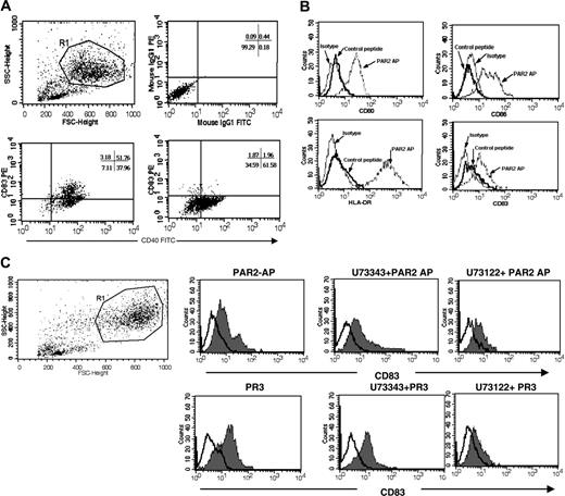
We next examined whether the maturation of DCs triggered by PR3 occurs through PAR-2. To examine the role of PAR-2 in PR3-induced DC maturation, affinity-purified anti–PAR-2 mAb (SAM11 raised against AAs 37-50 of human PAR-2) was used. The cells were pretreated with a monoclonal antibody directed against the PR3 interaction site in PAR-2 that blocks receptor cleavage and thereby PAR-2 activation. After the incubation, the cells were stimulated with or without PR3, and then the expression of CD83 and the costimulatory molecule CD40 was analyzed by flow cytometry. We found the effect of PR3 on maturation of DCs was inhibited by the anti–PAR-2 antibody (Figure 4A), suggesting that PR3-induced activation of DCs was in fact mediated by PAR-2.
PAR-2 agonist peptide and PR3 induce DC maturation with similar intensity
The involvement of PAR-2 in iDC maturation was further analyzed by using the PAR-2AP SLIGKV-NH2, corresponding to the PAR-2 tethered ligand, to induce DC maturation. As shown in Figure 4B, incubation of iDCs with PAR-2AP up-regulated the expression of HLA-DR, CD80, CD86, and CD83 on DCs with similar intensity compared with PR3. In contrast, the control peptide showed no effects (Figure 4B). These results indicated that PR3-induced activation of DCs was mediated by PAR-2.
Phospholipase C is involved in PR3-induced DC maturation
One of the ultimate tests for the role of PAR-2 in DC activation and function is the use of a specific inhibitor of PLC in combination with PR3 or PAR-2AP. The DCs treated with the specific inhibitor of PLC (U73122) and mock inhibitor (U73343), and subsequently stimulated with PR3 or with PAR-2AP, expressed the DC maturation markers CD83 and HLA-DR and costimulatory molecules. Expression of maturation markers on the DCs treated with PR3 and PAR-2AP was partially blocked by U73122, the specific inhibitor of PLC (Figure 4C).
Cleavage of human PAR-2 peptide by serine proteases
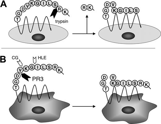
To examine the ability of PR3, HLE, and CG to activate or to inactivate PAR-2, a peptide corresponding to region surrounding the cleavage site of human PAR-2 was incubated with the serine proteases, and proteolytic fragments were analyzed by reverse chromatography. Cleavage of PAR-2 peptide by PR3 occurred after 90 minutes. The peptides corresponding to the cleavage product and the original PAR-2 peptide (AA 32-45) were subjected to N-terminal sequencing and mass spectrometry. Sequencing revealed that both peptides started with the N-terminus and the cleavage product ended with valine residue at position 42 (V42-D43). MALDI-TOF and MS/MS analyzing confirmed that the peptide contained the amino acid residues 32 to 42 (32SSKGRSLIGKV42; Figure 5). A peak corresponding to residues 43 to 45 could not be detected.
PR3 induces expression of PAR-2 on mDCs. (A) Immature DCs were incubated with PR3 or HLE and TNF-α for 48 hours. For PAR-2 staining, cells were incubated with FITC-conjugated mouse anti–human PAR-2 or IgG2a isotype control for 30 minutes at 4°C and then were analyzed by flow cytometry. The positive staining is expressed as mean percentage of positive cells and as a median fluorescence intensity (MFI) ratio described in “Patients, materials, and methods.” Values are the mean ± SEM (donors n = 16). (B) iDCs were stimulated with PR3 for 24 hours at 37°C. PR3 was pretreated with and without PMSF for 30 minutes at 37°C before use. Immature DCs were pretreated with and without cytochalasin B (CCB) for 30 minutes or CHX for 6 hours at 37°C. Then, the cells were stimulated with and without PR3 for 24 hours in the presence or absence of CCB or CHX. After the incubation, the cells were collected and the expression of PAR-2 on the cells was analyzed by flow cytometry. The positive staining is expressed as mean percentage of positive cells and as a median fluorescence intensity (MFI) ratio. *P < .01 compared with PR3 alone.
PR3 induces expression of PAR-2 on mDCs. (A) Immature DCs were incubated with PR3 or HLE and TNF-α for 48 hours. For PAR-2 staining, cells were incubated with FITC-conjugated mouse anti–human PAR-2 or IgG2a isotype control for 30 minutes at 4°C and then were analyzed by flow cytometry. The positive staining is expressed as mean percentage of positive cells and as a median fluorescence intensity (MFI) ratio described in “Patients, materials, and methods.” Values are the mean ± SEM (donors n = 16). (B) iDCs were stimulated with PR3 for 24 hours at 37°C. PR3 was pretreated with and without PMSF for 30 minutes at 37°C before use. Immature DCs were pretreated with and without cytochalasin B (CCB) for 30 minutes or CHX for 6 hours at 37°C. Then, the cells were stimulated with and without PR3 for 24 hours in the presence or absence of CCB or CHX. After the incubation, the cells were collected and the expression of PAR-2 on the cells was analyzed by flow cytometry. The positive staining is expressed as mean percentage of positive cells and as a median fluorescence intensity (MFI) ratio. *P < .01 compared with PR3 alone.
The maturation of DCs triggered by PR3 occurs through PAR-2. (A) Immature DCs were pretreated with SAM-11 at 10 μg/mL alone or with PR3 (10 μg/mL) for 24 hours. The cell surface expression of CD40 and CD83 was analyzed by flow cytometry. (B) DCs were stimulated with 100 μM PAR-2 agonist peptide (SLIKV-NH2, PAR-2AP) or a control peptide (100 μg/mL) for 48 hours and then were stained for CD80, CD83, CD86, and HLA-DR and analyzed by flow cytometry. The histograms show fluorescence intensity on gated cells. The thin lines represent staining with isotype-matched irrelevant mAb. (C) Effect of PLC inhibition on the expression of maturation marker on DCs. Immature DCs were treated with 400 nM U73122 or the control compound U73343 (mock inhibitor) 1 hour before exposure to PAR-2AP or PR3 for 24 hours. The expression of the DC maturation marker CD83 was analyzed by flow cytometry. Open curves indicate isotype Ab control; shaded areas, CD83. Tracings are representative of 3 distinct experiments.
The maturation of DCs triggered by PR3 occurs through PAR-2. (A) Immature DCs were pretreated with SAM-11 at 10 μg/mL alone or with PR3 (10 μg/mL) for 24 hours. The cell surface expression of CD40 and CD83 was analyzed by flow cytometry. (B) DCs were stimulated with 100 μM PAR-2 agonist peptide (SLIKV-NH2, PAR-2AP) or a control peptide (100 μg/mL) for 48 hours and then were stained for CD80, CD83, CD86, and HLA-DR and analyzed by flow cytometry. The histograms show fluorescence intensity on gated cells. The thin lines represent staining with isotype-matched irrelevant mAb. (C) Effect of PLC inhibition on the expression of maturation marker on DCs. Immature DCs were treated with 400 nM U73122 or the control compound U73343 (mock inhibitor) 1 hour before exposure to PAR-2AP or PR3 for 24 hours. The expression of the DC maturation marker CD83 was analyzed by flow cytometry. Open curves indicate isotype Ab control; shaded areas, CD83. Tracings are representative of 3 distinct experiments.
CG and HLE cleaved the PAR-2 peptide much faster than PR3, with considerable cleavage detectable after 30 minutes. N-terminal sequencing of the fragments revealed the cleavage sites for both enzymes: CG cleaved between lysine and valine (K41-V42) and HLE between isoleucine and glycine (I39-G40). Trypsin, a known activator of PAR-2, was used as a positive control. Trypsin treatment of PAR-2 peptide resulted in 2 fragments corresponding to a single cleavage site between residues R36-S37 (Figure 5). PR3 from 2 different sources showed identical results.
Discussion
In the present work, we investigated whether human neutrophil PR3 (Wegener autoantigen) could initiate immunity by inducing human DC maturation. We found the first evidence that PR3 induces in vitro the maturation of a fraction of monocyte-derived DCs, and these DCs become fully competent APCs for the stimulation of PR3-specific CD4+ T cells, based on the following results: (1) PR3 specifically up-regulates membrane expression of CD83, HLA-DR, and costimulatory molecules on DCs. (2) PR3-maturated DCs induce stimulation of PR3-specific CD4+ T cells, which produce IFN-γ, consistent with a dominant Th1 phenotype, and an increased T-cell stimulatory capacity was detected in WG. (3) The maturation program induced by PR3 uses the PAR-2 receptor and signaling pathway: PR3 induces PAR-2 expression on the surface of mDCs, and PR3-induced maturation of DCs was inhibited by a serine protease inhibitor, by an antibody directed against PAR-2, and by inhibition of phospholipase C. (4) The effect of PR3 on DC maturation is PR3 specific and cannot be generalized to other serine proteases (HLE and CG). (5) DC maturation was also induced by PAR-2 peptide agonist. Figure 6 shows a diagram summarizing the main findings of this study.
Cleavage of human PAR-2 peptide by serine proteases. A peptide corresponding to region surrounding the cleavage site of human PAR-2 (32SSKGRSLIGKVDGT45) was incubated with the serine proteases PR3, HLE, and CG, and proteolytic fragments were analyzed by reverse chromatography, N-terminal sequencing, and mass spectrometry. The cleavage product of PR3 ended with a valine residue at position 42 (V42-D43), CG cleaved between lysine and valine (K41-V42), and HLE between isoleucine and glycine (I39-G40). Trypsin, a known activator of PAR-2, was used as a positive control.
Cleavage of human PAR-2 peptide by serine proteases. A peptide corresponding to region surrounding the cleavage site of human PAR-2 (32SSKGRSLIGKVDGT45) was incubated with the serine proteases PR3, HLE, and CG, and proteolytic fragments were analyzed by reverse chromatography, N-terminal sequencing, and mass spectrometry. The cleavage product of PR3 ended with a valine residue at position 42 (V42-D43), CG cleaved between lysine and valine (K41-V42), and HLE between isoleucine and glycine (I39-G40). Trypsin, a known activator of PAR-2, was used as a positive control.
The involvement of proteases in DC antigen processing and presentation has been described.31 Recently, Fields et al15 demonstrated that murine serine proteases can act as danger signals, serving as a maturation stimulus for iDCs. Further, the authors showed that serine proteases can exert their effects via PAR. PR3 is an ideal candidate for this role because it is not expressed (or quickly inactivated by serine protease inhibitors) in the extracellular space of healthy tissue, but its level increases during infection, trauma, and tissue necrosis. A number of publications demonstrated that at sites of inflammation increased amounts of PR3 or HLE are detected in the extracellular space in patients with WG.9,32,33
Here, we extend the role of proteases in DCs by showing that PR3 can induce human DC development. We found that PR3 induces phenotypic and functional maturation of a fraction of DCs. Indeed, PR3-treated DCs up-regulate expression of CD83, a DC maturation-specific marker, HLA-DR molecules, and B7-1 and B7-2 costimulatory molecules. Mature DCs markedly reduced the surface expression of CD14, a molecule expressed mainly by monocyte and iDCs. The mechanism of this effect is still unclear: we cannot exclude that the down-regulation of CD14 is the consequence of the enzymatic degradation of the molecule or whether the loss of CD14 expression on DCs after PR3 treatment results from a combination of the cleavage and differentiation, since it has been demonstrated that HLE and CG are able to reduce the expression of CD14 on monocytes through enzymatic degradation.34,35 However, it was described that PR3 is able to modulate CD14 expression on monocytes, and the effects of PR3 on the cell surface CD14 expression are dependent on the monocyte subset: CD14 is up-regulated on CD16– monocytes and down-regulated on CD16+ monocytes.36 Thus, it seems that the cleavage of CD14 is cell specific and, at least for the cell population used in our study, does not have a fundamental role in the differentiation process of DCs. Altogether, these data demonstrate that the presence of PR3 without the addition of exogenous stimuli leads to the development of characteristic mDCs in vitro.
Model of induction of a Th1-cytokine response by proteinase 3 (PR3). In WG, expression of PR3 in the extracellular space is increased. PR3 stimulates the expression of PAR-2 on iDCs. PR3 activates PAR-2 resulting in maturation of DCs, as indicated by expression of CD80, CD83, CD86, and HLA-DR. These PR3-maturated DCs stimulate CD4+ T cells to generate increased expression of interferon-γ (IFN-γ). Expression of IFN-γ favors the development of a granulomatous inflammation as seen in WG. Hypothetically, chronic T-cell activation by PR3-maturated DCs may finally promote the development of B cells toward ANCA-producing plasma cells.
Model of induction of a Th1-cytokine response by proteinase 3 (PR3). In WG, expression of PR3 in the extracellular space is increased. PR3 stimulates the expression of PAR-2 on iDCs. PR3 activates PAR-2 resulting in maturation of DCs, as indicated by expression of CD80, CD83, CD86, and HLA-DR. These PR3-maturated DCs stimulate CD4+ T cells to generate increased expression of interferon-γ (IFN-γ). Expression of IFN-γ favors the development of a granulomatous inflammation as seen in WG. Hypothetically, chronic T-cell activation by PR3-maturated DCs may finally promote the development of B cells toward ANCA-producing plasma cells.
In contrast to PR3, we observed that HLE and CG had no effect on the maturation of DCs. This is in agreement with previous reports showing that HLE and CG do not activate PAR-2 in human lung cell lines, and HLE does not activate a PAR-2–expressing human microvascular endothelium cell line. A recent report demonstrates that HLE and CG do not activate PAR-2, but rather disarm the receptor, preventing activation by trypsin but not by SLIGKV-NH2.19 Although PR3 and HLE have very similar substrate specificity and share high sequence homology (56%), PR3 has different properties than HLE, especially being the main target antigen for ANCA in WG. The fact that PR3 but not HLE is a selective target of ANCA in WG cannot be explained by its abundance, as neutrophils contain 3 times more HLE than PR3,37 but might be a result of different structural or functional properties. We herein demonstrate that despite their close homology, PR3 and HLE are not targeted using a similar pathway. Thus, the presence of PR3 in the damaged tissue and its ability to influence DC development and maturation demonstrated in this report may represent a novel pathway of affecting immune responses in vivo.
As indicated in “Introduction,” necrotizing granulomatous inflammation in the respiratory tract is the defining feature of WG. Current evidence supports the hypothesis that the granulomatous inflammation in WG is driven by activated CD4+ T cells producing Th1 cytokines.6,38,39 To date, the nature of the immunoregulatory defect is unclear, but given the close association between ANCA and WG, an obvious possibility is that autoreactive CD4+ T cells specific for PR3 are also responsible for the pathogenic Th1 responses.
Thus, we asked whether PR3-treated DCs are able to prime T cells and whether they induced polarization of Th cells. To examine whether PR3-treated DCs were able to prime T cells, we measured the cytokine secretion of CD4+ T cells by an IFN-γ/IL-4 capture assay. Of interest, following either PR3- or TNF-α–induced DC activation, T cells differentiated only into IFN-γ–producing cells, not the IL-4–producing cells. Studies of the potential for peripheral blood T cells from patients with WG to respond to PR3 have given conflicting results.40 To date, the results of previous experiments have not demonstrated expansion of PR3-specific T cells in WG. Although, PR3 has modest effects on lymphocytic proliferation, these effects are seen in both patient and control samples. In the present study, we observed a statistically significant increase in the frequency of IFN-γ–secreting T cells from patients with WG, but there was no significant correlation between disease activity and T-cell responses.
Our present results strongly pointed out that DCs exposed to PR3 acquire the potency to activate autologous T cells in vitro, driving their polarization toward the Th1 phenotype.
We next examined whether the maturation of DCs triggered by PR3 could be mediated by the interaction of PR3 with PAR-2, that is, activated by proteolysis. PAR-2 is cleaved within its N-terminal extracellular domain by serine proteases such as trypsin, unmasking a new amino terminus starting with the sequence SLIGKV, which binds intramoleculary and activates the receptor. To study the cleavage profile of each serine protease, we used a classical approach: a synthetic peptide corresponding to a region spanning the cleavage site of the PAR-2, residues 32 to 45 (32SSKGRSLIGKVDGT45), was HPLC-separated after the cleavage and analyzed by amino acid sequencing and MALDI mass spectrometry. The results that were obtained show that PR3 can cleave the synthetic peptide after the valine residue at position 42 (V42-D43), which results in a C-terminal release of the activating peptide. Thus, PR3 has the potential to cleave the peptide on the opposite site of the tethered ligand (SLIGKV). In contrast, Uehara et al17,18 reported that PR3 cleaves the PAR-2 peptide at the site R36-S37. Differences in purity of the proteases may account for the divergent findings regarding the cleavage site of PR3. In our study, the purity of the serine proteases has been checked by SDS-PAGE, silver staining, and immunoblotting. In addition, the use of 2 different PR3 preparations gave identical results.
At this stage, we consider that the cleavage at site V42-D43 may lead to release of the tethered ligand, which can possibly act as an unbound agonist at the activation site of PAR-2, as it has been shown for other PAR-2 agonists such as SLIGRL-NH2 or tc-LIGRLO-NH2.41,42 Several lines of evidence suggest that the cleavage at the site V42-D43 by PR3 may be functionally relevant. First, a blocking antibody against PAR-2 (SAM11 directed against amino acids 37-5043,44 ) inhibits the PR3-induced maturation of dendritic cells. This effect would not be expected if PR3 would disarm PAR-2 rather than activating the receptor. Second, the principal mechanism of PAR-mediated activation is through Gαq proteins, resulting in activation of phospholipase C (PLC).44 Therefore, the involvement of PAR-2 in iDC maturation was further analyzed by addition of a specific inhibitor of PLC in combination with PR3 or PAR-2AP. The expression of maturation markers following treatment with PR3 and PAR-2AP was partially blocked by a specific inhibitor of PLC, but not by mock inhibitor. These findings demonstrate that the differentiation of iDCs by PR3 via PAR-2 activation uses the Gαq-protein signaling pathway only partially. Third, PR3, but not HLE and CG, induced the expression of PAR-2 on DCs, suggesting that this effect is PR3 specific and cannot be generalized to other serine proteases. Fourth, the PAR-2AP SLIGKV-NH2, corresponding to the PAR-2 tethered ligand, induced maturation of DCs. PAR-2AP up-regulated the expression of CD83, HLA-DR, and costimulatory molecules on DCs with similar intensity compared with PR3, suggesting a similar mode of action. Fifth, HLE and CG digestion of the PAR-2 peptide resulted in different cleavages, but not at the activating site of PAR-2, suggesting that only the cleavage induced by PAR-2 is functionally relevant. Actually, our data show that both serine proteases (HLE, cleavage after position 39-40; and CG, after position 41) destroy the activating peptide by cutting inside its ligand sequence. Elastase and cathepsin G do not activate PAR-2, and this is in agreement with previous reports.19,45
The present data show that PR3 has a dual effect on the PAR-2 expression and activation pathway. First, we demonstrated that PR3 induced the surface expression of PAR-2 on a small fraction of DCs via still-unknown mechanisms and that PAR-2 expression was inducible from intracellular storage. Second, PR3 cleaved PAR-2 and induced maturation of PAR-2–expressing DCs. Our observation that PR3 induced PAR-2 expression is in concordance with a recent study of Uehara et al17 that demonstrated that PR3 induced PAR-2 expression on the cell surface of oral epithelial cells. The biologic significance of the level of PAR-2 expression on DCs is supported (1) by the fact that an expression of similar intensity (MFI ratio) was shown to induce a significant cytokine response in another study with CD14+ monocytes46 ; and (2) by the relative difference in T-cell priming capacity of DCs between patients with WG and healthy controls or patients with CD in the present study. The central role of PAR-2 for DC development is delineated by the observation that PAR-2–/– mice failed to develop DCs in the absence of an exogenous stimulus.15
The mechanism by which PR3 initiates PAR-2 up-regulation in cells is still unknown. The involvement of cell surface PAR-2 seems less likely, since in our study the expression of PAR-2 on the iDC surface was below the detectable limits, and we speculate that another receptor may be targeted by PR3: DCs express the broadest repertoire of Toll-like receptors (TLRs), which can recognize a plethora of microbial compounds and endogeneous “danger/alarm” signals from injured cells, which can initiate immune responses by activating DCs. Therefore, it was demonstrated that HLE may activate TLR-4,47 and this finding raises important questions: Is PR3 able to modulate the expression of TLR on DCs? How does this process affect PAR-2 expression? The answers to these questions may elucidate the, as yet poorly defined, role of PARs and serine proteases in the complex inflammatory network of human immunity and diseases, particularly of WG. Recently, a number of studies speculated that autoantigens may serve as danger/alarm signals and suggested a “beneficial role” of autoimmunity in tissue repair processes. The failure of adequate immunoregulatory mechanisms promotes conversion of this physiologic autoreactivity to autoimmune disease. Studies with several autoantigens (ie, histidyl-transfer RNA synthetase, retinal S-antigen, etc) have demonstrated that these autoantigens can initiate an innate immune response and, in sensitive individuals, adaptive immune response by attracting iDCs and T and B cells expressing chemokine receptors.48 Although the precise (patho)physiologic role of PR3 in the immunopathogenesis and/or autoimmune reaction of WG has yet to be established with certainty, our data suggest that the proteolytic activation of PAR2 on DCs represents a new avenue in the direction to understand the early pathogenic events in WG: DCs can become stimulated to mature by PR3 released by damaged cells through the PAR-2 signaling pathway. These PR3-maturated DCs become fully competent APCs for the stimulation of autoreactive Th1-type CD4+ T cells. These findings will stimulate further work to characterize the role of PR3 and PAR-2 in the initiation of the main immune phenomena, granuloma and PR3-specific autoimmunity in WG, and may provide a new and more specific approach to anti-inflammatory therapy for this life-threatening disorder.
Prepublished online as Blood First Edition Paper, February 14, 2006; DOI 10.1182/blood-2005-05-1875.
Supported by BMBF grant no. 01 G1 9951, Competence network systemic inflammatory rheumatic diseases, and Verein zur Förderung der Erforschung und Bekämpfung rheumatischer Erkrankungen Bad Bramstedt e.V.
The publication costs of this article were defrayed in part by page charge payment. Therefore, and solely to indicate this fact, this article is hereby marked “advertisement” in accordance with 18 U.S.C. section 1734.
We thank Dr Martin Ernst, Research Institute Borstel, for his help with analysis of fluorescence-activated cell sorter (FACS) data and for helpful discussions; Dr Sussana Nikolaus, Department of Gastroenterology, University of Kiel, for providing blood samples from CD patients; and Monika Backes for the skillful technical assistance.


![Figure 2. DCs gain T-cell stimulatory capacity after exposure to PR3. Mature DCs obtained by TNF-α, PR3, and TNF-α+ PR3 exposure (from day 4 to day 8) were pulsed with PR3 on day 3 of their culture, and were plated at day 6 with autologous CD4+ T cells for 24 hours. Subsequently, the cells were subjected to the IFN-γ/IL-4 capture assay. The cells are labeled as described in “Patients, materials, and methods,” and then analyzed by flow cytometry. In order to confirm that the effect observed for PR3 is specific for this serine protease, similar experiments using HLE were also carried out. Data are plotted as the mean percentage of IFN-γ–producing cells ± SEM (healthy controls [HC], n = 5; WG, n = 10; CD, n = 6). *P < .05.](https://ash.silverchair-cdn.com/ash/content_public/journal/blood/107/11/10.1182_blood-2005-05-1875/4/m_zh80110696090002.jpeg?Expires=1770257147&Signature=BlOvbMO9uFkegJKkGOufm0bm82lDsTu887k~SPkh1H2bjm3xqYPjeeCwXtByXwKHg~fwf7lali6V0DfYaG9gLV2epFOrzJX9-X6VvpjH9KxSt3vnHgqWx0yp6QNEWB7LR4qOxZU0dG2RHKrjskPIg85uspxA4E8oeZVDeyIroqpq1Zg~Pgw2fqeJONPAYLDvvDZS8Nm-xLScg2C4eeHNhllnPvefZsz~EllYsxEIM5NX~2tdfs2mdb5Y~BIg9~0VP8ViGWPR6AvJDSMIxMAhH7L9bhxZFNw4GPe9Nl51d8DzlCckHAJTDZBlcQNc18MY6Gh~rl8evK9PRiR0kz7Kdw__&Key-Pair-Id=APKAIE5G5CRDK6RD3PGA)


This feature is available to Subscribers Only
Sign In or Create an Account Close Modal