Abstract
Severe malarial anemia (SMA) is the most frequent life-threatening complication of malaria and may contribute to the majority of malarial deaths worldwide. To explore the mechanisms of pathogenesis, we developed a novel murine model of SMA in which parasitemias peaked around 1.0% of circulating red blood cells (RBCs) and yet hemoglobin levels fell to 47% to 56% of baseline. The severity of anemia was independent of the level of peak or cumulative parasitemia, but was linked kinetically to the duration of patent infection. In vivo biotinylation analysis of the circulating blood compartment revealed that anemia arose from accelerated RBC turnover. Labeled RBCs were reduced to 1% of circulating cells by 8 days after labeling, indicating that the entire blood compartment had been turned over in approximately one week. The survival rate of freshly transfused RBCs was also markedly reduced in SMA animals, but was not altered when RBCs from SMA donors were transferred into naive recipients, suggesting few functional modifications to target RBCs. Anemia was significantly alleviated by depletion of either phagocytic cells or CD4+ T lymphocytes. This study demonstrates that immunologic mechanisms may contribute to SMA by promoting the accelerated turnover of uninfected RBCs.
Introduction
Plasmodium falciparum malaria infects 10% of the global population, causing 2 million fatalities annually.1 Severe malarial anemia (SMA) is the most prevalent serious complication of malaria, the commonest life-threatening syndrome, and may be the leading cause of malarial deaths worldwide.2 Despite high relevance to human welfare, the molecular and cellular basis of SMA remains obscure.
SMA is thought to arise from both decreased red blood cell (RBC) production and increased RBC destruction. Destruction of RBCs can occur as a result of parasite invasion and replication; however, in malaria-endemic areas SMA is consistently observed at relatively low parasite burdens. For example, in Gambian children SMA was associated with a geometric mean of parasite density of 10 470 parasites per μL, around 0.2% of circulating cells.3 Mathematic modeling of hematologic data from experimental human P falciparum infections,4 as well as analysis of clinical data from endemic areas,5 has suggested that up to 12 uninfected RBCs (uRBCs) are lost for every infected RBC. Thus it is widely accepted that the direct destruction of RBCs following parasitization cannot account for the degree of anemia observed during malaria infection, suggesting that the destruction of uRBCs is the major cause of hemoglobin (Hb) loss.6
Increased destruction of uRBCs is proposed to result from mechanisms such as bystander intravascular hemolysis or accelerated senescence, arising from lipid peroxidation,7 reduced red cell deformability,8 modification by surface-bound IgG or complement,9 up-regulation of host phagocytic function,10 and adsorption of parasite-derived antigens.11 Many of these proposed mechanisms are derived from observational studies of human malaria infection in endemic areas. Correlative clinical data often require further experimental hypothesis testing to determine casual processes, particularly within in vivo experimental models. However, the study of RBC destruction in rodent models of malaria is confounded by the development of hyperparasitemia, with levels of infection peaking between 25% to 75% of circulating RBCs,12 resulting in a predominantly hemolytic anemia. Hyperparasitemia in humans is uncommon in malaria holoendemic areas and is often defined as more than 5% infection of peripheral RBCs (more than 250 000 parasites/μL). Based on these definitions, almost all naive murine malaria infections can be classified as hyperparasitemic, and the associated hemolytic anemia may not be reflective of SMA in human populations.
Therefore, in this study, we sought to investigate the destruction of RBCs in rodent models of SMA that are uncomplicated by excessive parasite burdens. The World Health Organization (WHO) definition of SMA is hemoglobin (Hb) levels less than 50 g/L (5 g/dL) in the presence of parasitemia of at least 10 000/μL (0.2% of peripheral RBCs) and a normocytic blood film.13 We observed the development of this degree of anemia with low parasite burdens in semi-immune BALB/c mice and in naive Wistar rats infected with P berghei ANKA, with kinetic features similar to those observed in experimental malaria infections of semi-immune Aotus14,15 and naive humans.4,16 Using these models, we demonstrate that SMA arises from the accelerated turnover of uRBCs, and that phagocytic cells and CD4+ T lymphocytes are significantly involved in disease pathogenesis. This suggests that SMA may be mediated in part by immunopathologic processes.17
Materials and methods
Rodent malaria infections and profiles of SMA
BALB/c mice aged 7 to 8 weeks or 15-week-old Wistar rats were injected intraperitoneally with 104 or 106P berghei ANKA–infected RBCs, respectively. Parasite and reticulocyte levels were monitored every 2 days by Giemsa-stained thin blood film and are expressed as a percentage of more than 500 RBCs. The day of patency was determined following microscopic examination of 5000 RBCs. Hb was measured by absorbance at 540 nm of 4 μL tail-vein blood suspended in 1 mL Drabkin reagent (Sigma, St Louis, MO) and is expressed as a percentage of baseline levels. Hematocrit (Hct) was determined by diluting 2 μL tail-vein blood in 5 mL PBS and counting by hemocytometer. Hb and Hct were tightly correlated, and Hct was calculated using percentage change in Hb level and a resting Hct of 6 × 10⁹ cells/mL.
Generation of semi-immune mice
Cohorts of infected mice were treated at day 6 after infection with chloroquine (10 mg/kg intraperitoneally) and pyrimethamine (10 mg/kg intraperitoneally) daily for 5 days. Mice were rested for 2 weeks and rechallenged with 104P berghei ANKA. During subsequent rounds of infection, mice were monitored and drug-cured prior to parasitemias reaching 5%. Mice underwent 4 to 5 cycles of drug-cured infection before being challenged with 104P berghei parasites.
Phenylhydrazine treatment
Mice were injected intravenously with 60 mg/kg phenylhydrazine (PHZ; Sigma), or PBS, on 2 consecutive days. Hb from tail-vein blood was quantified in Drabkin reagent using a 3-point triangulation method (500, 540, and 600 nm).
In vivo biotinylation of RBCs
RBCs of semi-immune mice and naive rats were labeled in vivo by intravenous injection of EZ-Link Sulfo-NHS-Biotin (Pierce, Rockford, IL) in PBS at dosages of 0.75 to 1.0 mg per mouse and 3.0 to 4.5 mg per rat. For transfer experiments, RBCs were collected by heparinized cardiac puncture 4 hours after labeling, washed, and resuspended in PBS.
Carboxy-fluorescein succinimidyl ester (CFSE) labeling of RBCs
RBCs from naive mice were harvested by heparinized cardiac puncture and washed 3 times in warm PBS. Cells were incubated with 10 μM CFSE (Molecular Probes, Eugene, OR) for 30 minutes in the dark at 37°C, washed, and resuspended in PBS.
RBC transfers
Labeled RBCs were injected intravenously into naive and semi-immune mice (100-150 μL). The total number of labeled cells transferred was calculated from flow cytometry positive percentages multiplied by Hct. Survival rates of labeled cells are expressed as a percentage of the initial transferred population. Naive mice receiving RBCs from infected donors were maintained on pyrimethamine-treated drinking water (70 mg/L) to prevent the establishment of P berghei infection.
Flow cytometry of biotinylated RBCs
Heparinized tail-vein blood (2 μL) was washed with PBS, incubated with strepavidin-conjugated phycoerythrin (PE; Pharmingen, San Diego, CA) at room temperature in the dark for 30 minutes, washed again, resuspended in PBS, and analyzed by flow cytometry.
In vivo depletion of phagocytic cells
Clodronate (gift of Roche Diagnostics, Mannheim, Germany) was encapsulated in liposomes as described earlier.18 P berghei–infected semi-immune mice received 200 μL intravenously of clodronate-liposomes in PBS or PBS alone on day of patency and 150 μL intravenously 4 days after patency.
In vivo depletion of CD4+ T cells
P berghei–infected semi-immune mice received 300 μg rat antimouse CD4 mAb (GK1.5) or an isotype-matched control rat mAb (GL121) in PBS intravenously on day of patency, and 300 μg intraperitoneally on days 2, 4, 6, 8, and 10 after patency. Flow cytometry revealed less than 0.1% splenic CD4+ cells 24 and 48 hours after GK1.5 administration.
Statistical analysis
Data are expressed as the mean plus or minus the standard error about the mean (SEM) and Student unpaired t tests were performed. A permutation test devised by Dr Russell Thomson (WEHI Bioinformatics Group)19 was used to assess differences between Hb profiles over the course of infection.
Results
Semi-immune mouse model of SMA at low parasite burden
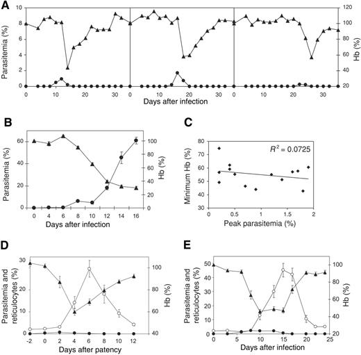
Semi-immune BALB/c mice were generated by successive cycles of infection with 104P berghei followed by antimalarial drug-cure. When challenged with 104P berghei, the kinetics of infection in the semi-immune cohort varied considerably between individuals (Figure 1A) and the day of patency occurred between day 10 to day 22 after infection. Independent of the time-to-patency, parasitemias resolved rapidly, without chemotherapy, within 6 days of patency. The peak values and kinetics experienced by individual animals were averaged (Table 1). In semi-immune mice, the average peak parasitemia was 0.94%, which occurred approximately 2 days after patency. Naive, age-matched BALB/c inoculum controls developed parasite burdens in excess of 60% by day 16 after infection (Figure 1B). At this time, naive mice were severely anemic, with Hb levels at 30% of baseline. Naive animals displayed a high incidence and severity of hematuria, and anemia was attributed to the hemolysis of infected RBCs. In contrast, semi-immune mice experienced severe anemia in response to a self-resolving, low burden of parasitemia (< 1%), with rapid decreases in Hb following the peak in parasitemia (Figure 1A). Minimum Hb levels were seen 4.1 days after patency, 2 days after the peak in parasitemia, representing a loss of 45% of baseline Hb in 96 hours. Hb levels were strongly correlated with Hct (R2 = 0.835), consistent with the normochromic profile of SMA in humans. Reticulocyte levels peaked at 35% of circulating cells at 5.9 days after patency, 2 days after the anemic crisis, and returned to baseline upon the resolution of anemia. The correlation between minimum Hb and maximum parasitemia was very poor (R2 = 0.073, Figure 1C), indicating that the degree of anemia was independent of the level of peak parasitemia. Although the day of patency was highly variable, it did not impact the development of anemia as minimum Hb levels were independent of the time to patency (R2 = 0.054). Following patency, however, the subsequent kinetics of infection, anemia, and compensatory reticulocytosis were very tightly linked (Table 1). The data from individual mice were normalized to the day of patency (Figure 1D), revealing very low variance in the data set and establishing a novel murine model of SMA at low parasite burden.
The average kinetics and magnitude of peak parasitemia, minimum Hb, and peak reticulocyte levels during P berghei infection of semi-immune mice
. | Peak day after patency . | Peak value, % . |
|---|---|---|
| Parasitemia | 1.7 ± 0.40 | 0.94 ± 0.17 |
| Minimum Hb | 4.1 ± 0.25 | 55.2 ± 2.2 |
| Reticulocytes | 5.9 ± 0.39 | 34.8 ± 3.9 |
. | Peak day after patency . | Peak value, % . |
|---|---|---|
| Parasitemia | 1.7 ± 0.40 | 0.94 ± 0.17 |
| Minimum Hb | 4.1 ± 0.25 | 55.2 ± 2.2 |
| Reticulocytes | 5.9 ± 0.39 | 34.8 ± 3.9 |
n = 14 ± SEM.
Severe malarial anemia at low parasite burden in rodent malaria infections. (A) Representative data from 3 infected semi-immune BALB/c mice showing parasitemia (•) and Hb levels (▴) following infection with 104P berghei. (B) Parasitemia (•) and Hb levels (▴) of naive inoculum control BALB/c mice. (C) Lack of correlation between peak parasitemia and minimum Hb levels in infected semi-immune mice. (D) Parasitemia (•), reticulocyte levels (○), and Hb (▴) of semi-immune mice normalized to the day of patency. (E) Mean parasitemias (•), reticulocyte levels (○), and Hb (▴) in naive adult rats infected with P berghei. n = 14 ± SEM for immune mice; n = 6 ± SEM for naive mice and naive rats.
Severe malarial anemia at low parasite burden in rodent malaria infections. (A) Representative data from 3 infected semi-immune BALB/c mice showing parasitemia (•) and Hb levels (▴) following infection with 104P berghei. (B) Parasitemia (•) and Hb levels (▴) of naive inoculum control BALB/c mice. (C) Lack of correlation between peak parasitemia and minimum Hb levels in infected semi-immune mice. (D) Parasitemia (•), reticulocyte levels (○), and Hb (▴) of semi-immune mice normalized to the day of patency. (E) Mean parasitemias (•), reticulocyte levels (○), and Hb (▴) in naive adult rats infected with P berghei. n = 14 ± SEM for immune mice; n = 6 ± SEM for naive mice and naive rats.
Naive rat model of SMA at low parasite burden
We sought to determine whether SMA at low parasite burden could be replicated in naive rodent hosts. Naive Wistar rats aged 15 weeks were challenged with 106P berghei, and parasitemia, Hb, and reticulocyte levels were determined over the course of infection (Figure 1E) as well as mean minimum and maximum values (Table 2). The period of patent infection was protracted, beginning on days 6 to 8 after infection, peaking at 3.4% iRBC and resolving by days 15 to 17 after infection. Following the onset of patency, there was a rapid and considerable loss of Hb beginning on days 8 to 10 after infection, with prolonged anemia reaching an average nadir of 39% of baseline on day 13.0 after infection. Sustained reticulocyte production occurred in response to anemia, with a sharp escalation in circulating reticulocytes at day 13, which remained high until day 17. This facilitated an increase in Hb levels and reticulocyte levels returned to basal levels as rats recovered from anemia.
Magnitude and kinetics of individual peak parasitemia, minimum Hb, and peak reticulocyte levels during P berghei infection of naive rats
. | Peak day after patency . | Peak value, % . |
|---|---|---|
| Parasitemia | 9.6 ± 0.6 | 3.4 ± 0.5 |
| Minimum Hb | 13.0 ± 0.9 | 39.0 ± 1.4 |
| Reticulocytes | 16.0 ± 0.3 | 52.0 ± 4.2 |
. | Peak day after patency . | Peak value, % . |
|---|---|---|
| Parasitemia | 9.6 ± 0.6 | 3.4 ± 0.5 |
| Minimum Hb | 13.0 ± 0.9 | 39.0 ± 1.4 |
| Reticulocytes | 16.0 ± 0.3 | 52.0 ± 4.2 |
n = 6 ± SEM.
The period and duration of patent infection was linked to the period and duration of anemia in both the semi-immune mouse model and the naive rat model of SMA. Semi-immune mice experienced an average of 4.6 ± 0.5 days of patent parasitemia and 5.1 ± 0.5 days of anemia (< 75% of baseline Hb). In naive rats, patent infections lasted an average of 10.3 ± 0.2 days and the corresponding period of anemia was 9.9 ± 0.2 days. The cumulative parasitemias over the course of infection could not account for the magnitude of anemia experienced, and while the degree of anemia was independent of peak parasitemias (Figure 1C), the duration of patency and duration of anemia were closely associated in both rodent models.
Reticulocyte production during SMA
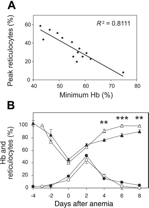
To assess the responsiveness of the erythropoietic compartment, reticulocyte production during SMA was examined in semi-immune mice. The peak level of circulating reticulocytes correlated with the degree of anemia (Figure 2A). To determine whether the magnitude of the erythropoietic response was physiologically appropriate, reticulocyte production during SMA was compared with that in mice with similar losses of Hb due to nonmalarial causes. The administration of PHZ in vivo results in the hemolysis of RBCs, inducing a dose-dependent anemia with appropriate compensatory erythropoiesis. PHZ-treated mice experienced a reduction of Hb to 40% of baseline levels, and a subset of mice with comparable levels of SMA was selected from a semi-immune cohort and data were normalized to the day of minimum Hb (Figure 2B). Intravascular hemolysis caused pronounced hematuria in PHZ-treated mice, as was observed in naive hyperparasitemic animals. However, this condition was not observed in the semi-immune SMA model despite a similar magnitude of anemia, indicating that intravascular hemolysis was not a major contributory mechanism to SMA in the semi-immune model. The kinetics and magnitude of reticulocyte production were similar in both PHZ-treated and SMA mice, with reticulocyte responses peaking 2 days after minimum Hb levels at 45% and 50%, respectively. Thus, the erythropoietic response to SMA was intact and appropriate for the degree of anemia experienced. Hb levels in PHZ-treated mice returned to baseline 7.0 ± 1.0 days after reaching minimum levels. Despite comparable levels of severe anemia and compensatory reticulocytosis, malaria-infected mice had significantly reduced Hb levels on days 4, 6, and 8 (P < .01) (Figure 2B) and took 12 ± 1.3 days to return to baseline (not shown). This reduced capacity to restore Hb suggests ongoing destruction or compromised survival of RBCs during recovery from SMA.
Erythropoietic response to severe malarial anemia in semi-immune mice. (A) Correlation between peak reticulocyte levels and minimum Hb levels during SMA in semi-immune mice (n = 14). (B) Hb (▴ and ▵) and reticulocyte (• and ○) levels ± SEM in mice with SMA (▴ and •, n = 4) or nonmalarial PHZ-induced anemia (▵ and ○, n = 6). Mice were selected for equivalent levels of anemia and data are normalized to the day of peak anemia (day = 0). **P < .01; ***P < .001.
Erythropoietic response to severe malarial anemia in semi-immune mice. (A) Correlation between peak reticulocyte levels and minimum Hb levels during SMA in semi-immune mice (n = 14). (B) Hb (▴ and ▵) and reticulocyte (• and ○) levels ± SEM in mice with SMA (▴ and •, n = 4) or nonmalarial PHZ-induced anemia (▵ and ○, n = 6). Mice were selected for equivalent levels of anemia and data are normalized to the day of peak anemia (day = 0). **P < .01; ***P < .001.
Accelerated RBC turnover during SMA
To determine the survival rates of RBCs in the semi-immune model of SMA, in vivo biotinylation of the circulating blood compartment was undertaken on the day of patency and cell survival was traced by flow cytometry (Figure 3A). Biotinylation of RBCs is widely used in experimental and clinical settings20,21 and revealed no impact on RBC survival in vivo (Figure 3A, inset). During the course of SMA, there was a 50% reduction in labeled RBCs between day 0 and day 4 after patency, with a concurrent 49% reduction in Hb levels (Figure 3A), demonstrating that the clearance of RBCs from circulation accounts for the severity of anemia. Between days 4 and 8 after patency, Hb levels stabilized and began to rise; however, the level of labeled RBCs continued to decrease, falling to 1% of circulating cells by day 8 after patency. This indicated that the entire initial blood compartment (9 × 10⁹ cells) had been turned over in approximately one week (Figure 3B). Animals were followed for 20 days after patency till baseline Hb levels were restored, and labeled RBCs did not reappear in circulation throughout this time (data not shown). This demonstrates that labeled RBCs did not participate in the restoration of Hb levels and suggests that the RBCs had been destroyed.
The extent and rate of RBC clearance in the naive Wistar rat model of SMA was determined by in vivo biotinylation of circulating RBCs at different time points following P berghei infection (Figure 3C). Cells were labeled on day 6, the day of patency; day 10, the day of peak anemia; and day 13, the midpoint of the anemic crisis, and showed similar rates of clearance from circulation (Figure 3C). A 50% reduction in the total number of labeled RBCs in circulation was observed 4.3, 3.2, and 4.0 days from days 6, 10, and 13 after infection, respectively, indicating that accelerated RBC turnover occurred at similar rates throughout the course of infection. Of interest, the level of biotinylated RBCs continued to decrease between days 17 and 24 after infection (Figure 3C) when parasites were no longer detectable in circulation. This suggests that the mechanism driving RBC clearance remained active after patent parasitemias had resolved.
Accelerated RBC turnover during severe malarial anemia. (A) Survival of the circulating RBC population biotinylated on the day of patency in semi-immune mice infected with P berghei (▪) and Hb levels (▵). Basal turnover of biotinylated RBCs in noninfected mice is shown in inset (□). Cell survival was monitored by flow cytometry and is expressed as a percentage change in total number of initial biotinylated RBCs. n = 4 ± SEM. (B) Representative flow cytometry profile of biotinylated RBCs in (i) an infected semi-immune mouse and (ii) naive resting control mouse on days 0 (day of labeling) and 8 after patency. (C) Survival of circulating RBC populations in naive rats, biotinylated on day 6 (•), day 10 (□), and day 13 (▴) after patency. Basal turnover of biotinylated RBCs in uninfected rats is shown in inset (○). Cell survival was monitored by flow cytometry and is expressed as a percentage change in total number of initial biotinylated RBCs. n = 3 ± SEM per group.
Accelerated RBC turnover during severe malarial anemia. (A) Survival of the circulating RBC population biotinylated on the day of patency in semi-immune mice infected with P berghei (▪) and Hb levels (▵). Basal turnover of biotinylated RBCs in noninfected mice is shown in inset (□). Cell survival was monitored by flow cytometry and is expressed as a percentage change in total number of initial biotinylated RBCs. n = 4 ± SEM. (B) Representative flow cytometry profile of biotinylated RBCs in (i) an infected semi-immune mouse and (ii) naive resting control mouse on days 0 (day of labeling) and 8 after patency. (C) Survival of circulating RBC populations in naive rats, biotinylated on day 6 (•), day 10 (□), and day 13 (▴) after patency. Basal turnover of biotinylated RBCs in uninfected rats is shown in inset (○). Cell survival was monitored by flow cytometry and is expressed as a percentage change in total number of initial biotinylated RBCs. n = 3 ± SEM per group.
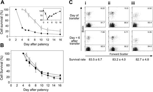
RBC fate is not determined by changes to target cells. (A) Naive RBCs were labeled with CSFE (•) or biotin (○) and adoptively transferred into infected semi-immune mice on day 2 or day 4 after patency, respectively. Survival was monitored by flow cytometry and is expressed as a percentage change in total number of transferred RBCs; n = 6 ± SEM. The inset shows the percentage change in Hb (▴) in semi-immune mice over the same period of days after patency. (B) Comparison of rate of the survival of naive CSFE-labeled (•), naive biotin-labeled (○), and resident host biotin-labeled (▪) cells in semi-immune mice. Values are expressed as percentage change in the total number of cells present from day 4 after patency (100%). n = 6 ± SEM. (C) RBCs from (i) noninfected naive mice, (ii) nonanemic semi-immune mice on the day of patency, and (iii) anemic semi-immune mice on day 4 after patency were biotinylated in vivo and adoptively transferred into naive recipients. Survival was monitored by flow cytometry. Plots show circulating RBC populations in the recipients on the day of transfer (day 0) and day 6 after transfer. Survival rate values show the mean percentage survival of cells from each donor group on day 6 after transfer. n = 5 ± SEM.
RBC fate is not determined by changes to target cells. (A) Naive RBCs were labeled with CSFE (•) or biotin (○) and adoptively transferred into infected semi-immune mice on day 2 or day 4 after patency, respectively. Survival was monitored by flow cytometry and is expressed as a percentage change in total number of transferred RBCs; n = 6 ± SEM. The inset shows the percentage change in Hb (▴) in semi-immune mice over the same period of days after patency. (B) Comparison of rate of the survival of naive CSFE-labeled (•), naive biotin-labeled (○), and resident host biotin-labeled (▪) cells in semi-immune mice. Values are expressed as percentage change in the total number of cells present from day 4 after patency (100%). n = 6 ± SEM. (C) RBCs from (i) noninfected naive mice, (ii) nonanemic semi-immune mice on the day of patency, and (iii) anemic semi-immune mice on day 4 after patency were biotinylated in vivo and adoptively transferred into naive recipients. Survival was monitored by flow cytometry. Plots show circulating RBC populations in the recipients on the day of transfer (day 0) and day 6 after transfer. Survival rate values show the mean percentage survival of cells from each donor group on day 6 after transfer. n = 5 ± SEM.
Accelerated turnover of transfused RBCs during SMA
We sought to compare the clearance rates of freshly transfused RBCs with those of resident host RBCs during SMA. Differentially labeled RBCs from naive noninfected mice were transfused into infected semi-immune mice at day 2 after patency (CFSE-labeled) and day 4 after patency (biotin-labeled). Transfer populations were limited to 5% to 10% of circulating cells to minimize the impact on recipient Hct. To control for the efficiency of RBC transfer between groups, cell survival was expressed as the percentage change in the total number of transferred cells in circulation, as monitored by flow cytometry (Figure 4A). When transferred into noninfected naive controls, CFSE- and biotin-labeled RBCs showed survival rates of 66% and 65%, respectively, by day 14 after transfer (not shown). However, when transferred into SMA mice on day 2 after patency, naive CSFE-labeled RBCs underwent rapid clearance with a 68% decrease in cell number within 48 hours of transfer. This corresponded with the period of anemic crisis of the host (Figure 4A, inset). Naive biotin-labeled cells were then transferred into semi-immune mice on day 4 after patency, the point of minimum Hb. These transferred cells were 50% cleared from circulation 4.5 days after transfer, corresponding to day 8.5 after patency. The clearance rates of naive cells transferred on days 2 and 4 after patency were compared with the clearance rate of labeled host RBCs (Figure 4B). Cell survival is expressed as the percentage change of total cell number from day 4 after patency onward. Clearance rates were not significantly different, regardless of whether RBCs were long-term host residents or had been transferred at different time points after patency. Thus during SMA, RBCs transferred from naive donors were cleared concurrently with resident host cells, and the length of time RBCs were exposed to the host environment in infected anemic animals did not influence cell survival rates.
Normal survival of RBCs from SMA donors in resting recipients
Next, we sought to determine whether RBCs taken from semi-immune mice undergoing SMA were subject to accelerated clearance when transferred into naive resting recipients. RBCs were taken from 3 donor groups: noninfected, nonanemic controls (Figure 4Ci); infected semi-immune mice on the day of patency (Figure 4Cii); and infected semi-immune mice 4 days after patency, during the peak of anemic crisis (Figure 4Ciii). Donor RBCs were biotinylated in vivo, harvested, transferred into naive recipients, and traced by flow cytometry to determine cell survival. RBCs from the 3 donor groups showed no difference in their rates of clearance in naive recipients (Figure 4C). On day 6 after transfer, the survival of RBCs was 83%, 84%, and 83% for control, day of patency, and day 4 after patency donors, respectively (P = .46). Therefore, RBCs from SMA donors displayed normal survival in naive resting recipients, suggesting no intrinsic changes to the RBCs themselves. Thus, alterations to the host environment may be responsible for the accelerated RBC turnover clearance during SMA.
Depletion of phagocytic cells alleviates SMA
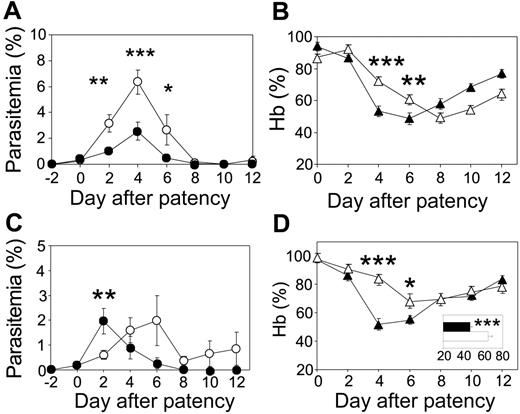
The contribution of host phagocytic cells to the development of SMA was investigated by liposome-encapsulated clodronate depletion of macrophage populations.22 Clodronate depletion of phagocytic macrophages resulted in significantly higher peak parasitemias, 6.3% ± 0.9% by day 4 after patency versus 2.5% ± 0.7% for PBS-treated controls (P < .001) (Figure 5A). Thus, macrophages contribute significantly to effector mechanisms controlling P berghei parasitemia in semi-immune mice. However, despite increased parasite burdens, the depletion of phagocytic cells alleviated the severity of SMA, with higher Hb levels on day 4 (P < .001) and day 6 (P < .01) after patency (Figure 5B). Protection from SMA after phagocyte depletion was transient, with the anemic crisis delayed by 4 days. The Hb profile of mice depleted of phagocytic macrophages was significantly different to that of controls across the entire curve (P < .001), implicating this population in the pathogenesis of SMA.
Depletion of immune effector cells alleviates SMA in semi-immune mice. (A) Parasitemia and (B) Hb levels, in phagocyte-depleted semi-immune mice (open symbols) and control semi-immune mice (closed symbols), normalized to the day of patency. n = 9 ± SEM. (C) Parasitemia and (D) Hb levels, in CD4+ cell–depleted (open symbols) and control (closed symbols) mice, normalized to day of patency. Mean individual minimal Hb levels for CD4+ cell–depleted (open bars) and controls (closed bars) are shown in the inset. n = 8 ± SEM for the CD4+ T-cell–depleted and n = 7 ± SEM for control groups. *P < .05; **P < .01; ***P < .001.
Depletion of immune effector cells alleviates SMA in semi-immune mice. (A) Parasitemia and (B) Hb levels, in phagocyte-depleted semi-immune mice (open symbols) and control semi-immune mice (closed symbols), normalized to the day of patency. n = 9 ± SEM. (C) Parasitemia and (D) Hb levels, in CD4+ cell–depleted (open symbols) and control (closed symbols) mice, normalized to day of patency. Mean individual minimal Hb levels for CD4+ cell–depleted (open bars) and controls (closed bars) are shown in the inset. n = 8 ± SEM for the CD4+ T-cell–depleted and n = 7 ± SEM for control groups. *P < .05; **P < .01; ***P < .001.
CD4+ T cells contribute to the severity of SMA
As T lymphocytes are key regulators of macrophage function, we sought to determine whether CD4+ T cells play a role in the etiology of SMA of low parasite burden. CD4+ T cells of infected semi-immune mice were depleted by in vivo administration of lytic monoclonal antibody from the day of patency. Controls received a nonlytic antibody of the same isotype. The average peak parasitemias in the 2 groups were similar at 2.0% (Figure 5C). Depletion of CD4+ T cells modestly influenced the kinetics of infection with a delay in peak parasitemia and an inability to completely resolve infection (Figure 5C). Despite this, mice depleted of CD4+ T cells showed delayed kinetics in the onset of the anemic crisis (Figure 5D) with significantly higher Hb levels on day 4 (P < .001) and day 6 (P < .05) after patency. Across the cohort, anemia was significantly alleviated in CD4-depleted mice with average minimum Hb levels of 65% of baseline compared with 47% in controls (P < .001; Figure 5D, inset).
Discussion
It has long been proposed that the degree of anemia experienced during clinical malaria infection cannot be completely accounted for by the loss of RBCs due to the level of parasitemia, leading to the idea that SMA arises in part from the destruction of nonparasitized cells.4,16 Clinical and experimental malaria literature has acknowledged that acute infections in naive mouse models do not accurately recapitulate this phenomenon, due to the high burdens of parasitemia experienced.23,24 The novel semi-immune BALB/c model developed in this study, however, shows the development of SMA at low parasite burden. Following normalization to the day of patency, hematologic profiles displayed highly correlated and predictable parameters of infection, anemia, and recovery, thus establishing the semi-immune BALB/c mouse as a robust and amenable experimental model of SMA.
The profiles of SMA from individual semi-immune BALB/c mice bear a striking resemblance to those of semi-immune Aotus monkeys challenged with P falciparum,14 with the onset of anemia at low parasite burden closely following the development of a patent infection. Of importance, similar profiles of SMA at low parasite burden are seen in both experimental P falciparum infections in naive humans4 and in naturally acquired infections in endemic regions.3 SMA of low parasite burden is consistently observed in multiple host-Plasmodium combinations: semi-immune mouse-berghei (Figure 1D), naive rat-berghei (Figure 1E), semi-immune Aotus-falciparum,14,15 semi-immune and naive human-falciparum,4 and naive human-vivax,16 suggesting that the mechanisms of pathogenesis are conserved across taxa. The conclusion of Jakeman et al4 and Egan et al14 regarding the pathogenesis of SMA in P falciparum infections of Aotus and humans was that anemia must result from the accelerated destruction of uRBCs; however, no experimental evidence of this phenomenon has been presented. The semi-immune BALB/c mouse model and the naive Wistar rat model of SMA at low parasite burden provide experimental demonstration of the accelerated destruction of uRBCs during malaria infection.
In the semi-immune mouse model, severe anemia develops in response to malaria infection at low parasite burden in the presence of appropriate erythropoietic responses. Therefore, this model allows assessment of the contribution of increased RBC destruction to the development of anemia, as confounding factors such as decreased RBC production and RBC loss due to parasite invasion have been minimized. In semi-immune mice, the clearance of RBCs from circulation accounted for the severity of Hb loss at the onset of SMA; however, accelerated turnover of RBCs continued even while Hb levels were restored (Figure 3A). The extent of RBC loss during the anemic crisis included the entire initial blood compartment of 9 × 10⁹ cells and may indeed be greater due to ongoing RBC loss during the recovery phase. In semi-immune mice, cumulative parasitemias were calculated to be approximately 3% of circulating cells over the course of infection, which suggests a ratio of more than 30:1 loss of nonparasitized–parasitized RBCs during SMA. Previously calculated ratios of approximately 10:1 during human SMA4,5 may therefore be underestimates, as RBC losses were deduced from net minimum Hb values without adjusting for ongoing cell turnover during recovery. There is evidence for continuing RBC turnover during human SMA, as recovery rates of Hb are reduced compared with anemia due to other causes (eg, blood loss trauma),5 and anemia is known to persist after clearance of parasites.25 This phenomenon was recapitulated in the murine model, with delayed recovery from SMA compared with PHZ-induced hemolysis (Figure 2B). Together, the data support a protracted period after peak anemia, in which clearance of RBCs continues in the absence of a patent infection, accounting for the impeded return to baseline Hb levels during SMA.
Accelerated RBC turnover in human SMA is proposed to result from acquired changes to the surface or structure of noninfected RBCs that target them for destruction either by intravascular hemolysis or immune-mediated clearance.6 High levels of intravascular hemolysis induce hematuria in both mice26 and humans,27 but hematuria is uncommon in clinical malaria infections in endemic areas28 and was not observed in the SMA models elucidated here. Hb balance studies of malaria patients suggest that extravascular clearance is the major mechanism of RBC destruction during infection,29 and therefore it is unlikely that intravascular hemolysis is a major contributory mechanism to SMA at low parasite burden.
RBC transfer experiments revealed that cells fated for destruction in hosts experiencing SMA did not express sufficient changes to cause accelerated clearance in a normal resting animal, and, upon transfusion, fresh cells from naive donors were cleared at the same rate as resident RBCs in an SMA host (Figure 4). These findings suggest that changes to target RBCs play a minimal role in the etiology of SMA in semi-immune mice, and, conversely, mice experiencing SMA express a physiologic status sufficient to cause accelerated destruction of RBCs independent of the target cell history. This is consistent with the previously reported lack of detectable modifications such as immunoglobulin or complement deposition on RBCs in the semi-immune Aotus model15 and the lack of a consistent association of positive direct antiglobulin test (DAT) and SMA in humans.30,31 The survival curves of naive RBCs transferred into infected semi-immune mice showed strong similarities to the survival curves of compatible donor RBCs transferred into the circulation of patients recovering from P falciparum and P vivax infection.10,32 These studies demonstrated that during human malaria infection RBCs have a significantly shortened mean cell half-life and that both autologous and compatible donor RBCs experience clearance from circulation at equal rates following successful treatment with antimalarial drugs. This provides a clinical validation of the processes occurring in rodent models and the combined transfer data suggest that changes extrinsic to the RBCs themselves may be responsible for their accelerated clearance. Further to this, transfusion studies conducted during the treatment of anemic Kenyan children showed that 25% of patients receiving blood transfusions experienced rises in Hb of less than 20 g/L (2 g/dL), and that 25% of severely anemic children maintained Hb less than 50 g/L (5 g/dL) after transfusion.33 A continuing destruction of RBCs, independent of their source, may account for exacerbated anemia in the face of appropriate therapeutic intervention and explain the relatively poor ability of transfusion to elevate Hb levels in these patients.
The significant alleviation of SMA by depletion of host CD4+ T cells or phagocytic cells (Figure 5) establishes the contribution of immune mechanisms to the pathogenesis of SMA. Hb levels were 35% greater in phagocyte-depleted and 64% greater in CD4+-depleted semi-immune mice on day 4 after patency compared with nontreated controls. The data presented here strongly support the notion that, akin to other life-threatening malarial pathologies,17 SMA is mediated in part by immunopathogenic mechanisms. In particular, we propose that the destruction of uRBCs may result from a hyperactivated phagocytic system. Phagocytosis of noninfected RBCs has been documented during human infections,34,35 and hyperphagocytosis has been implicated in malarial thrombocytopenia.36 Constitutive or basal homeostatic RBC clearance is predominantly mediated by splenic red pulp macrophages, and increased recruitment or activation of this population during malaria infection may accelerate splenic RBC clearance. SMA in humans is associated with high serum levels of neopterin, a marker of macrophage activation, induced particularly by IFN-γ.37 These observations have led to the proposal that SMA has an inflammatory etiology.38 Counterregulatory TH2 cytokines such as IL-437 and IL-1039,40 are inversely associated with malarial anemia, suggesting that a loss of host cytokine regulation contributes to the severity of disease. Thus, the accelerated turnover of RBCs during SMA may be regulated by diverse factors controlling macrophage phagocytic activity and recruitment, such as host CD4+ T cells, cytokine/chemokine cascades, and bioactive parasite products such as hemozoin41 and glycosylphosphatidylinositol.42,43 Significantly, adoptive transfer of parasite-specific, TH1 CD4+ T-cell lines promotes anemia during naive P berghei infections,44 and this may relate to the capacity of CD4+ T cells to up-regulate macrophages. As SMA is pronounced in Aotus immunized with P falciparum antigens,14,15 blood-stage vaccines that suppress parasitemias may not necessarily protect against the development of anemia. Whether T-cell priming by these experimental vaccines promotes SMA is not clear. Rodent models of SMA may provide useful preclinical test systems to assess further the efficacy of vaccines in prevention or promotion of this disease state.
SMA does not result in fatality in the animal models presented here, as physiologically appropriate reticulocytosis eventually restored Hb to basal levels. In rodents, the erythropoietic response to acute anemia occurs predominantly in the spleen, and it is likely that extramedullary erythropoiesis facilitates the recovery from SMA in these models. However, in humans, SMA could be exacerbated or even fatal should erythropoietic suppression prevent adequate reticulocyte compensation. P falciparum infection is often associated with impaired erythropoietic responses, with patients displaying suboptimal reticulocyte levels for the degree of malarial anemia experienced.45,46 Malaria-induced erythropoietic suppression has been studied in acute infections of experimental animal models, which have provided insights into the mechanisms of decreased RBC production during infection.47-50 During malaria infection, a decreased responsiveness to adequate levels of erythropoietin may lead to suppressed development of erythroid precursors, resulting in an inappropriately low level of production of new RBCs, as recently reviewed.24 Therefore, during SMA, erythropoietic suppression may overlap with the accelerated destruction of RBCs, leading to an exacerbated syndrome and fatality. The quantitative contribution of each mechanism may vary across differing clinical and epidemiologic settings, contributing to varying patterns of malarial pathology.51 As rodents are tractable models amenable to experimental perturbation, it may now be possible in both acute and semi-immune infections to investigate more closely the multiple host and parasite factors proposed to influence these 2 contributory mechanisms to SMA in vivo. These findings thus extend further the use of rodent malarias as models for disease processes in humans.
Prepublished online as Blood First Edition Paper, October 6, 2005; DOI 10.1182/blood-2005-08-3460.
Supported by NH&MRC, NIH, HFSP, and the UNDP/World Bank/WHO Program (TDR). L.S. is an International Research Scholar of the Howard Hughes Medical Institute.
An Inside Blood analysis of this article appears at the front of this issue.
The publication costs of this article were defrayed in part by page charge payment. Therefore, and solely to indicate this fact, this article is hereby marked “advertisement” in accordance with 18 U.S.C. section 1734.


This feature is available to Subscribers Only
Sign In or Create an Account Close Modal