The marrow microenvironment consists of several different interacting cell types, including hematopoietic-derived monocyte/macrophages and nonhematopoietic-derived stromal cells. Gene-expression profiles of stromal cells and monocytes cultured together differ from those of each population alone. Here, we report that CXCL7 gene expression, previously described as limited to the megakaryocyte lineage, is expressed by monocytes cocultured with stromal cells. CXCL7 gene expression was confirmed by quantitative reverse transcriptase–polymerase chain reaction (RT-PCR), and secretion of protein was detected by enzyme-linked immunosorbent assay (ELISA) and Western blot. At least 2 stromal-derived activities, one yet to be identified, were required for optimal expression of CXCL7 by monocytes. NAP-2, the shortest form of CXCL7 detected in the coculture media, was confirmed to decrease the size and number of CFU-Meg colonies. The propeptide LDGF, previously reported to be mitogenic for fibroblasts, was not secreted by stimulated monocytes. The re-combinant form of LDGF produced in a prokaryotic expression system did not have biologic activity in our hands. The monocytic source of CXCL7 was also detected by immunohistochemistry in normal bone marrow biopsies, indicating an in vivo function. We conclude that stromal-stimulated monocytes can serve as an additional source for CXCL7 peptides in the microenvironment and may contribute to the local regulation of megakaryocytopoiesis.
Introduction
Hematopoietic regulation takes place within the marrow microenvironment (ME), which is defined by function as a complex of cells and cell products critical for the maintenance and regulation of stem cells and their progeny. The ME contains mesenchymal cells (eg, fibroblasts, adipocytes, and smooth muscle, all referred to as stromal cells); it also contains hematopoietic-derived monocytes/macrophages. Primary long-term cultures (LTCs) of marrow stroma have been used to approximate the ME in vitro.1 The model has proven useful for identifying cells and factors that play a role in supporting hematopoiesis. However, the heterogeneity of the cell types that comprise a functional LTC have led many investigators to generate stromal cell lines to dissect ME functional components.2-4 We have previously reported on immortalized stromal lines generated by transducing human stromal cells with E6/E7 genes from human papilloma virus (HPV).5,6 Two of these lines have been studied and characterized in detail. One, designated HS27a, supports maintenance of early hematopoietic progenitors in “cobblestone areas” and does not secrete growth factors. The other, designated HS5, does not support cobblestone areas but does secrete copious amounts of growth factors. These lines have hence been hypothesized to represent cell types that contribute to functionally distinct niches.
Monocyte-derived macrophages have long been known as critical components of both the in vivo and in vitro ME. In vivo, the macrophage can function as the “nurse cell” of the erythropoietic islands in bone marrow. In vitro, the monocytes, which arise from the hematopoietic stem cell, can form up to 40% of total cells in LTCs despite serial passages.7 Monocytes/macrophages are known to secrete large quantities of several cytokines, including IL-1, IL-6, G-CSF, TNF, PDGF, TGF-β, and M-CSF, all of which can affect both stromal cells and hematopoietic cells.8-13 Increasing proportions of macrophages in primary LTCs are associated with decreasing numbers of hematopoietic progenitors.14,15 It is unclear in this case whether the monocyte/macrophage directly affects progenitors or stromal function, or both. There is also evidence that monocyte function is altered by exposure to stromal cells.16
Our laboratory has used microarray technology to compare changes in gene expression that occur when monocytes and stromal cells are cultured together compared with both cell types cultured alone.16 Several genes are up-regulated in the cocultures. One that was unexpected is CXCL7, a chemokine previously thought to be expressed only within the megakaryocyte lineage. CXCL7 is translated as a propeptide, then cleaved to several smaller forms each reported to have specific functions. The longest form, called PPBP (pro-platelet basic protein) or LDGF (leukocyte-derived growth factor), is reported to be a fibroblast mitogen.17 The shortest form, called NAP-2 (neutrophil activating peptide-2), is reported to inhibit megakaryocytopoiesis.18 Thus, the CXCL7 peptides, if derived from stroma-stimulated monocytes in vivo, have potential functional significance within the ME.
Materials and methods
Cell culture
All cells were cultured at 37°C under 95% air, 5% CO2. Human marrow stromal cell lines HS5 and HS27a were grown in RPMI 1640 medium (Gibco, Carlsbad, CA), supplemented with l-Glutamine (0.4 mg/mL), sodium pyruvate (1 mM), and 10% fetal calf serum (FCS). After reaching confluency, cells were trypsinized and transferred at a cell density of 40 × 103/cm2 to multiwell plates for coculture studies. At this density, cultures were about 80% confluent and reached complete confluency at 3 days.
NIH/3T3 cells were obtained from ATCC (Manassas, VA) and were cultured in DMEM supplemented with 10% FCS and penicillin (100 IU/mL), streptomycin (100 μg/mL). U937 and HEK 293 cells were also obtained from ATCC and cultured in RPMI 1640 medium supplemented with 10% FCS and penicillin (100 IU/mL), streptomycin (100 μg/mL).
Bone marrow aspirates and peripheral-blood samples were obtained from healthy donors after obtaining written informed consent using forms approved by the Institutional Review Board (IRB) of the Fred Hutchinson Cancer Research Center (FHCRC). Primary LTCs from marrow of healthy donors were prepared using methods previously described.19 To obtain primary stromal fibroblasts, 50 to 75 × 106 bone marrow mononuclear cells were plated in T-75 flasks in Alpha-MEM media with 10% FCS for 20 to 24 hours. The nonadherent cells were washed off, and the adherent cells were collected with 0.05% trypsin. The collected cells were incubated with CD14 and CD45 microbeads (Miltenyi Biotec, Auburn, CA) at a concentration of 20 μL/107 cells for 20 minutes. The CD45–/CD14– cells were negatively selected with the AutoMACS (magnetic cell sorting) apparatus. The final product, containing less than 1% CD14+ and 5% CD45+ cells, was plated in Alpha-MEM supplemented with 10% FCS and grown to confluence.
Normal peripheral-blood mononuclear cells (PBMCs) were isolated over a Ficoll step gradient, hemolyzed, and washed 3 times in Hanks Balanced salt solution (HBSS). CD14+ cells were isolated by immunomagnetic bead separation as previously described.16 Briefly, PBMCs were incubated with monoclonal antibody against CD14 followed by an anti–mouse immunoglobulin–conjugated magnetic beads followed by magnetic separation, done twice. Purity of CD14+ cells from healthy donors was greater than 95%. In some experiments, CD14+ cells were cultured in a transwell insert (Millicell-CM, 0.4-μm mesh, 30-mm diameter; Millipore, Billerica, MA), and the nonadherent cells were collected for RNA isolation.
CD34-enriched cells from G-CSF–mobilized blood mononuclear cells were obtained through the Cell Processing Shared Resource of the FHCRC, as previously described.20
ELISA and SDS–polyacrylamide gel electrophoresis (PAGE)
CXCL7 protein concentration in conditioned media was measured by enzyme-linked immunosorbent assay (ELISA) using Human CXCL7/NAP-2 DuoSet (R&D Systems, Minneapolis, MN). Measurements were done at least in duplicate for 2 dilutions. For sodium dodecyl sulfate–polyacrylamide electrophoresis (SDS-PAGE), precast gels (Invitrogen, Carlsbad, CA) with 16% acrylamide were used. Immunoblots of these gels were stained with Ponceau S (Sigma, St Louis, MO) for proteins, blocked in 5% nonfat milk in TBS, and probed sequentially with polyclonal rabbit antibodies against NAP-2 (PeproTech, Rocky Hill, NJ) and horseradish peroxidase (HRP)–conjugated secondary antibodies (Amersham Biosciences, Piscataway, NJ). Bound HRP activity was detected by an enhanced chemiluminescent method (Amersham). Prestained molecular weight marker proteins (Invitrogen) were used to calculate molecular weight of proteins.
Syber green real-time polymerase chain reaction (RT-PCR)
Expressed levels of CXCL7 mRNA were quantitated using real-time PCR. Total RNA was purified using RNeasy spin columns (Qiagen, Valencia, CA) per the manufacturer's specifications followed by reverse transcription into cDNA using oligo dT12-18 primer and superscript II reverse transcriptase (Invitrogen). RT-PCR was performed after mixing the cDNA with SYBR Green PCR master mix (Applied Biosystems, Foster City, CA) and appropriate primers. Products were quantitated with an ABI prism 7700 sequence detector (Applied Biosystems). Conditions for PCR were 95 degrees for 15 seconds and 68 degrees for 2 minutes for 40 cycles. Primers for CXCL7 were 5′-ACCATGAGCCTCAGACTTGATACC and 3′-TTAATCAGCAGATTCATCACCTGCC; primers for G3PDH were 5′-ATCGCTCTGAAATTAGCCTACTGCC and 3′-TAAGGCTGGCAAATA-CACCAACACC. Data shown are the mean ± SE for at least 2 samples.
Production of recombinant LDGF and PBP
CXCL7 peptides are encoded by the PPBP gene (Unigene Cluster Hs.2164). Posttranslational cleavage of the full-length peptide (LDGF) gives rise to the smaller forms. The full coding sequence of LDGF, including the kozak motif, was duplicated by RT-PCR from a cDNA template of stromal stimulated monocytes. The 5′ primer was CCC TGA GGCGGCCGC TTC TCC ACC ATG AGC CTC AGA CTT G with a NotI digestion site 5′ of the start codon and the 3′ primer was CCA GCG CAATTG TTA ATC AGC AGA TTC ATC ACC TGC TGC C with an MfeI site inserted 3′ of the stop codon (both restriction sites underlined). This product was cloned into TOPO-TA vector (Invitrogen), sequenced, and transferred to the expression cassette of pIRES-EGFP (Clonetech, Mountainview, CA). HEK 293 cells were transfected with this LDGF-pIRES-EGFP vector using Lipofectamine 2000 transfection agent (Invitrogen). Transfection efficiency as confirmed by GFP expression was greater than 80% at 48 hours. Transfected cells were cultured in serum-free suspension media CD293 (Invitrogen) for 5 days, and conditioned media were clarified by centrifugation. The conditioned media were found to have large quantities of CXCL7 peptide. The Proteomics Shared resource facility at the FHCRC confirmed by mass spectrometry that the peptide was identical to the known human CXCL7 sequence. Both the molecular weight and sequence analysis indicated that this peptide was not the full-length LDGF, but rather the largest cleaved form designated platelet basic protein (PBP).
PBP was purified with Hi-trap heparin Apharose columns (Amersham Biosciences, Uppsala, Sweden) using methods similar to those previously described.17 Briefly, the conditioned media were dialyzed against an equilibration buffer (50 mM NaCl, 50 mM Tris-Cl, pH 7.4) and then chromatographed on heparin Apharose columns also equilibrated with the same buffer. After further washes, PBP was eluted with 2.0 M NaCl and then dialyzed against the equilibration buffer and tested for the presence of the protein by Western blot analyses.
As the HEK 293 cells secreted the smaller PBP but not the full-length peptide (LDGF), we decided to use a bacterial expression system to make recombinant human LDGF (rhLDGF) using methods described by Iida et al.17 The coding sequence of LDGF was PCR-amplified from cDNA template using a 5′ primer CCC TGA GCA TAT GAG CCT CAG ACT TGA TAC CAC CCC (NdeI site immediately 5′ of the start codon) and 3′ primer CCA GCG GGA TCC TTA ATC AGC AGA TTC ATC ACC TGC (BamHI site immediately 3′ of the stop codon) and cloned into a pET3a vector (Stratagene, La Jolla, CA) which uses the T7 RNA polymerase promoter to drive the protein expression. BL21-Gold (DE3) expression strain of Escherichia coli (Stratagene) was then chemically transformed with the LDGF-pET3a vector using the manufacturer's protocol. Transformed strains were grown in L-B broth and induced with IPTG to produce the recombinant LDGF. After confirming that the recombinant protein was mostly in inclusion bodies, the inclusion bodies were isolated using chemical cell lysis and solubilized with 8 M urea using standard protocols. The extract was then purified for rhLDGF as described for rhPBP. The molecular weight of this product was ascertained by SDS-PAGE and Western blot, and the sequence was verified by mass spectrometry. Protein concentrations and purity of the recombinant products were verified by spectrophotometry and Coomassie blue staining after gel electrophoresis.
Immune histochemistry (IHC) for CXCL7 peptides in normal bone marrow
IHC for CXCL7 was performed by the Experimental Histopathology Laboratory at the FHCRC. Paraffin-embedded, B5-fixed marrow biopsies from healthy donors were sectioned at 4 μm and deparaffinized. Antigen retrieval was performed by steaming the slides in Dako Target Retrieval Solution, Citrate pH6 (DakoCytomation, Carpintera, CA) for 20 minutes and then cooling for 20 minutes. Biotin Blocking System (DakoCytomation) was applied to each slide for 10 minutes, followed by Alkaline Phosphatase Peroxidase Block (BioFX, Owings Mills, MD) for 10 minutes. Nonspecific protein blocking was accomplished using 15% swine serum and 5% human serum (Jackson ImmunoResearch Laboratories, West Grove, PA) diluted in Dako Wash Buffer with 1% bovine serum albumin (Sigma-Aldrich, St Louis, MO). For single antigen staining, the slides were incubated with 0.5 μg/mL anti–human polyclonal rabbit antibodies against CXCL7/NAP-2 (PeproTech) or 0.1 μg/mL anti–human monoclonal mouse antibody against CD68 (clone PG-M1; DakoCytomation) at room temperature for 30 minutes. Appropriate isotype controls (polyclonal rabbit and monoclonal IgG3, respectively) were also used alongside. Slides were then washed and incubated with HRP-conjugated Envision Plus (DakoCytomation) for 30 minutes. The HRP was visualized with 3,3-diaminobenzidine (DAB; DakoCytomation) for 7 minutes and counterstained with hematoxylin for 2 minutes. For double staining with both CD68 and CXCL7, the anti-CD68 antibody was applied a concentration of 1 μg/mL and incubated at room temperature for 30 minutes. CD68 staining was detected using biotinylated goat anti–mouse F(ab′)2 secondary antibody (Vector Laboratories, Burlingame, CA), Vectastain ABC-AP kit (Vector Laboratories), and Vector Blue Substrate Kit (Jackson ImmunoResearch Laboratories). The slides were blocked again with 15% swine serum and 5% human serum diluted in Dako Wash Buffer with 1% bovine serum albumin. The slides were then incubated with anti–CXCL7/NAP-2 antibody at a concentration of 0.5 μg/mL for 30 minutes at room temperature. NAP-2 staining was detected using EnVision Plus/HRP, Rabbit (DakoCytomation) and the NovaRED Substrate Kit (Vector Laboratories). The slides were cover-slipped using Faramount aqueous mounting media (DakoCytomation).
CFU-Meg assays
CFU-Meg assays were performed using MegaCult-C kit (Stem Cell Technologies, Vancouver, BC, Canada) per the manufacturer's instructions. Briefly, 5 × 10³ G-CSF–mobilized CD34+ cells were plated in a serum-free medium with thrombopoietin (50 ng/mL), IL-3 (10 ng/mL), and IL-6 (10 ng/mL) mixed with collagen, with or without the CXCL7 peptides. The colonies were dehydrated and fixed after adequate colony maturation (10-14 days) and stained with a primary antibody against CD41 (GpIIb-IIIa) linked to a secondary biotinylated antibody-alkaline phosphatase–conjugated detection system to amplify the signal. The stained slides are scored for both the size and number of colonies. Small colonies had 3 to 20 cells, medium had 21 to 49 cells, and large had 50 or more cells.
Fibroblast mitogenic assays
Mitogenic assays were performed with both NIH/3T3 fibroblasts and primary stromal fibroblasts using the MTT (3-(4,5 dimethylthiazol-2-ly) 2,5 diphenyltetrozolium bromide) assay as described.21 NIH/3T3 fibroblasts were plated at 20 × 103 cells per well in 24-well plates in DMEM supplemented with 10% FCS. After 24 hours, the cells were washed with HBSS, and the medium was changed to DMEM with 0.1% FCS for 48 hours. Fresh medium was added, with or without cytokines, 24 to 28 hours before the MTT (Sigma) was added. Primary marrow-derived fibroblasts were plated in 24-well plates at a density of 20 × 103 cells/well in Alpha-MEM with 10% FCS. After 24 hours, the cells were washed with HBSS, and the medium was changed to Alpha-MEM with 1% FCS. After 48 hours, fresh medium was added with or without cytokines, and the MTT assay was carried out at specified times.
Results
Stromal cells and stromal cell–conditioned media induce expression of CXCL7 in CD14+ cells
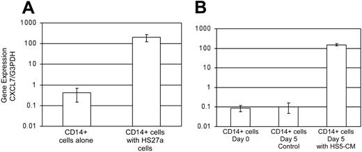
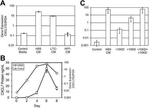
As reported previously, microarray data revealed that CXCL7 gene expression is increased 13.7-fold in CD14+/HS27a cocultures compared with HS27a cultured alone.16 This was confirmed by RT-PCR in CD14+ cells from 8 healthy donors (Figure 1A). Microarray analysis of cocultures of CD14+ cells with HS5 cells compared with HS5 alone indicated that CXCL7 was significantly up-regulated in this coculture system as well. The results of these microarray experiments as well as others exploring the monocyte-stromal interactions in the ME are now available in the GEO database (http://www.ncbi.nlm.nih.gov/geo/) as series GSE 3472. Additional experiments indicated that contact between stromal cells and monocytes was not required and that CXCL7 was expressed by the monocytes. This was verified by PCR detection of transcripts in CD14+ cells cultured in stromal cell–conditioned medium (CM) shown in Figure 2B. Figure 2A compares expression data from CD14+ cells cultured with HS5 CM or primary LTCs, both of which induce CXCL7. In contrast, CXCL7 gene expression is not detected in cells cultured in control medium or in CM from human foreskin fibroblasts. The time course for CXCL7 mRNA expression induced by HS5 CM is shown in Figure 2B, which also shows that the protein concentration, quantitated using ELISA, reached the maximal concentration by day 6. Comparable data were also obtained from the conditioned media obtained from primary LTCs established from 4 different healthy bone marrow donors (data not shown).
The HS5 CM–derived stimulating activity was found to have at least 2 components, one larger than 10 kDa and one smaller than 10 kDa (Figure 2C). Given that HS5 CM contains several cytokines with known effects on monocytes and that most of them are more than 10 kDa in size, we investigated the effects of each of these cytokines alone on the up-regulation of CXCL7 gene in monocytes, in combination, with or without the addition of the smaller than 10-kDa fraction of HS5 CM. The tested cytokines included GM-CSF, G-CSF, IL-1β, IL-6, M-CSF, Flt-3 ligand, IFN-γ, MCP-1, and SDF-1. The cytokines were tested in concentrations known to be present in the HS5 CM. Results of these experiments indicated that a combination of GM-CSF (2 ng/mL), IL-1β (0.2 ng/mL), and M-CSF (0.25 ng/mL), plus the smaller than 10-kDa fraction of HS5 CM could induce CXCL7 up-regulation comparable to that of the whole unfractionated HS5 CM. Thus, the stromal signal responsible for the gene up-regulation is a complex one, requiring a combination of cytokines as well as an undefined component smaller than 10 kDa in size. Studies are under way to characterize this component of the stromal signal.
Gene expression of CXCL7 in response to stromal signals. (A) CD14+ cells from 8 different healthy donors were cultured alone or cocultured with HS27a cells for 3 days. Nonadherent cells were harvested, and CXCL7 gene expression was determined by Syber Green real-time PCR and normalized to G3PDH gene expression. Data represent mean ± SE; P = .015 by Student t test. (B) CXCL7 gene expression in CD14+ cells from 10 healthy donors at day 0, day 5 in control media (RPMI with 10% FCS), and day 5 in culture in media conditioned by HS5 cells (HS5 CM). Data represent mean ± SE; P < .001 by Student t test comparing HS5 CM to either day 5 control or day 0.
Gene expression of CXCL7 in response to stromal signals. (A) CD14+ cells from 8 different healthy donors were cultured alone or cocultured with HS27a cells for 3 days. Nonadherent cells were harvested, and CXCL7 gene expression was determined by Syber Green real-time PCR and normalized to G3PDH gene expression. Data represent mean ± SE; P = .015 by Student t test. (B) CXCL7 gene expression in CD14+ cells from 10 healthy donors at day 0, day 5 in control media (RPMI with 10% FCS), and day 5 in culture in media conditioned by HS5 cells (HS5 CM). Data represent mean ± SE; P < .001 by Student t test comparing HS5 CM to either day 5 control or day 0.
CXCL7 gene expression and protein secretion in CD14+ cells. (A) cDNA was prepared from CD14+ cells after 5 days of culture in control media (RPMI with 10% FCS), media conditioned by HS5 cells, media conditioned by primary long-term cultures (LTC CMs), or media conditioned by human foreskin fibroblasts (HFFs). CXCL7 gene expression was measured by Syber Green real-time PCR as in “Materials and methods”. Data represent mean ± SE of 2 experiments. Both HS5 CM and LTC CM differ from control and HFF CM by P = .004 and .003, respectively, using Student t test. (B) To measure the time course of CXCL7 up-regulation, cDNA was prepared from CD14+ cells cultured in HS5 CM for up to 8 days. CXCL7 gene expression, measured by real-time PCR, is shown on the y-axis on the right. The media isolated from the same cultures were assayed for CXCL7 protein content using ELISA, shown on the y-axis to the left. (C) Conditioned media obtained from HS5 cultures was fractionated by filtration with a 10-kDa cutoff. CD14+ cells were cultured for 3 days in control media, HS5 CM, the smaller than 10-kDa fraction, the larger than 10-kDa fraction, and a combination of both fractions. Results are shown as mean of at least 2 samples ± SE.
CXCL7 gene expression and protein secretion in CD14+ cells. (A) cDNA was prepared from CD14+ cells after 5 days of culture in control media (RPMI with 10% FCS), media conditioned by HS5 cells, media conditioned by primary long-term cultures (LTC CMs), or media conditioned by human foreskin fibroblasts (HFFs). CXCL7 gene expression was measured by Syber Green real-time PCR as in “Materials and methods”. Data represent mean ± SE of 2 experiments. Both HS5 CM and LTC CM differ from control and HFF CM by P = .004 and .003, respectively, using Student t test. (B) To measure the time course of CXCL7 up-regulation, cDNA was prepared from CD14+ cells cultured in HS5 CM for up to 8 days. CXCL7 gene expression, measured by real-time PCR, is shown on the y-axis on the right. The media isolated from the same cultures were assayed for CXCL7 protein content using ELISA, shown on the y-axis to the left. (C) Conditioned media obtained from HS5 cultures was fractionated by filtration with a 10-kDa cutoff. CD14+ cells were cultured for 3 days in control media, HS5 CM, the smaller than 10-kDa fraction, the larger than 10-kDa fraction, and a combination of both fractions. Results are shown as mean of at least 2 samples ± SE.
Because CXCL7 was previously thought to be a megakaryocytic lineage-specific cytokine, platelet contamination of CD14+ cells can be invoked as a potential source of CXCL7 transcripts. However, this is unlikely for several reasons: First, platelets are devoid of transcriptional machinery and therefore cannot increase the number of transcripts over 5 days. Second, the purity of the CD14+ cells was greater than 95% for all of the healthy donors, making contamination by other cell types an unlikely source for the 14-fold increase in transcripts. We also looked at the transcript levels of other genes thought to be megakaryocyte lineage–specific (Glycoprotein IIb and platelet factor 4) in stromal-stimulated CD14+ cell cultures. The transcript levels of these genes were not up-regulated (data not shown), providing further evidence that the up-regulation of CXCL7 gene products is not due to platelets, but is indeed from CD14+ cells.
All media, culture supernatants, and cell lines used for the experiments were tested for endotoxin (LPS), and Mycoplasma and were found to be negative. This was done to rule out the possibility of endotoxin or Mycoplasma-induced up-regulation of CXCL7 gene expression.
CD14+ cells secrete 2 forms of CXCL7 peptides in response to stromal stimulation
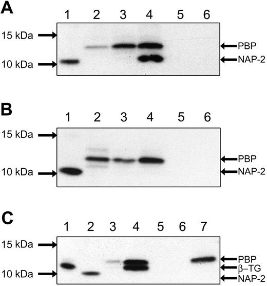
We verified that the increased level of mRNA detected is translated into protein by Western blot analyses and ELISA. CD14+ cells were cocultured with HS27a, HS5, or alone, and the CM were collected after 3 to 6 days. CXCL7 protein was concentrated using Sepharose CM beads, then run on SDS-PAGE, transferred to nitrocellulose, and probed with rabbit anti-CXCL7 antibodies and HRP-conjugated secondary antibodies. Results (Figure 3A) show that 2 distinct forms of CXCL7, PBP and NAP-2, were detected in the CM from the HS27a coculture, whereas only PBP was detected in the HS5 cocultures (Figure 3B). This suggests that the larger form may be processed by an activity that is only induced in monocyte-HS27a cocultures.
Previous reports had indicated that the full-length LDGF propeptide was detectable from LPS-stimulated PBMCs.17 To verify the size of the protein secreted by LPS stimulation, PBMCs were prepared as in “Materials and methods” and stimulated with LPS endotoxin for 24 hours. The conditioned media and whole-cell lysates were assayed by Western blot for CXCL7 peptides (Figure 3C). The LPS-stimulated medium was shown to contain a CXCL7 peptide corresponding in size to PBP, whereas the cell lysate contained 2 distinct bands, presumably PBP and β-thromboglobulin (or β-TG, another CXCL7 peptide). Both these peptides were significantly smaller than 14 kDa, the size of recombinant LDGF. We could not detect any CXCL7 peptides similar in size to LDGF in any of the cell preparations or culture media from CD14+ cells or PBMCs, suggesting that this propeptide is cleaved and therefore does not have a function outside of the cell.
Western blot of CXCL7 peptides. (A) Western analyses of culture media from CD14+ and HS27a cells. Culture media were collected, and Western analyses were performed for CXCL7 peptides. Lanes 1 and 2 represent positive controls NAP-2 and PBP, respectively. Culture conditions for the other lanes are as follows: CD14+ cells cocultured with HS27a cells for 3 days (lane 3), CD14+ cells cocultured with HS27a cells for 6 days (lane 4), CD14+ cells cultured alone for 6 days (lane 5), and HS27a cells cultured alone for 6 days (lane 6). At day 3, the CXCL7 peptides secreted are of comparable size to PBP, whereas by day 6, two distinct peptides are detected, PBP and NAP-2. (B) Western analyses of CD14+ coculture with HS5 cells. As in panel 3A, lanes 1 and 2 show NAP-2 and PBP. Culture conditions for the rest of the lanes are as follows: CD14+ cells cocultured with HS5 cells for 3 days (lane 3); CD14+ cells cocultured with HS5 cells for 6 days (lane 4); CD14+ cells cultured alone for 6 days (lane 5); and HS5 cells cultured alone for 6 days (lane 6). Here, only the PBP peptide is detected at days 3 and 6 of coculture. (C) CXCL7 peptides secreted by PBMCs in response to LPS stimulation. PBMCs were stimulated by 1 μg/mL LPS for 24 hours. Positive controls are β-TG, recombinant NAP-2, and recombinant PBP (lanes 1, 2, and 7). Conditions for the rest of the lanes are as follows: media from PBMCs cultured with LPS (lane 3), cell extract from PBMCs with LPS (lane 4), media and cell extract from PBMCs cultured without LPS (lanes 5 and 6). PBP is detectable in the culture media stimulated by LPS; both PBP and β-TG are detectable in the cellular extract. No CXCL7 peptides are seen when no LPS is added.
Western blot of CXCL7 peptides. (A) Western analyses of culture media from CD14+ and HS27a cells. Culture media were collected, and Western analyses were performed for CXCL7 peptides. Lanes 1 and 2 represent positive controls NAP-2 and PBP, respectively. Culture conditions for the other lanes are as follows: CD14+ cells cocultured with HS27a cells for 3 days (lane 3), CD14+ cells cocultured with HS27a cells for 6 days (lane 4), CD14+ cells cultured alone for 6 days (lane 5), and HS27a cells cultured alone for 6 days (lane 6). At day 3, the CXCL7 peptides secreted are of comparable size to PBP, whereas by day 6, two distinct peptides are detected, PBP and NAP-2. (B) Western analyses of CD14+ coculture with HS5 cells. As in panel 3A, lanes 1 and 2 show NAP-2 and PBP. Culture conditions for the rest of the lanes are as follows: CD14+ cells cocultured with HS5 cells for 3 days (lane 3); CD14+ cells cocultured with HS5 cells for 6 days (lane 4); CD14+ cells cultured alone for 6 days (lane 5); and HS5 cells cultured alone for 6 days (lane 6). Here, only the PBP peptide is detected at days 3 and 6 of coculture. (C) CXCL7 peptides secreted by PBMCs in response to LPS stimulation. PBMCs were stimulated by 1 μg/mL LPS for 24 hours. Positive controls are β-TG, recombinant NAP-2, and recombinant PBP (lanes 1, 2, and 7). Conditions for the rest of the lanes are as follows: media from PBMCs cultured with LPS (lane 3), cell extract from PBMCs with LPS (lane 4), media and cell extract from PBMCs cultured without LPS (lanes 5 and 6). PBP is detectable in the culture media stimulated by LPS; both PBP and β-TG are detectable in the cellular extract. No CXCL7 peptides are seen when no LPS is added.
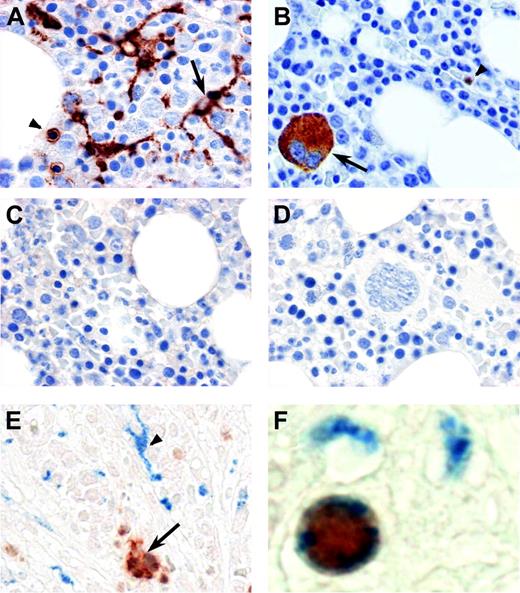
Immune histochemical analysis of normal bone marrow for CD. Serial sections from B5-fixed bone marrow biopsies from healthy donors were subjected to antigen retrieval and incubated with a monoclonal antibody against CD68 (A). The arrow shows macrophages with long cellular processes staining positive for CD68, whereas the arrowhead shows a smaller positively stained mononuclear cell with no cellular processes (similar to the cell shown in panel F). Panel B shows staining with rabbit polyclonal antibody against CXCL7. The arrow points to a positively stained megakaryocyte, also identifiable by large size and multiple nuclei. The arrowhead shows a smaller mononuclear cell positive for CXCL7. The bound antibodies were detected with the HRP-conjugated Envison Plus system (DakoCytomation). Sections stained with appropriate isotype control antibodies are shown in panel C (monoclonal mouse IgG3 antibody) and panel D (polyclonal rabbit antibody). Panels E and F show double immune histochemical staining for CXCL7 and CD68 in bone marrow biopsies from healthy donors. CD68 (blue) is visualized with the Vectastain ABC-AP kit and Vector Blue, and CXCL7 (red) by EnVision Plus/HRP and NovaRED substrate. As shown in panel E, distinct patterns of staining for both CD68 and CXCL7 are observed as in panels A and B, but a small proportion of mononuclear cells without extensive cellular processes also stain for both of the antigens, suggesting that a subset of bone marrow monocytes also express CXCL7. No counterstain was used for panels E and F. All images were acquired using a Nikon Eclipse E800 microscope (Nikon Instruments, Kanagawa, Japan), Coolsnap CF Color Camera (Photometrics, Tucson, AZ), and Metamorph software (Molecular Devices, Sunnyvale, CA). For panels A-D and F, a 100 ×/1.3 NA objective with oil immersion was used; for panel E, a 60 ×/1.4 NA objective with oil immersion was used. Images were adjusted for brightness and contrast with Adobe Photoshop 7.0 (Adobe, San Jose, CA). Panel F was magnified 3 times after acquisition for better cellular detail.
Immune histochemical analysis of normal bone marrow for CD. Serial sections from B5-fixed bone marrow biopsies from healthy donors were subjected to antigen retrieval and incubated with a monoclonal antibody against CD68 (A). The arrow shows macrophages with long cellular processes staining positive for CD68, whereas the arrowhead shows a smaller positively stained mononuclear cell with no cellular processes (similar to the cell shown in panel F). Panel B shows staining with rabbit polyclonal antibody against CXCL7. The arrow points to a positively stained megakaryocyte, also identifiable by large size and multiple nuclei. The arrowhead shows a smaller mononuclear cell positive for CXCL7. The bound antibodies were detected with the HRP-conjugated Envison Plus system (DakoCytomation). Sections stained with appropriate isotype control antibodies are shown in panel C (monoclonal mouse IgG3 antibody) and panel D (polyclonal rabbit antibody). Panels E and F show double immune histochemical staining for CXCL7 and CD68 in bone marrow biopsies from healthy donors. CD68 (blue) is visualized with the Vectastain ABC-AP kit and Vector Blue, and CXCL7 (red) by EnVision Plus/HRP and NovaRED substrate. As shown in panel E, distinct patterns of staining for both CD68 and CXCL7 are observed as in panels A and B, but a small proportion of mononuclear cells without extensive cellular processes also stain for both of the antigens, suggesting that a subset of bone marrow monocytes also express CXCL7. No counterstain was used for panels E and F. All images were acquired using a Nikon Eclipse E800 microscope (Nikon Instruments, Kanagawa, Japan), Coolsnap CF Color Camera (Photometrics, Tucson, AZ), and Metamorph software (Molecular Devices, Sunnyvale, CA). For panels A-D and F, a 100 ×/1.3 NA objective with oil immersion was used; for panel E, a 60 ×/1.4 NA objective with oil immersion was used. Images were adjusted for brightness and contrast with Adobe Photoshop 7.0 (Adobe, San Jose, CA). Panel F was magnified 3 times after acquisition for better cellular detail.
Immune histochemical localization of CXCL7 proteins in healthy human bone marrow biopsies
Immune histochemical analysis of bone marrow biopsies from healthy donors were done for both a monocyte/macrophage lineage-specific marker (CD68) and NAP-2 alone and in combination. As show in Figure 4A, mononuclear cells staining positive for CD68 are present widely in the bone marrow with long cellular processes presumably communicating with maturing hematopoietic cells; these are likely resident macrophages. Also present are smaller CD68+ cells without cellular processes which are likely monocytes not differentiated to macrophages. Megakaryocytes, identified by large size and multiple nuclei, stain positive for CXCL7 as shown in Figure 4B. Also positive are smaller mononuclear cells. To ascertain whether any of the CD68+ monocytes/macrophages expressed CXCL7, double staining for CD68 and NAP-2 was done, as shown in Figure 4E-F. A small percentage of the mononuclear cells coexpressed CD68 and CXCL7. An example of a double-positive cell is shown in Figure 4F. This provides an in vivo correlate to the induced expression of CXCL7 in CD14+ cells in vitro.
CXCL7 peptides are not directly mitogenic to fibroblasts
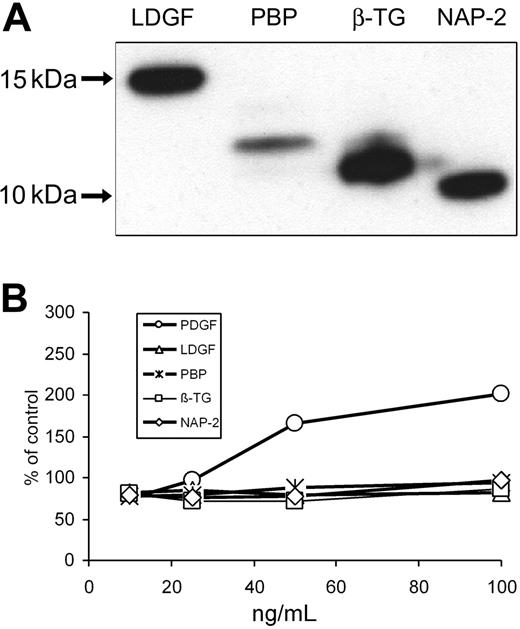
Recombinant peptides LDGF and PBP were made and their sequences confirmed as described in “Materials and methods,” and shown in Figure 5A. As previously reported, eukaryotic cells processed the propeptide to form PBP, the largest of the secreted peptides. To study the full-length peptide, we had to synthesize it in a prokaryotic system. We then tested the mitogenic effects of all the CXCL7 peptides (LDGF, PBP, β-TG, and NAP-2) as well as PDGF (as a positive control) in primary marrow-derived fibroblasts as well as in mouse NIH/3T3 cells. Results show that none of the CXCL7 peptides have any effect on proliferation of primary marrow-derived fibroblasts (Figure 5B). Comparable results were also obtained with murine NIH/3T3 fibroblasts (data not shown).
NAP-2 can inhibit both the size and number of megakaryocyte colonies
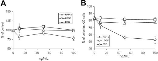
It has been reported that NAP-2, which is produced by megakaryocytes and stored in platelets, functions in an autocrine fashion to inhibit thrombopoiesis. We tested the ability of 3 CXCL7 peptides (LDGF, β-TG, and NAP-2) to inhibit megakaryocyte growth using standard CFU-Meg assays. Although there was a modest inhibition of CFU-Meg when NAP-2 was added to the CFU-Meg assays, there was no significant change with the addition of either LDGF or β-TG (Figure 6A). However, we found that the CFU-Meg colonies that did form in the presence of NAP-2 were significantly smaller than control colonies (Figure 6B). As CFU-Meg colony size has been correlated to platelet numbers in vivo,22 NAP-2 may act to limit platelet production by decreasing the number of megakaryocyte precursors capable of division as well as the number of divisions they undergo.
Recombinant CXCL7 peptides and their mitogenic properties on fibroblasts. (A) Recombinant CXCL7 peptides LDGF and PBP were prepared and purified as described in “Materials and methods.” Western analysis of recombinant LDGF and PBP along with positive controls β-TG and NAP-2 are shown. (B) Proliferative response of primary marrow-derived fibroblasts to CXCL7 peptides (LDGF, PBP, β-TG, and NAP-2) as well as PDGF was measured using the MTT assay. Results are shown as a percentage of control cultures with no added peptides. Although there was increased proliferation evident with increasing concentration of PDGF, none of the CXCL7 peptides tested stimulated proliferation. Results are shown as mean of 3 samples ± SE.
Recombinant CXCL7 peptides and their mitogenic properties on fibroblasts. (A) Recombinant CXCL7 peptides LDGF and PBP were prepared and purified as described in “Materials and methods.” Western analysis of recombinant LDGF and PBP along with positive controls β-TG and NAP-2 are shown. (B) Proliferative response of primary marrow-derived fibroblasts to CXCL7 peptides (LDGF, PBP, β-TG, and NAP-2) as well as PDGF was measured using the MTT assay. Results are shown as a percentage of control cultures with no added peptides. Although there was increased proliferation evident with increasing concentration of PDGF, none of the CXCL7 peptides tested stimulated proliferation. Results are shown as mean of 3 samples ± SE.
CXCL7 peptides and megakaryocyte colony growth. (A) G-CSF–mobilized CD34+ cells were cultured in serum-free culture media using the Megac-ult-C kit (Stem Cell Technologies), fixed, stained after 10 to 14 days with anti-CD41 antibodies, and scored for colony number and size (small colonies had 3-20 cells, medium 21-50, and large more than 50 cells), all per the manufacturer's instructions. Results are shown as a percentage of control colonies. Colony numbers were significantly smaller than control colony numbers when NAP-2 was added at concentrations of either 50 ng/mL (P = .010) or 100 ng/mL (P = .018). Colony numbers were not affected by LDGF or β-TG. (B) Percentage of CFU-Meg colonies larger than 20 cells in the same assays as in panel A. NAP-2 significantly decreases the size of the colonies at concentrations of 50 ng/mL and 100 ng/mL (P values of .001 and .006, respectively). For the CFU-Meg assays, G-CSF–mobilized CD34+ cells from 4 different donors were plated in duplicate sets. Results shown are mean values ± SE. P values were calculated using Student t test.
CXCL7 peptides and megakaryocyte colony growth. (A) G-CSF–mobilized CD34+ cells were cultured in serum-free culture media using the Megac-ult-C kit (Stem Cell Technologies), fixed, stained after 10 to 14 days with anti-CD41 antibodies, and scored for colony number and size (small colonies had 3-20 cells, medium 21-50, and large more than 50 cells), all per the manufacturer's instructions. Results are shown as a percentage of control colonies. Colony numbers were significantly smaller than control colony numbers when NAP-2 was added at concentrations of either 50 ng/mL (P = .010) or 100 ng/mL (P = .018). Colony numbers were not affected by LDGF or β-TG. (B) Percentage of CFU-Meg colonies larger than 20 cells in the same assays as in panel A. NAP-2 significantly decreases the size of the colonies at concentrations of 50 ng/mL and 100 ng/mL (P values of .001 and .006, respectively). For the CFU-Meg assays, G-CSF–mobilized CD34+ cells from 4 different donors were plated in duplicate sets. Results shown are mean values ± SE. P values were calculated using Student t test.
Discussion
The CXCL7 gene is translated as a 14-kDa propeptide, designated LGDF, which is cleaved into several smaller forms, PBP, CTAP-III, β-TG, and NAP-2.23 Until recently, CXCL7 gene expression was thought to be restricted to cells within the megakaryocytic lineage.24,25 Recent reports have suggested that other cell types may produce this chemokine as well.17,26-28 In this report, we have shown that monocytes are capable of secreting CXCL7 peptides in response to stromal stimuli. Interestingly, the specific forms of the peptide detected in conditioned media from stroma/monocyte cocultures can differ depending on the stromal cells present. HS27a cells stimulate monocytes to secrete both PBP and NAP-2, whereas HS5 stimulates the production of PBP only. The stimulation of CXCL7 gene expression in monocytes can be mediated by primary marrow fibroblasts but not primary foreskin fibroblasts. This supports the hypothesis that stroma-stimulated, monocyte-derived CXCL7 may have a specific function in the ME.
Peptides of the CXCL7 family have diverse biologic activities attributed to them, but some of these functions are controversial. Fibroblast mitogenic activity has been attributed to the 3 larger peptides (LDGF, PBP, and CTAP-III),17,29-32 although some investigators have questioned this activity for both PBP and CTAP-III.17 NAP-2 and β-TG, the smallest 2 forms, were reported to be inhibitors of megakaryocytopoiesis in a lineage-specific fashion. Our data are in keeping with later reports suggesting that NAP-2 alone has this activity.18,33 NAP-2 is also a potent chemotactic agent for neutrophils.34 Other functions of importance have been attributed to various CXCL7 peptides: CTAP-III may be responsible for intracellular transport of sphingomyelin,35 β-TG may be chemotactic to fibroblasts.36 Other less defined functions like heparinase activity of PBP and CTAP-III may be important for the modification of the matrix proteins.37 Thus, CXCL7 represents a protein derived from a single gene but exerting different biologic activities depending on its cleavage, which appears to be dependent directly or indirectly on signals from other cells. The ME is a system in which several of these functions could be of importance.
Among the several functions attributed to CXCL7 peptides, we chose to investigate 2 that could be particularly relevant to the regulation of the ME. First, we focused on LDGF, the longest peptide, which was reported to function as a mitogen for fibroblasts and to be secreted by monocytes in response to endotoxin stimulation. Similar bioactivity has been reported for PBP and CTAP-III. Because recombinant versions of these proteins were not available, we prepared recombinant human LDGF and PBP and verified their protein sequences before proceeding with testing their biologic function. Of note, in our hands, stimulation of monocytes with either endotoxin or stromal CM did not result in LDGF secretion. Furthermore, only prokaryotic expression systems could produce the full-length protein from the transduced gene. In contrast, eukaryotic cell lines and endotoxin-stimulated monocytes did secrete PBP, presumably after cleaving the initial 34-kDa signal peptide. However, although LDGF was prepared exactly as previously described,17 our functional studies indicated that neither LDGF nor PBP had mitogenic activity for primary marrow-derived fibroblasts or NIH/3T3 fibroblasts.
NAP-2, the smallest CXCL7-encoded peptide, can inhibit megakaryocyte proliferation when added to CFU-Meg assays and therefore may contribute to a negative feedback mechanism for regulating platelet counts.18,38 We tested the effect of CXCL7 peptides (LDGF, β-TG, and NAP-2) on the in vitro production of megakaryocytes from CFU-Megs. In our studies, NAP-2 not only decreased megakaryocyte colony numbers but also reduced the number of cells per colony, confirming the reports that NAP-2 limits megakaryocyte growth in vitro. The potential in vivo relevance for monocyte-derived CXCL7 is suggested by the detection of CXCL7-positive, CD68+ mononuclear cells in marrow biopsies.
The monocyte/macrophage components of the ME are unique in that they arise from hematopoietic stem cells and are not fixed in place like other stromal elements. They are uniquely equipped to home to a variety of niches in response to chemoattractants, respond to localized signals from stromal cells, and modify their transcriptional profile accordingly. One example is the stromal-stimulated production of monocyte-derived osteopontin, which in turn reduces the expression of Notch on CD34+ progenitor cells.16 The production of CXCL7 peptides by stromal-stimulated monocytes adds to the functional repertoire of these cells. As this repertoire broadens, the manner in which abnormal monocytes may adversely affect the hematopoietic ME also broadens. One could reasonably speculate that abnormal monocytes may contribute to dysplasias via modification of the ME. Given the presence of monocytes, normal or otherwise, in all tissues, it is also reasonable to speculate that they can modify the ME of other tissue systems. In fact, their role in promoting or limiting the tumor-supportive ability of nonhematopoietic MEs is already gaining more attention.39 Understanding the role of the monocyte/macrophage in various MEs should provide opportunities for development of new treatments.
Prepublished online as Blood First Edition Paper, January 3, 2006; DOI 10.1182/blood-2005-10-4285.
Supported by the National Institutes of Health (grant T32CA09515) (M.M.P.), (grants R01HL62923 and P30DK56465) (B.T.-S.), and by the Ladies Auxiliary to the Veterans of Foreign Wars of the United States (M.M.P.).
M.M.P. designed and performed the experiments, analyzed the data, and wrote the manuscript; M.I. helped design and perform some of the experiments and analyze the data and edited the manuscript; N.A. performed the microarray analysis; L.G. performed the microarray analysis and edited the manuscript; B.T.-S. designed the experiments, provided critical oversight and research support, and edited the manuscript.
The publication costs of this article were defrayed in part by page charge payment. Therefore, and solely to indicate this fact, this article is hereby marked “advertisement” in accordance with 18 U.S.C. section 1734.
We thank Dr Michael A. Harkey at the Center for Excellence in Molecular Hematology (CCEMH) at the FHCRC for help with cloning and recombinant protein expression; Dr Julie Randolph-Habecker at the Experimental Histopathology at the FHCRC for preparing and evaluating the immune histochemical sections; Ludmila Golubev, Gretchen Johnson, and Abigail Cruz for maintaining tissue culture; and Bonnie Larson and Helen Crawford for preparing the manuscript.

This feature is available to Subscribers Only
Sign In or Create an Account Close Modal