The biologic hallmark of polycythemia vera (PV) is the formation of endogenous erythroid colonies (EECs) with an erythropoietin-independent differentiation. Recently, it has been shown that an activating mutation of JAK2 (V617F) was at the origin of PV. In this work, we studied whether the STAT5/Bcl-xL pathway could be responsible for EEC formation. A constitutively active form of STAT5 was transduced into human erythroid progenitors and induced an erythropoietin-independent terminal differentiation and EEC formation. Furthermore, Bcl-xL overexpression in erythroid progenitors was also able to induce erythroid colonies despite the absence of erythropoietin. Conversely, siRNA-mediated STAT5 and Bcl-xL knock-down in human erythroid progenitors inhibited colony-forming unit-erythroid (CFU-E) formation in the presence of Epo. Altogether, these results demonstrate that a sustained level of the sole Bcl-xL is capable of giving rise to Epo-independent erythroid colony formation and suggest that, in PV patients, JAK2V617F may induce EEC via the STAT5/Bcl-xL pathway.
Introduction
In contrast with secondary erythrocytosis, progenitor cells from polycythemia vera (PV) patients can undergo in vitro erythroid differentiation despite the absence of Epo,1,2 and presence of such endogenous erythroid colonies (EECs) is routinely used as a diagnostic assay.1,3,4 To this date, mechanisms implicated in EEC formation are poorly understood. Recently, an acquired mutation in the JAK2 kinase leading to its constitutive phosphorylation has been described in PV patients. This mutation leads to a constitutive activation of JAK2 that seems to play a crucial role in the onset of the disease.5-7 During erythropoiesis, JAK2 activates many transduction pathways, which can be implicated in terminal maturation. For example, recent data showed that activation of AKT was sufficient for Epo-independent colony-forming unit-erythroid (CFU-E) formation in mice.8 During erythropoiesis, one of the principal targets of JAK2 is the signal transducer and activator of transcription STAT5. After phosphorylation by JAK2, STAT5 dimerizes and translocates into the nucleus where it regulates transcription of target genes implicated in cell proliferation and survival, among which that of the antiapoptotic protein Bcl-xL.9 The JAK2/STAT5/Bcl-xL pathway is crucial during erythropoiesis9-12 : JAK2-/- mice die in utero from severe anemia,10 and inactivation of STAT5 leads to a severe defect in erythropoiesis.9,11 In the present work, using both an siRNA and an overexpression strategy, we investigated whether the STAT5/Bcl-xL pathway was implicated in EEC formation. In particular, we overexpressed Bcl-xL or a constitutively active form of STAT5 in human normal erythroid cells and observed that the transduced cells could undergo an Epo-independent erythroid differentiation and in this way mimic PV phenotype.
Study design
Cell culture
UT7 clone expressing Mpl (UT7 5.3) was maintained in the presence of 5 ng/mL GM-CSF and differentiated toward erythropoiesis in the presence of 2 UI/mL human recombinant erythropoietin (Epo; OrthoBiotech, Paris, France).13 After informed consent, cells obtained from peripheral blood (PB) of patients treated with G-CSF were separated over a Ficoll-metrizoate gradient, and CD34+ cells were purified and cultured in a serum-free medium in the presence of Epo, SCF, IL-3, and dexamethasone (DXM) as previously described.14 For colony assays, CD36+/GpA- cells were plated in H4100 Medium (Stem Cell Technologies, Vancouver, BC, Canada) supplemented with 10% FCS, 25 ng/mL SCF, with or without Epo 3 UI/mL. CFU-E numbers were counted at day 7. For colony assays without Epo, 5000 cells were plated per dish, instead of 1000 in normal conditions.
Retroviral constructs, retrovirus production, and cell infection
The STAT5 cDNA with an activating mutation15 (gift from F. Gouilleux, INSERM E0351, Amiens, France) and the human Bcl-xL cDNA were cloned upstream from the IRES-GFP sequence in the MIGR plasmid. Retrovirus particle production was achieved as previously described.14 Infection of human CD34+ cells was performed at days 4 and 5 in the presence of Epo, SCF, IL-3, and DXM as described earlier.14 At day 6, CD36+/GpA-/GFP+ cells were sorted and cultured in a serum-free medium supplemented with 25 ng/mL SCF with or without Epo or plated in methylcellulose as described in “cell culture”.
SiRNA experiments
Results and discussion
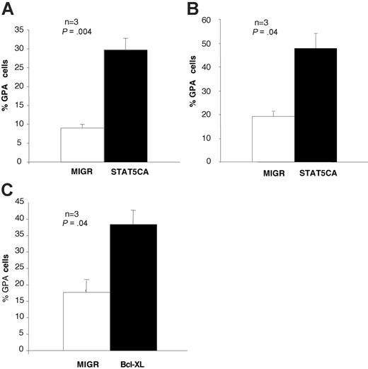
Recent focus on the JAK2V617F mutation in PV patients argues for a direct implication of JAK2-dependent signaling pathways in EEC formation.5,6 Because STAT5 is the principal JAK2 target in erythroid cells, we investigated whether EEC formation was dependent only on STAT5 activation or required other signaling pathways that would be activated by JAK2. For this purpose, we used a retroviral vector coding for a constitutively active form of STAT5 (STAT5CA), which is spontaneously translocated into the nucleus.15 After transduction in UT7 cells, a leukemic cell line with erythroid properties, this vector led to a spontaneous induction of GpA expression despite the absence of Epo (Figure 1A). We next investigated effects of STAT5CA on erythroid differentiation of human primary progenitors. Purified PB CD34+ cells were cultured in a serum-free medium as described in “Materials and methods.” After transduction with the STAT5CA vector, CD36+/GpA-/GFP+ cells were sorted and cultured in the presence of SCF alone. As shown previously, these cells correspond to erythroid progenitors at a CFU-E stage and are dependent on Epo for their survival and terminal differentiation.14 After STAT5CA expression, they could undergo erythroid terminal differentiation despite the absence of Epo (Figure 1B).
STAT5 and Bcl-xL can induce erythroid differentiation. (A) A constitutively active form of STAT5 induced erythroid differentiation in UT7 cells. UT7 cells were transduced either with the empty retrovirus MIGR or with the retrovirus coding for a constitutively phosphorylated STAT5 (STAT5CA). After cell sorting according to GFP expression, cells were cultured in the presence of GM-CSF, without Epo. GpA expression was monitored by flow cytometry at day 4 after retrovirus infection. In the cells expressing STAT5CA, the percentage of GpA+ cells was much greater than in UT7 transduced with the empty vector (MIGR: 9% ± 1%; STAT5CA: 30% ± 3%; n = 3; Student t test: P = .004). (B) STAT5CA expression in human primary progenitors induced Epo-independent terminal erythroid differentiation. CD34+ cells from PB were cultured in the presence of Epo, SCF, IL-3, and DXM and then transduced twice with the different retroviral vectors (MIGR or STAT5CA). Transduced CD36+/GpA- cells were sorted at day 6 and cultured in the presence of SCF without Epo. Flow cytometry analysis showed 48 hours later that the percentage of GpA+ cells was higher with STAT5CA (48% ± 6%) than with the empty vector MIGR (19% ± 2%; n = 3; Student t test: P = .04). Determination of GpA expression was done using a more sensitive cell analyzer than the one used for cell sorting, and this explains why around 20% of the MIGR-transduced cells were found GpA positive. Moreover, cells transduced with the empty vector died after 48 hours of Epo removal, whereas STAT5CA-expressing cells further survived and proliferated (data not shown). (C) Bcl-xL overexpression in human primary progenitors induced GpA expression despite the absence of Epo. Primary cells were transduced either with the empty vector MIGR or with a vector coding for the Bcl-xL cDNA. Two days after cell sorting and Epo removal, 38% ± 4% of Bcl-xL-overexpressing cells were GpA positive, whereas only 18% ± 4% of cells infected with the empty vector MIGR were GpA positive (n = 3; Student t test: P = .04). Data are represented as mean ± SEM. As observed in STAT5CA-expressing cells, Bcl-xL-overexpressing cells could further proliferate and differentiate. When transduced with the empty vector, all cells died 48 hours after EPO removal.
STAT5 and Bcl-xL can induce erythroid differentiation. (A) A constitutively active form of STAT5 induced erythroid differentiation in UT7 cells. UT7 cells were transduced either with the empty retrovirus MIGR or with the retrovirus coding for a constitutively phosphorylated STAT5 (STAT5CA). After cell sorting according to GFP expression, cells were cultured in the presence of GM-CSF, without Epo. GpA expression was monitored by flow cytometry at day 4 after retrovirus infection. In the cells expressing STAT5CA, the percentage of GpA+ cells was much greater than in UT7 transduced with the empty vector (MIGR: 9% ± 1%; STAT5CA: 30% ± 3%; n = 3; Student t test: P = .004). (B) STAT5CA expression in human primary progenitors induced Epo-independent terminal erythroid differentiation. CD34+ cells from PB were cultured in the presence of Epo, SCF, IL-3, and DXM and then transduced twice with the different retroviral vectors (MIGR or STAT5CA). Transduced CD36+/GpA- cells were sorted at day 6 and cultured in the presence of SCF without Epo. Flow cytometry analysis showed 48 hours later that the percentage of GpA+ cells was higher with STAT5CA (48% ± 6%) than with the empty vector MIGR (19% ± 2%; n = 3; Student t test: P = .04). Determination of GpA expression was done using a more sensitive cell analyzer than the one used for cell sorting, and this explains why around 20% of the MIGR-transduced cells were found GpA positive. Moreover, cells transduced with the empty vector died after 48 hours of Epo removal, whereas STAT5CA-expressing cells further survived and proliferated (data not shown). (C) Bcl-xL overexpression in human primary progenitors induced GpA expression despite the absence of Epo. Primary cells were transduced either with the empty vector MIGR or with a vector coding for the Bcl-xL cDNA. Two days after cell sorting and Epo removal, 38% ± 4% of Bcl-xL-overexpressing cells were GpA positive, whereas only 18% ± 4% of cells infected with the empty vector MIGR were GpA positive (n = 3; Student t test: P = .04). Data are represented as mean ± SEM. As observed in STAT5CA-expressing cells, Bcl-xL-overexpressing cells could further proliferate and differentiate. When transduced with the empty vector, all cells died 48 hours after EPO removal.
Because STAT5 has been shown to play a crucial role in erythropoiesis through induction of the antiapoptotic protein Bcl-xL,9,11 we next investigated whether effects of STAT5CA were dependent on Bcl-xL induction. We cloned the coding sequence of human Bcl-xL in the retroviral vector MIGR. As shown in Figure 1C, overexpression of Bcl-xL in CD36+/GpA- cells induced GpA expression despite the absence of Epo. Thus, both constitutive activation of STAT5 and Bcl-xL overexpression could substitute for Epo to induce terminal differentiation of primary erythroid progenitor cells. Of interest, an immunostaining experiment showed that these 2 vectors increased levels of Bcl-xL protein in erythroid progenitors (data not shown). These levels, however, did not exceed those observed in physiological conditions after Epo exposure. This is in agreement with recent data showing an effect of Bcl-xL on erythroid differentiation, either in the FDCP cell line18 or in primary mouse erythroblasts.19 This effect may be partly independent of Bcl-xL antiapoptotic properties because Bcl2, another antiapoptotic factor, is not able to induce EEC in transgenic mice,20 and its overexpression induces granulocytic but not erythroid differentiation in FDCP cells.18
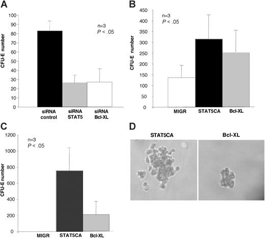
In order to investigate the importance of the STAT5/Bcl-xL pathway in CFU-E formation, we transduced CD36+ progenitor cells with an siRNA targeted on STAT5 or Bcl-xL, and plated them in methylcellulose for CFU-E assays. We observed with either STAT5 or Bcl-xL siRNA a drastic reduction of cloning efficiency (Figure 2A). Deregulation of these proteins could therefore be implicated in EEC formation observed in PV cells. Since a high Bcl-xL expression has been described in PV erythroid progenitors,21 we investigated whether STAT5CA or Bcl-xL overexpression could reproduce the malignant phenotype (ie, formation of EEC) as do PV cells. PB CD34+ cells were cultured in a serum-free medium and transduced with the different retroviruses at days 4 and 5. CD36+/GpA-/GFP+ cells were sorted at day 7 and plated in methylcellulose with or without Epo. In the presence of Epo, total CFU-E number was higher with the STAT5CA or the Bcl-xL vector than with the control (Figure 2B). Without Epo, Bcl-xL as well as STA5CA vectors could induce EEC (Figure 2C). However, STAT5CA expression led to a greater number of colonies, whereas Bcl-xL overexpression induced smaller ones similar to those observed in PV patients (Figure 2D). These differences could be due to recruitment of other targets activated by STAT5, especially genes implicated in the cell cycle.
STAT5 and Bcl-xL are implicated in EEC formation. (A) Inhibition of Bcl-xL and STAT5 in primary cells using an siRNA strategy decreased CFU-E formation. Day-5 CD36+ cells cultured in the presence of Epo, DXM, IL-3, and SCF were electroporated with siRNA targeted on either STAT5 or Bcl-xL, or with a nonspecific control sequence. GpA- cells were plated at day 6 in methylcellulose in the presence of Epo and SCF. CFU-Es were counted 7 days later. Number of CFU-Es was significantly reduced after knock-down of either STAT5 or Bcl-XL compared with the control (n = 3; Student t test: P < .05). (B-C) A constitutively active form of STAT5 as well as Bcl-xL overexpression induced EEC formation in methylcellulose assays. PB CD34+ cells were cultured in the presence of Epo, SCF, IL-3, and DXM, transduced at days 4 and 5 either with the MIGR, STAT5CA, or Bcl-xL vectors. At day 7, CD36+/GpA-/GFP+ cells were sorted, and 5000 cells were plated in methylcellulose in the presence of SCF alone. As a positive control, 1000 cells were plated in parallel in the presence of SCF and Epo. Histograms represent the total number of CFU-Es at day 7. In the presence of Epo, the CFU-E number was higher with the STAT5CA and the Bcl-xL vectors than with the control (B). In the absence of Epo (C), whereas MIGR-transduced cells did not give rise to a significant number of CFU-Es, either STAT5CA or Bcl-xL vectors could induce EEC formation (n = 3, each in triplicate; Student t test: STAT5CA vs MIGR, P < .05; Bcl-xL vs MIGR, P < .05). Data are represented as mean ± SEM. (D) Qualitative differences between STAT5CA-induced (left) and Bcl-xL-induced (right) EECs. Bcl-xL-induced EECs were not only less numerous (Student t test: P = .001), but also contained a lower number of cells than the STAT5CA-induced CFU-Es. These Bcl-xL-induced EECs were very similar to those routinely observed in PV patients. CFUs were counted using a Zeiss Telaval 31 microscope (Zeiss, Oberkochen, Germany) and a 20×/0.35 numeric aperture objective (Micromecanique, Evry, France). Images were captured using a Nikon Eclipse TE300 microscope (Nikon, Tokyo, Japan) connected to a Zeiss Axiocam digital camera. Images were acquired using Zeiss Axiovision 4 software.
STAT5 and Bcl-xL are implicated in EEC formation. (A) Inhibition of Bcl-xL and STAT5 in primary cells using an siRNA strategy decreased CFU-E formation. Day-5 CD36+ cells cultured in the presence of Epo, DXM, IL-3, and SCF were electroporated with siRNA targeted on either STAT5 or Bcl-xL, or with a nonspecific control sequence. GpA- cells were plated at day 6 in methylcellulose in the presence of Epo and SCF. CFU-Es were counted 7 days later. Number of CFU-Es was significantly reduced after knock-down of either STAT5 or Bcl-XL compared with the control (n = 3; Student t test: P < .05). (B-C) A constitutively active form of STAT5 as well as Bcl-xL overexpression induced EEC formation in methylcellulose assays. PB CD34+ cells were cultured in the presence of Epo, SCF, IL-3, and DXM, transduced at days 4 and 5 either with the MIGR, STAT5CA, or Bcl-xL vectors. At day 7, CD36+/GpA-/GFP+ cells were sorted, and 5000 cells were plated in methylcellulose in the presence of SCF alone. As a positive control, 1000 cells were plated in parallel in the presence of SCF and Epo. Histograms represent the total number of CFU-Es at day 7. In the presence of Epo, the CFU-E number was higher with the STAT5CA and the Bcl-xL vectors than with the control (B). In the absence of Epo (C), whereas MIGR-transduced cells did not give rise to a significant number of CFU-Es, either STAT5CA or Bcl-xL vectors could induce EEC formation (n = 3, each in triplicate; Student t test: STAT5CA vs MIGR, P < .05; Bcl-xL vs MIGR, P < .05). Data are represented as mean ± SEM. (D) Qualitative differences between STAT5CA-induced (left) and Bcl-xL-induced (right) EECs. Bcl-xL-induced EECs were not only less numerous (Student t test: P = .001), but also contained a lower number of cells than the STAT5CA-induced CFU-Es. These Bcl-xL-induced EECs were very similar to those routinely observed in PV patients. CFUs were counted using a Zeiss Telaval 31 microscope (Zeiss, Oberkochen, Germany) and a 20×/0.35 numeric aperture objective (Micromecanique, Evry, France). Images were captured using a Nikon Eclipse TE300 microscope (Nikon, Tokyo, Japan) connected to a Zeiss Axiocam digital camera. Images were acquired using Zeiss Axiovision 4 software.
Considering these results, we hypothesized that the EEC formation observed in myeloproliferative disorders could be partially due to the JAK2-dependent activation of the STAT5/Bcl-xL pathway. In agreement with this, JAK2 inhibitors have been shown to inhibit Epo-independent terminal differentiation of PV cells.22 Moreover, when JAK2V617F was expressed in the IL-3-dependent cell line BaF3, a spontaneous STAT5 phosphorylation could be detected in the absence of cytokine.6,7 Whether this mutation requires a functional Epo-R and STAT5 to induce EEC formation via an Epo-independent Bcl-xL induction in PV erythroid cells is still under investigation.
Prepublished online as Blood First Edition Paper, May 9, 2006; DOI 10.1182/blood-2005-10-009514.
Supported by grants from INSERM and the Institut Gustave Roussy.
An Inside Blood analysis of this article appears at the front of this issue.
The publication costs of this article were defrayed in part by page charge payment. Therefore, and solely to indicate this fact, this article is hereby marked “advertisement” in accordance with 18 U.S.C. section 1734.
We are grateful to Frederic Larbret for cell sorting experiments, and to Dr Virginie Moucadel and to Prof Nicole Casadevall for their kind assistance.


This feature is available to Subscribers Only
Sign In or Create an Account Close Modal