Using fluorescence in situ hybridization we investigated amplification of chromosome band 1q21 (Amp1q21) in more than 500 untreated patients with monoclonal gammopathy of undetermined significance (MGUS; n = 14), smoldering multiple myeloma (SMM; n = 31), and newly diagnosed MM (n = 479) as well as 45 with relapsed MM. The frequency of Amp1q21 was 0% in MGUS, 45% in SMM, 43% in newly diagnosed MM, and 72% in relapsed MM (newly diagnosed versus relapsed MM, P < .001). Amp1q21 was detected in 10 of 12 patients whose disease evolved to active MM compared with 4 of 19 who remained with SMM (P < .001). Patients with newly diagnosed MM with Amp1q21 had inferior 5-year event-free/overall survival compared with those lacking Amp1q21 (38%/52% versus 62%/78%, both P < .001). Thalidomide improved 5-year EFS in patients lacking Amp1q21 but not in those with Amp1q21 (P = .004). Multivariate analysis including other major predictors revealed that Amp1q21 was an independent poor prognostic factor. Relapsed patients who had Amp1q21 at relapse had inferior 5-year postrelapse survival compared with those lacking Amp1q21 at relapse (15% versus 53%, P = .027). The proportion of cells with Amp1q21 and the copy number of 1q21 tended to increase at relapse compared with diagnosis. Our data suggest that Amp1q21 is associated with both disease progression and poor prognosis.
Introduction
High-dose therapy with autologous stem cell transplantation has become the standard of care in multiple myeloma (MM), especially for relatively young patients, and can prolong survival1-4 ; however, survival ranges from a few months to more than 10 years.1-4 Monoclonal gammopathy of undetermined significance (MGUS), an asymptomatic plasma-cell dyscrasia seen in approximately 3% of persons over 60 years of age, and smoldering MM (SMM) share virtually all the genetic lesions seen in MM, yet MGUS rarely converts to overt MM requiring therapy.5-7
Karyotype abnormalities in plasma-cell dyscrasias are common and highly complex and contain random events that might have little effects on tumor pathogenesis. Cytogenetic analyses, including fluorescence in situ hybridization (FISH), have contributed to identification of nonrandom chromosomal aberrations in plasma-cell dyscrasias8-12 and to a better understanding of the clinical implications of these chromosomal aberrations in MM.13-15 Deletion of chromosome 13 (del(13)), hypodiploidy, and t(4;14), which leads to ectopic overexpression of FGFR3 and MMSET, are known to be associated with a poor prognosis.
Previous studies based on comparative genomic hybridization (CGH) have revealed that unbalanced chromosomal structural changes were present in almost all plasma-cell dyscrasias, and chromosomal gains of 1q consistently involved the 1q21 region in MM.16-18 We have reported previously that the gain of 1q21 can occur as isochromosomes, duplications, or jumping translocations in MM.19,20
The gain of the 1q21 region, which is one of the most recurrent chromosomal aberrations in MM and other cancers,21-24 has been speculated to be linked to a poor prognosis in MM.25 However, a comprehensive analysis of the prognostic significance of gains and/or amplifications of 1q21 (henceforth referred to as Amp1q21) has never been evaluated in a large cohort of newly diagnosed MM. In addition, the association of the genetic events underlying the conversion of MGUS and SMM to MM and its eventual fatal progression are not completely understood.
Here we provide evidence that Amp1q21 is associated with a poor prognosis in newly diagnosed MM and a shortened postrelapse survival and may be central to progression of plasma-cell dyscrasias.
Patients, materials, and methods
Patients
Samples for this study were obtained from patients with MGUS (n = 14), SMM (n = 31), newly diagnosed MM (n = 479), and relapsed MM (n = 45). The baseline characteristics of 524 untreated patients with MGUS, SMM, and newly diagnosed MM are summarized in Table 1. Newly diagnosed and relapsed MM patients in this study were all enrolled in University of Arkansas research protocol UARK 98-026 (Total Therapy 226,27 ), which is a melphalan-based tandem autologous stem cell transplantation protocol that randomized 668 patients to receive or not receive thalidomide. A total of 479 of 668 patients with newly diagnosed MM had interphase FISH results of 1q21 available at baseline; 189 of the 668 patients enrolled on the Total Therapy 2 trial were not analyzed due to either lack of sufficient samples and/or poor quality after hybridization.
Baseline patient characteristics
. | No. of patients/total no. (%) . | . | . | ||
|---|---|---|---|---|---|
| Characteristics . | MGUS . | SMM . | MM . | ||
| Age 65 y or older | 5/14 (36) | 7/31 (23) | 115/479 (24) | ||
| Female sex | 6/14 (43) | 17/31 (55) | 205/479 (43) | ||
| White race | 12/14 (86) | 29/31 (94) | 429/479 (90) | ||
| IgA subtype | 0/8 (0) | 6/26 (23) | 120/475 (25) | ||
| B2M level 4 mg/L or more | 1/14 (7) | 0/31 (0) | 146/479 (30) | ||
| CRP level 4 mg/L or more | 0/14 (0) | 3/31 (10) | 43/479 (9) | ||
| Creatinine level 2 mg/dL or more | 2/13 (15) | 0/30 (0) | 51/475 (11) | ||
| LDH level 190 IU/L or more | 5/14 (36) | 3/31 (10) | 148/479 (31) | ||
| ALB level less than 3.5 g/dL | 0/12 (0) | 3/31 (10) | 157/463 (34) | ||
| HGB level less than 10 g/dL | 0/14 (0) | 3/31 (10) | 135/479 (28) | ||
| CA | 1/13 (8) | 1/29 (3) | 148/440 (34) | ||
| Hyperdiploidy* | 0/13 (0) | 1/29 (3) | 79/440 (18) | ||
| Hypodiploidy* | 1/13 (8) | 0/29 (0) | 52/440 (12) | ||
| PC in BM aspirate 33% or more | 0/14 (0) | 6/28 (21) | 183/402 (46) | ||
| PC in BM biopsy 33% or more | 0/14 (0) | 0/28 (0) | 193/402 (48) | ||
To convert β2-microglobulin level from milligrams per liter to nanomoles per liter, multiply milligrams per liter by 85. To convert creatinine level from milligrams per deciliter to micromoles per liter, multiply milligrams per liter by 88.4. To convert albumin and hemoglobin levels from grams per deciliter to grams per liter, multiply grams per deciliter by 10.
B2M indicates serum β2-microglobulin; CRP, C-reactive protein; LDH, serum lactate dehydrogenase; ALB, serum albumin; HGB, hemoglobin; CA, cytogenetic abnormalities defined by G banding; PC, plasma cells; BM, bone marrow
Ploidy was defined by G banding
At the time of analysis, in the 479 newly diagnosed patients we analyzed, the median follow-up of surviving patients was 53 months (range, 25 to 89) and there were 297 events and 207 deaths in this subset. In 45 relapsed patients who had interphase FISH results of 1q21 available at relapse, the median follow-up of postrelapse survival was 17 months (range, 0.3 to 78). The median follow-up of patients with MGUS and SMM was 22 months (range, 1 to 88) and 47 months (range, 1 to 123), respectively. The Institutional Review Board of the University of Arkansas for Medical Sciences approved the research studies, and all subjects provided written informed consent approving use of their samples for research purposes.
Cell lines
Human myeloma cell lines NCU-MM1, OCI-MY5, U266, EJM, SK-MM1, JJN3, H929, ANBL6, RPMI8226, KMS12PE, MM_M1, OCI-MY1, XG7, Delta47, L363, XG1, OPM1, CAG, UTMC2, OPM2, ARP1, KMS11, and FR4 were cultured in RPMI 1640 with 10% fetal bovine serum (FBS), 100 IU/mL penicillin, and 100 mg/mL streptomycin at 37°C in 5% CO2 and 10 ng/mL human recombinant interleukin-6 (R&D Systems, Minneapolis, MN) when needed.
Fluorescence in situ hybridization
Bacterial artificial chromosomes (BACs) at 1q21 (RP11-307C12) and at 1q31 (RP11-32D17) were purchased from BAC/PAC Resources (Oakland, CA). Chromosome 13 ploidy was evaluated with a BAC clone for marker D13S31 mapping to 13q14. IGH break-a-part probe (Vysis, Downers Grove, IL) was used to analyze for the presence of 14q32 translocations. All probes were directly labeled by nick translation with Spectrum-Green and/or Spectrum-Red (Vysis). BAC and P1 artificial chromosome (PAC) clone information was obtained through the website of the National Center for Biotechnology Information28 (NCBI), and the probes were confirmed to map to the precise chromosome bands using metaphase spreads from the peripheral-blood lymphocytes from healthy donors.
Interphase FISH with cytoplasmic immunoglobulin light chain staining was performed according to the procedure described previously.13 A detailed protocol for probe production and hybridization can be found in Documents S1-S3 (available on the Blood website; see the Supplemental Materials link at the top of the online article). Briefly, the probes were hybridized along with AMCA-labeled antibodies to κ or λ immunoglobulin light chains (Vector Laboratories, Burlingame, CA) to cytospin preparations of mononuclear cells from bone marrow aspirates by Ficoll separation fixed with ethanol (Figure S1A). The slides were stored at -20°C until FISH analyses were performed. Interphase FISH signals were evaluated in at least 100 clonal plasma cells (mainly 100) in each patient. If at least 3 copies were seen in at least 20% of clonal plasma cells, it was considered evidence of gain/amplification. To investigate effects of the magnitude of Amp1q21 on clinical outcomes and each category, Amp1q21 was divided into 2 categories: (1) 3 copies of 1q21 (the percentage of cells with at least 4 copies was seen in less than 20% of clonal plasma cells) and (2) at least 4 copies of 1q21 (the percentage of cells with at least 4 copies was seen in at least 20% of clonal plasma cells.
A detailed account of the FISH data for 1q21 on all samples presented in this study can be found in Table S4.
Chromosome preparation for metaphase FISH analysis in human myeloma cell lines was performed according to the standard procedure as described previously.29 Briefly, cells were cultured in log phase, treated with colcemid (final concentration, 005 mg/mL) for 30 minutes, harvested with hypotonic potassium chloride (0.075M KCl), and fixed with methanol/glacial acetic acid (3:1; cornoy solution). Metaphase FISH analyses were performed according to the manufacturer's protocol (Vysis) (Figure S1B-C).
Statistical analyses
Event-free survival (EFS) and overall survival (OS) distributions were estimated using the Kaplan-Meier method, and differences among survival curves were analyzed by the log-rank test. The χ2 test and the Fisher exact test were used to test for the independence of categories. Multivariate analysis of EFS and OS adjusted for the effects of Amp1q21 for other predictors was performed using the Cox proportional hazards regression model. P values below .05 were considered significant.
Gene-expression profiling
Gene-expression profiling using the Affymetrix U133 Plus 2.0 microarray (Affymetrix, Santa Clara CA), was performed on CD138-purified plasma cells from bone marrow aspirates of newly diagnosed patients as described previously.30 Chromosomal translocations involving the IGH locus result in transcriptional hyperactivation of protooncogenes such as CCND1, CCND3, FGFR3/MMSET, c-MAF, and MAFB.31 These translocation events are detectable as “spiked” expression by microarray analysis.30
Results
Correlation of Amp1q21 and clinical and biologic features in newly diagnosed MM
Table 2 summarizes patient characteristics according to Amp1q21 in newly diagnosed MM. Interphase FISH analysis for 1q21 was successful in 479 of 668 patients who enrolled in Total Therapy 2. Patient characteristics of 189 newly diagnosed patients who did not have FISH results of 1q21 at baseline were similar to those of 479 patients who had FISH results (data not shown). In 479 patients with newly diagnosed MM, there were 7 patients with 1 copy of 1q21, 267 with 2 copies of 1q21, and 205 with at least 3 copies of 1q21 (Amp1q21) (Tables 3, 4). Patients with Amp1q21 tended to have a high incidence of IgA subtype (P < .001), high level of B2M (P = .002), high level of LDH (P < .001), low level of HGB (P = .004), high incidence of abnormal karyotypes defined by G banding (P < .001), high incidence of hyperdiploidy (P = .009), high incidence of hypodiploidy (P < .001), high percentage of plasma cells in bone marrow by biopsy (P = .007), high incidence of del(13) defined by FISH (P < .001), low frequency of stage 1 (P < .001), and high frequency of stage 3 (P = .002) based on the International Staging System (ISS) compared with those lacking Amp1q21 in newly diagnosed MM (Table 2).
Baseline patient characteristics according to Amp1q21 in newly diagnosed MM
. | No. of patients/total no. (%) . | . | . | ||
|---|---|---|---|---|---|
| Characteristics . | No Amp1q21* . | Amp1q21 . | P . | ||
| Age, 65 y or older | 62/274 (23) | 53/205 (26) | .413 | ||
| Female sex | 107/274 (39) | 98/205 (48) | .055 | ||
| White race | 241/274 (88) | 188/205 (92) | .184 | ||
| IgA subtype | 53/274 (19) | 67/201 (33) | < .001 | ||
| B2M level 4 mg/L or more | 68/274 (25) | 78/205 (38) | .002 | ||
| CRP level 4 mg/L or more | 24/274 (9) | 19/205 (9) | .847 | ||
| Creatinine level 2 mg/dL or more | 24/272 (9) | 27/203 (13) | .119 | ||
| LDH level 190 IU/L or more | 67/274 (24) | 81/205 (40) | < .001 | ||
| ALB level less than 3.5 g/dL | 80/265 (30) | 77/198 (39) | .050 | ||
| HGB level less than 10 g/dL | 63/274 (23) | 72/205 (35) | .004 | ||
| CA | 66/253 (26) | 82/187 (44) | < .001 | ||
| Hyperdiploidy† | 35/253 (14) | 44/187 (24) | .009 | ||
| Hypodiploidy† | 16/253 (6) | 36/187 (19) | < .001 | ||
| PC in BM aspirate 33% or more | 107/251 (43) | 76/151 (50) | .133 | ||
| PC in BM biopsy 33% or more | 105/246 (43) | 88/156 (56) | .007 | ||
| del(13)‡ | 37/252 (15) | 67/184 (36) | < .001 | ||
| Focal bone lesions by MRI, 3 or more | 140/265 (53) | 107/196 (55) | .708 | ||
| ISS stage 1 | 140/265 (53) | 73/202 (36) | < .001 | ||
| ISS stage 2 | 83/265 (31) | 73/202 (36) | .274 | ||
| ISS stage 3 | 42/265 (16) | 56/202 (28) | .002 | ||
MRI indicates magnetic resonance imaging; other abbreviations are explained in Table 1.
To convert β2-microglobulin level from milligrams per liter to nanomoles per liter, multiply milligrams per liter by 85. To convert creatinine level from milligrams per deciliter to micromoles per liter, multiply milligrams per liter by 88.4. To convert albumin and hemoglobin levels from grams per deciliter to grams per liter, multiply grams per deciliter by 10
Amp1q21: at least 3 copies of 1q21 defined by interphase FISH
Ploidy was defined by G banding
del(13): at least 80% of cells with up to 1 copy of D13S31 by interphase FISH
Summary of interphase FISH results of Amp1q21 in patients with MGUS, SMM, newly diagnosed MM, and relapsed MM
. | Amp1q21 (%) . |
|---|---|
| MGUS | |
| Total | 0/14 (0) |
| Progression, + | 0/1 (0) |
| Progression, – | 0/13 (0) |
| SMM | |
| Total | 14/31 (45) |
| Progression, + | 10/12 (83) |
| Progression, – | 4/19 (21) |
| MM | |
| At diagnosis | 205/479 (43) |
| At relapse | 33/45 (72) |
. | Amp1q21 (%) . |
|---|---|
| MGUS | |
| Total | 0/14 (0) |
| Progression, + | 0/1 (0) |
| Progression, – | 0/13 (0) |
| SMM | |
| Total | 14/31 (45) |
| Progression, + | 10/12 (83) |
| Progression, – | 4/19 (21) |
| MM | |
| At diagnosis | 205/479 (43) |
| At relapse | 33/45 (72) |
The median follow-up of patients with MGUS and SMM was 22 months (range, 1 to 88) and 47 months (range, 1 to 123), respectively. P < .001 for SMM progression, +, versus SMM progression, –. P < .001 for MM at diagnosis versus MM at relapse. P = .002 for total MGUS versus total MM
NA indicates not applicable
Summary of interphase FISH results for 1q21 in patients with newly diagnosed and relapsed MM according to the copy number of 1q21
. | No. of patients/total no. (%) . | . | . | |
|---|---|---|---|---|
. | At diagnosis . | At relapse . | P . | |
| 1 copy of 1q21 | 7/479 (0.01) | 0/45 (0) | ND | |
| 2 copies of 1q21 | 267/479 (56) | 12/45 (27) | < .001 | |
| 3 copies of 1q21 | 117/479 (24) | 13/45 (29) | .507 | |
| At least 4 copies of 1q21 | 88/479 (18) | 20/45 (44) | < .001 | |
ND indicates not done
Frequency of Amp1q21 in MGUS and SMM and transition from SMM to MM
Amp1q21 was detected in 0 of 14 (0%) patients with MGUS compared with 14 of 31 (45%) patients with SMM (P = .002) (Table 3). Among patients with SMM, Amp1q21 was detected in 10 of 12 (83%) patients who evolved to having active MM compared with 4 of 19 (21%) patients who remained with SMM (P < .001) (Table 3). Frequency of Amp1q21 in MGUS was significantly low compared with that in newly diagnosed MM (MGUS versus newly diagnosed MM, 0 of 14 [0%] versus 205 of 479 [43%], P = .001), and frequency of Amp1q21 in SMM was similar with newly diagnosed MM (SMM versus newly diagnosed MM, 14 of 31 [45%] versus 205 of 479 [43%], P = .797) (Table 3).
Prognostic relevance of Amp1q21 in newly diagnosed MM
Among newly diagnosed MM, the estimated median time to complete remission (CR) or near CR (nCR) was 9 months in those lacking Amp1q21. The estimated 2-year CR or nCR rate in this group was 73%. In patients with Amp1q21, the median time to CR or nCR was 8¼ months and the 2-year CR or nCR rate was 67% (Figure 1A). The log-rank P value was .11, and the hazard ratio for CR or nCR among those with Amp1q21 compared with those lacking Amp1q21 was 1.2, likely reflecting the modestly shorter estimated median time to CR or nCR among those with Amp1q21.
The 5-year EFS in patients lacking Amp1q21 was 62% compared with 38% in patients with Amp1q21 (P < .001), and 5-year OS in patients lacking Amp1q21 was 78% compared with 52% in patients with Amp1q21 (P < .001) (Figure 1B).
In a multivariate analysis, including abnormal karyotypes detected by G banding, B2M 297.5 nM (3.5 mg/L) or more, ALB less than 35 g/L (3.5 g/dL), LDH 190 IU/L or more, and del(13) defined by interphase FISH, Amp1q21 represented an independent poor prognostic factor for both EFS and OS in newly diagnosed MM (Table 5).
Multivariate Cox proportional hazards analysis in newly diagnosed MM
. | . | Event-free survival . | . | . | Overall survival . | . | . | ||||
|---|---|---|---|---|---|---|---|---|---|---|---|
. | % . | HR . | 95% CI . | P . | HR . | 95% CI . | P . | ||||
| Amp1q21 | 42 | 1.86 | 1.34, 2.57 | < .001 | 1.78 | 1.19, 2.68 | .005 | ||||
| del(13)* | 24 | 1.49 | 1.03, 2.14 | .034 | 1.64 | 1.05, 2.56 | .031 | ||||
| CA | 34 | 1.51 | 1.09, 2.09 | .014 | 1.82 | 1.22, 2.71 | .003 | ||||
| B2M level, 3.5 mg/L or more | 34 | 1.35 | 0.96, 1.88 | .081 | 1.35 | 0.90, 2.04 | .145 | ||||
| LDH level, 190 IU/L or more | 29 | 1.39 | 0.99, 1.95 | .057 | 1.46 | 0.96, 2.22 | .074 | ||||
| ALB level, less than 3.5 g/dL | 34 | 1.32 | 0.96, 1.81 | .083 | 1.45 | 0.99, 2.13 | .059 | ||||
A total of 389 of the 479 newly diagnosed myeloma patients with 1q21 FISH results had complete data for multivariate proportional hazards regression analysis in this study.
To convert β2-microglobulin level from milligrams per liter to nanomoles per liter, multiply milligrams per liter by 85. To convert albumin level from grams per deciliter to grams per liter, multiply grams per deciliter by 10
HR indicates hazard ratio; other abbreviations are explained in Table 1
del(13) was defined by interphase FISH using a D13S31 probe. If at least 80% of clonal plasma cells had at least 1 signal of the D13S31 probe, the tumor was considered positive for deletion in this analysis. We also provide a multivariate analysis using a 20% cut point in Table S3
Among newly diagnosed patients with Amp1q21, there were 117 patients with 3 copies of 1q21 and 88 with at least 4 copies of 1q21 (Table 4). Patients with 3 copies of 1q21 had inferior EFS and OS compared with those with up to 2 copies of 1q21 (5-year EFS/OS, 3 copies versus up to 2 copies, 40%/53% versus 62%/78%, both P < .001) (Figure S2). The 5-year EFS and OS were similar among patients with 3 copies and at least 4 copies of 1q21 (5-year EFS/OS, 3 copies versus at least 4 copies, 40%/53% versus 38%/50%, P = .344 for EFS and P = .453 for OS).
Incidence and rates of remission and EFS and OS based on the presence or absence of Amp1q21. (A) The proportion of cases within the group with no Amp1q21 (n = 267) and Amp1q21 (n = 201) achieving CR or nCR is plotted over time. (B) A Kaplan-Meier analysis of EFS (left) and OS (right) is displayed in relation to no Amp1q21 (n = 274) or Amp1q21 (n = 205). CI indicates confidence interval.
Incidence and rates of remission and EFS and OS based on the presence or absence of Amp1q21. (A) The proportion of cases within the group with no Amp1q21 (n = 267) and Amp1q21 (n = 201) achieving CR or nCR is plotted over time. (B) A Kaplan-Meier analysis of EFS (left) and OS (right) is displayed in relation to no Amp1q21 (n = 274) or Amp1q21 (n = 205). CI indicates confidence interval.
Adding thalidomide to control therapy improved 5-year EFS in patients lacking Amp1q21 but did not in those with Amp1q21 (5-year EFS, patients lacking Amp1q21 treated with thalidomide versus those treated without thalidomide, 73% versus 54%, P = .004; patients with Amp1q21 treated with thalidomide versus those treated without thalidomide, 42% versus 37%, P = .392) (Figure 2A). Adding thalidomide did not improve OS in either patients lacking Amp1q21 or those with Amp1q21 (P = .226, P = .638, respectively) (Figure 2B).
Correlation between Amp1q21, 14q32 translocations, oncogene spikes, and cytogenetic abnormalities
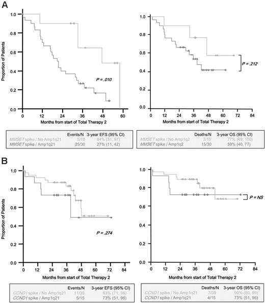
We also observed that the incidence of t(14q32) detected by interphase FISH was similar among patients with Amp1q21 and lacking Amp1q21 in patients who had FISH results for 1q21 and t(14q32) (P = .155, n = 127, Table 6). On the other hand, in patients who had both 1q21 FISH results and microarray data (n = 253), those with Amp1q21 had a high incidence of spiked expression of c-MAF or FGFR3/MMSET compared with those lacking Amp1q21 (P = .002, P < .001, respectively), whereas an incidence of spiked expression of CCND1 in patients with Amp1q21 was low compared with those lacking Amp1q21 (P < .001) (Table 7). Among the 40 patients with FGFR3/MMSET spikes, the 3-year EFS was 64% in those lacking Amp1q21 and 27% in those with Amp1q21 (P = .010) (Figure 3A). The 3-year OS in those lacking Amp1q21 was 77% and, in those with Amp1q21, 59% (P = .212) (Figure 3A). Among the 54 patients with CCND1 spikes, the presence or absence of Amp1q21 did not significantly alter the 3-year EFS or OS (Figure 3B). The lack of sufficient numbers of cases with CCND3, MAF, and MAFB spikes precluded their analysis here.
Correlation of t(14q32) and Amp1q21 in newly diagnosed MM
. | No. of patients/total no. (%) . | . | . | |
|---|---|---|---|---|
. | No Amp1q21 . | Amp1q21 . | P . | |
| t(14q32) | 45/80 (56) | 35/80 (44) | .155 | |
| No t(14q32) | 33/47 (70) | 14/47 (30) | < .001 | |
| Total | 78/127 (61) | 49/127 (39) | NA | |
. | No. of patients/total no. (%) . | . | . | |
|---|---|---|---|---|
. | No Amp1q21 . | Amp1q21 . | P . | |
| t(14q32) | 45/80 (56) | 35/80 (44) | .155 | |
| No t(14q32) | 33/47 (70) | 14/47 (30) | < .001 | |
| Total | 78/127 (61) | 49/127 (39) | NA | |
A total of 127 patients had both FISH results for 1q21 and IGH translocations in newly diagnosed MM enrolled in Total Therapy 2. The presence of t(14q32) was defined by interphase FISH using the commercial IGH break-a-part probes (Vysis). If at least 20% of clonal plasma cells had split signals of IGH probes, it was considered the presence of t(14q32)
NA indicates not applicable
Correlation of spiked expression of 5 protooncogenes associated with recurrent translocations in MM according to Amp1q21 in newly diagnosed MM
. | . | No. of patients/total no. (%) . | . | . | |
|---|---|---|---|---|---|
| Spikes . | Frequency, % . | No Amp1q21 . | Amp1q21 . | P . | |
| CCND1 | 21.3 | 39/54 (72) | 15/54 (28) | < .001 | |
| CCND3 | 2.4 | 3/6 (50) | 3/6 (50) | ND | |
| MAFB | 2.4 | 2/6 (33) | 4/6 (67) | .567 | |
| c-MAF | 2.4 | 0/6 (0) | 6/6 (100) | .002 | |
| MMSET | 15.8 | 10/40 (25) | 30/40 (75) | < .001 | |
| Others | 55.7 | 85/141 (60) | 56/141 (40) | ND | |
| Total | NA | 139/253 (55) | 114/253 (45) | NA | |
ND indicates not done; NA, not applicable
The presence of CAs in an MM karyotype is a powerful negative predictor of survival in MM. Patients with no CA but having Amp1q21 had an inferior outcome relative to those with no CA and lacking Amp1q21 (P < .001) (Figure 4). This effect was more pronounced in those patients with CA. Those with Amp1q21 in the context of CA had an inferior far worse outcome compared with those with CA but lacking this abnormality (P < .001) (Figure 4).
Correlation of Amp1q21 between newly diagnosed and relapsed MM and postrelapse survival
The frequency of Amp1q21 at relapse was significantly higher than in newly diagnosed MM (relapsed versus newly diagnosed MM, 33 of 45 [72%] versus 205 of 479 [43%], P < .001) (Table 3).
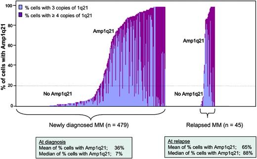
The mean and median percentage of myeloma cells with Amp1q21 at relapse tended to be higher than those in newly diagnosed MM (mean/median, relapsed versus newly diagnosed MM, 65%/88% versus 36%/7%) (Figure 5). The frequency of patients with at least 4 copies of 1q21 at relapse was significantly higher than that at diagnosis (20 of 45 [44%] versus 88 of 479 [8%], P < .001), whereas the frequency of patients with 3 copies of 1q21 was similar at relapse and diagnosis (13 of 45 [29%] versus 117 of 479 [24%], P = .507) (Table 4). The patients with at least 4 copies of 1q21 tended to have a high percentage of myeloma cells with Amp1q21 compared with those with 3 copies of 1q21 at both diagnosis and relapse (at diagnosis, 73% versus 92%; at relapse, 72% versus 97%; Table 8).
The mean percentage of cells with Amp1q21 according to the copy number of 1q21 at diagnosis and relapse
. | No. . | Mean % of cells with Amp1q21 . |
|---|---|---|
| At diagnosis | ||
| Up to 2 copies of 1q21 | 274 | 3 |
| 3 copies of 1q21 | 117 | 73 |
| At least 4 copies of 1q21 | 88 | 92 |
| Total | 479 | 36 |
| At relapse | ||
| Up to 2 copies of 1a21 | 12 | 3 |
| 3 copies of 1q21 | 13 | 72 |
| At least 4 copies of 1q21 | 20 | 97 |
| Total | 45 | 65 |
EFS and OS among patients with newly diagnosed MM according to Amp1q21 and adding thalidomide or not. A Kaplan-Meier analysis of EFS (left) and OS (right) is displayed in relation to no Amp1q21 (up to 2 copies of 1q21, n = 124) or Amp1q21 (3 or more copies of 1q21, n = 102) in patients treated with a regimen containing thalidomide and in relation to no Amp1q21 (n = 150) or Amp1q21 (n = 103) in patients treated with a regimen without thalidomide. Thal+ indicates patients treated with thalidomide; Thal-, patients treated without thalidomide.
EFS and OS among patients with newly diagnosed MM according to Amp1q21 and adding thalidomide or not. A Kaplan-Meier analysis of EFS (left) and OS (right) is displayed in relation to no Amp1q21 (up to 2 copies of 1q21, n = 124) or Amp1q21 (3 or more copies of 1q21, n = 102) in patients treated with a regimen containing thalidomide and in relation to no Amp1q21 (n = 150) or Amp1q21 (n = 103) in patients treated with a regimen without thalidomide. Thal+ indicates patients treated with thalidomide; Thal-, patients treated without thalidomide.
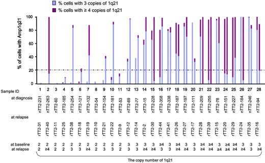
Twenty-eight patients had 1q21 FISH results available both at diagnosis and relapse. In these 28 patients, as shown in Figure 6, 27 had the same or greater copy numbers of 1q21 at relapse compared with those at diagnosis.
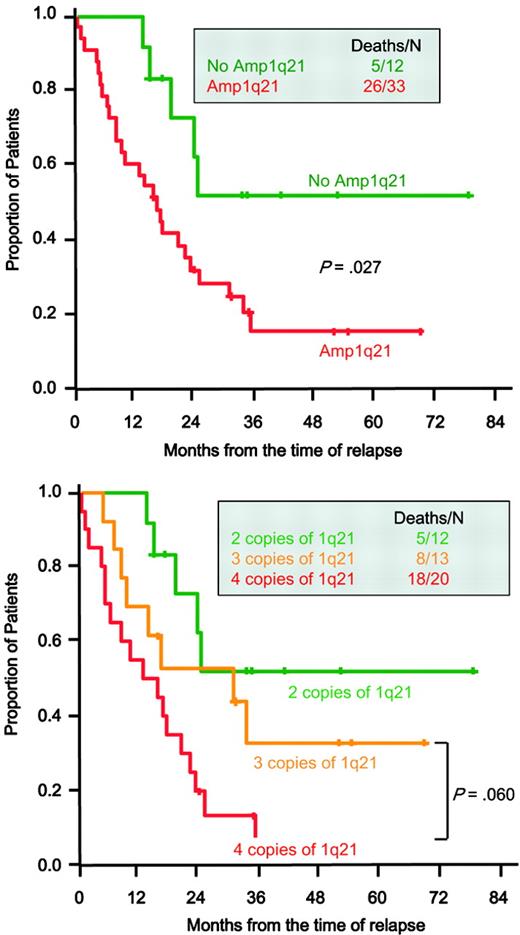
Among 45 relapsed patients who had 1q21 FISH results at relapse and receiving various salvage therapies, patients with Amp1q21 at relapse had inferior 5-year postrelapse survival compared with those lacking Amp1q21 at relapse (15% versus 53%, P = .027) (Figure 7). Among 33 relapsed patients with Amp1q21 at relapse, 5-year postrelapse survival in patients with at least 4 copies of 1q21 at relapse was 0% compared with 32% in those with 3 copies of 1q21 at relapse (P = .060) (Figure 7).
Incidence of Amp1q21 in human myeloma cell lines
Amp1q21 was detected by metaphase FISH in 21 of 23 (91%) human myeloma cell lines. The copy number of 1q21 was the same or greater compared with that of 1q31 in all 23 human myeloma cell lines tested (Figure S1B-C; Table S1). Importantly, the cell line OCI-MY5, which has 2 copies of the 1q21 region, harbors a deletion of the p arm and much of the q arm and juxtaposition of the 1q21 region to a nonhomologous chromosome (data not shown).
Discussion
MM is a plasma-cell malignancy that can sometimes be preceded by MGUS or SMM, and MM mostly is incurable, which can be linked to drug resistance. The precise mechanisms of initiation and progression of MM are poorly understood; however, MM is thought to develop through a multistep process, including a genomic instability, through which clonal evolution may occur. It has been suggested that gain of the 1q21 region, which is one of the most recurrent chromosomal aberrations in MM, is related to an advanced phenotype of MM and therefore may be associated with disease progression.25 Support for this concept is borne by our observation of an increased incidence of Amp1q21 in disease progression. Amp1q21 was observed in 0% in MGUS, 45% in SMM, 43% in newly diagnosed MM, 72% in relapsed MM, and 91% in human myeloma cell lines. Amp1q21 was associated with inferior EFS and OS in MM, a higher risk of transition of SMM to active MM, and a shortened postrelapse survival. Our data are consistent with previous reports on frequency of Amp1q21 in newly diagnosed MM based on FISH and CGH25,32-34 and with a report showing positive correlation between presence of Amp1q21 by CGH and transition from SMM to active MM.35
Among 479 patients with newly diagnosed MM, 205 patients (43%) with Amp1q21 had a high incidence of abnormal, hypodiploid, and hyperdiploid karyotypes and del(13) by FISH, IgA predominance, high-level B2M and LDH, a low level of HGB and, finally, a poor prognosis compared with those lacking Amp1q21. Patients with Amp1q21 seem to constitute a unique subgroup of MM.
We also observed that the incidence of Amp1q21 was similar to that of no Amp1q21 in patients with t(14q32), which is not consistent with a previous report.32 Interestingly, while Amp1q21 was only present in 28% of cases with CCND1 spikes, this anomaly was present in 100% of cases with c-MAF spikes and 75% of cases with FGFR3/MMSET spikes. These findings are similar to those seen with del(13) in which there is a strong positive correlation with MM with t(14;16) or t(4;14), whereas del(13) is infrequent in those with t(11;14).5,31 Together these data would indicate that Amp1q21 tends to occur in MM cells with dysregulated expression of c-MAF or FGFR3/MMSET after illegitimate IGH translocations, probably as a progression event. Survival analysis of the effect of Amp1q21 was evaluated in patients with FGFR3/MMSET spikes, a known high-risk feature. Although Amp1q21 was found to negatively affect EFS in this group, OS was not significantly different between those with and without Amp1q21. However, the effect on OS is consistent with the results for EFS, and analysis of a larger sample might reveal a significant difference. An important reason that this happens is that less than one quarter of patients with FGFR3/MMSET spikes lack Amp1q21, so it is hard to get a large enough sample to estimate their lower death rate. The fact that most FGFR3/MMSET spike-positive cases harbor Amp1q21 is highly significant and seems to be in need of a biologic explanation. Perhaps the cases lacking Amp1q21 represent an early stage of the disease and Amp1q21 will emerge at relapse. Longitudinal studies will be needed to address this hypothesis. Consistent with this concept, the proportion of MM cells with Amp1q21 ranged widely at diagnosis. The copy number of 1q21 in each patient and the mean and median percentage of cells with Amp1q21 all tended to increase at relapse compared with diagnosis, which may suggest that the clonal evolution of Amp1q21 and/or the clonal selection and expansion of MM cells with Amp1q21 occur during therapy. An important aim of future research in our group is to understand if genes located in the 1q21 region, whose expression is gene dosage dependent, promote resistance to chemotherapy.
EFS and OS among patients with newly diagnosed MM according to the presence or absence of common recurrent translocations and Amp1q21. A Kaplan-Meier analysis of EFS (left) and OS (right) in patients with MMSET spikes (A) or CCND1 spikes (B) according to the presence or absence of Amp1q21. Note that the difference in median follow-up of this group of patients is different from the overall group because microarray profiling was initiated after Total Therapy 2 had started.
EFS and OS among patients with newly diagnosed MM according to the presence or absence of common recurrent translocations and Amp1q21. A Kaplan-Meier analysis of EFS (left) and OS (right) in patients with MMSET spikes (A) or CCND1 spikes (B) according to the presence or absence of Amp1q21. Note that the difference in median follow-up of this group of patients is different from the overall group because microarray profiling was initiated after Total Therapy 2 had started.
OS among patients with newly diagnosed MM according to cytogenetic abnormalities and Amp1q21. A Kaplan-Meier analysis of OS in 440 patients who had both 1q21 FISH results and karyotype data by G banding according to the presence or absence of cytogenetic abnormalities and Amp1q21.
OS among patients with newly diagnosed MM according to cytogenetic abnormalities and Amp1q21. A Kaplan-Meier analysis of OS in 440 patients who had both 1q21 FISH results and karyotype data by G banding according to the presence or absence of cytogenetic abnormalities and Amp1q21.
The proportion of cells with Amp1q21 at diagnosis and relapse. The proportion of cells with Amp1q21 is indicated by the height of the bar on the y-axis. The proportion of cells with 3 and with 4 or more copies of 1q21 in each sample is indicated by blue and red, respectively. A total of 479 newly diagnosed MM and 45 relapsed MM samples are ordered from the lowest to highest proportion of cells with Amp1q21 from left to right in each group on the x-axis. The mean/median percentages of cells with Amp1q21 at diagnosis and relapse were 36%/65% and 7%/88%, respectively.
The proportion of cells with Amp1q21 at diagnosis and relapse. The proportion of cells with Amp1q21 is indicated by the height of the bar on the y-axis. The proportion of cells with 3 and with 4 or more copies of 1q21 in each sample is indicated by blue and red, respectively. A total of 479 newly diagnosed MM and 45 relapsed MM samples are ordered from the lowest to highest proportion of cells with Amp1q21 from left to right in each group on the x-axis. The mean/median percentages of cells with Amp1q21 at diagnosis and relapse were 36%/65% and 7%/88%, respectively.
Patients with at least 4 copies of 1q21 at diagnosis tended to have a higher percentage of cells with Amp1q21 (Table S2) and showed an initially more aggressive clinical course than those with 3 copies of 1q21. However, patients with at least 4 copies of 1q21 at diagnosis had similar 5-year EFS and OS compared with those with 3 copies of 1q21 (Figure S2), whereas patients with at least 4 copies of 1q21 at relapse had an inferior postrelapse survival compared with those with 3 copies of 1q21. This may suggest that myeloma cells with at least 4 copies of 1q21 are potentially associated with a more malignant and drug-resistant phenotype than those with 3 copies of 1q21 but that, during therapy, residual cells with 3 copies of 1q21 can continue to increase 1q21 copy numbers and thus become as malignant as those with at least 4 copies of 1q21 at diagnosis.
The proportion of cells with Amp1q21 and the copy number of 1q21 in each patient at diagnosis and relapse in paired patients. Twenty-eight patients had 1q21 FISH results available at both diagnosis and relapse. The proportion of cells with Amp1q21 is indicated by the height of the bar on the y-axis. Each sample is represented by a left and right bar indicating the proportion of cells with Amp1q21 at diagnosis and relapse, respectively. The proportion of cells with 3 and with 4 or more copies of 1q21 in each sample is indicated by blue and red, respectively. The numbers below each patient number indicate the copy number of 1q21 at diagnosis and relapse. Sample ID is same as that in Table S4.
The proportion of cells with Amp1q21 and the copy number of 1q21 in each patient at diagnosis and relapse in paired patients. Twenty-eight patients had 1q21 FISH results available at both diagnosis and relapse. The proportion of cells with Amp1q21 is indicated by the height of the bar on the y-axis. Each sample is represented by a left and right bar indicating the proportion of cells with Amp1q21 at diagnosis and relapse, respectively. The proportion of cells with 3 and with 4 or more copies of 1q21 in each sample is indicated by blue and red, respectively. The numbers below each patient number indicate the copy number of 1q21 at diagnosis and relapse. Sample ID is same as that in Table S4.
Postrelapse survival of MM patients enrolled in Total Therapy 2 according to Amp1q21 and the copy number of 1q21 at relapse. Kaplan-Meier analysis of postrelapse survival is shown in relation to no Amp1q21 (up to 2 copies of 1q21, n = 12) or Amp1q21 (3 or more copies of 1q21, n = 33) at relapse (top) and up to 2 copies (n = 12), 3 copies (n = 13), or 4 or more copies (n = 20) of 1q21 at relapse (bottom) determined by interphase FISH. At the time of analysis, the median follow-up of a postrelapse survival was 17 months (range, 0.3 to 78) in this analysis.
Postrelapse survival of MM patients enrolled in Total Therapy 2 according to Amp1q21 and the copy number of 1q21 at relapse. Kaplan-Meier analysis of postrelapse survival is shown in relation to no Amp1q21 (up to 2 copies of 1q21, n = 12) or Amp1q21 (3 or more copies of 1q21, n = 33) at relapse (top) and up to 2 copies (n = 12), 3 copies (n = 13), or 4 or more copies (n = 20) of 1q21 at relapse (bottom) determined by interphase FISH. At the time of analysis, the median follow-up of a postrelapse survival was 17 months (range, 0.3 to 78) in this analysis.
Despite the fact that Amp1q21 is present in up to 50% of newly diagnosed patients, we have shown that in multivariate analysis including other major predictors Amp1q21 was an independent adverse prognostic factor. However, among patients with ISS stage 3, OS was similar between those with Amp1q21 and lacking Amp1q21, whereas patients with Amp1q21 had inferior OS among patients with ISS stage 1 and 2 (Figure S3); therefore, Amp1q21 is a poor prognostic marker in patients whose B2M is less than 467.5 nM (5.5 mg/L).
We have reported that adding thalidomide to high-dose melphalan-based tandem autotransplantations improved EFS but did not improve OS due to a shorter postrelapse survival in the thalidomide group.27 In this study, we demonstrated that adding thalidomide improved EFS in patients lacking Amp1q21, whereas 5-year EFS in patients with Amp1q21 was similar between those who were treated with thalidomide and without thalidomide. Adding thalidomide did not improve OS either in patients lacking Amp1q21 or with Amp1q21. The molecular reason underlying this finding is currently not clear, but the finding suggests that the presence of Amp1q21 might be an important determinant for stratifying patients to receive or not receive thalidomide.
The cellular and molecular mechanisms of Amp1q21 are not well understood but are thought to involve decondensation of pericentromeric heterochromatin in MM and lymphoma.19,20,36 There also remains a question as to whether Amp1q21 is a cause or a consequence of disease progression. Amp1q21 may reflect chromosomal instability, which is related to tumor progression, which is usually through silencing tumor suppressor genes.37 However, activation of a protooncogene caused by increased gene dosage accompanying chromosomal instability can also play an important role in tumor development and progression. Evidence of high-level amplification of 1q21 in MM20,38 suggests that the amplification may be of pathogenic significance related to increased copy number and a concomitant increase in the expression of gene(s) within this region. We recently reported on the high-resolution mapping of a 10 Mb amplicon at 1q21,39 and we and others have reported on deregulated expression of candidate genes in the 1q21 region in MM and other cancers.38-51 However, none of these candidate genes, with the exception of RAB25, has been investigated in animal models. Moreover, the possible combined effect of multiple candidate genes has not been demonstrated or considered. In the future, precise direct functional analyses of each candidate gene using animal models and analyses of mass effects of several genes in the same amplicon on tumor development will be required to establish the association of genes within the amplicon as causal in MM progression.
In conclusion, we have demonstrated that Amp1q21 was an independent adverse prognostic marker in newly diagnosed MM treated with autotransplantations, associated with a shortened postrelapse survival and a higher risk of transition from SMM to MM. In addition, we have demonstrated that the proportion of cells with Amp1q21 and the copy number of 1q21 tended to increase at relapse compared with diagnosis, and adding thalidomide improved EFS in patients lacking Amp1q21 at diagnosis but did not in patients with Amp1q21 at diagnosis. Our data suggest that Amp1q21 is associated with a malignant phenotype in MM and may be central to progression of plasma-cell dyscrasias. Clarification of the mechanism(s) that cause Amp1q21 and identification of possible responsible gene(s) in this region might provide more precise insight into the natural history of myeloma and possible novel targets for future therapies in MM and in other cancers.
Prepublished online as Blood First Edition Paper, May 16, 2006; DOI 10.1182/blood-2006-03-009910.
Supported by National Institutes of Health grants CA55819 (J.D.S., B.B.) and CA97513 (J.D.S.) by the Fund to Cure Myeloma.
The online version of this article contains a data supplement.
An Inside Blood analysis of this article appears at the front of this issue.
The publication costs of this article were defrayed in part by page charge payment. Therefore, and solely to indicate this fact, this article is hereby marked “advertisement” in accordance with 18 U.S.C. section 1734.
We thank members of the Donna D. and Donald M. Lambert Laboratory of Myeloma Genetics for technical assistance: Ermin Tian, Christopher Adams, Christopher Crane, Adam Hicks, Bob Kordsmeier, Christopher Randolph, Steven McMorran, Owen Stephens, Ryan Williams, Yan Xaio, and Hongwei Xu. We thank Dr Michael Kuehl for providing many of the myeloma cell lines. We extend a special thanks to all patients, their caregivers, and referring physicians for making this work possible.


This feature is available to Subscribers Only
Sign In or Create an Account Close Modal