Falini et al1 first reported exon 12 mutations in the nucleophosmin (NPM1) gene in approximately 35% of patients with adult acute myeloid leukemia (AML). These mutations disrupt the nucleolar-localization signal and acquire a nuclear export signal motif at the C-terminus leading to abnormal cytoplasmic accumulation of NPM.2 NPM1-mutated AMLs frequently have CD34-negative blasts, a normal karyotype, and FLT3 gene mutations, and patients have good response to induction therapy. Subsequent studies have confirmed these features, and also have shown that NPM1 gene mutations are most common in AMLs with monocytic differentiation (French-American-British [FAB]-M4/M5).3,4 NPM1-mutated AMLs have a distinctive gene expression profile characterized by up-regulation of genes involved in stem-cell maintenance.5
We have observed rare cases of AML in which the blasts have prominent nuclear invaginations (so-called cuplike nuclei) similar to cases previously reported as AML with cuplike nuclear invaginations6 or “myeloid/natural killer cell” acute leukemia.7 AML with cuplike nuclei cases we have seen have immunophenotypic and cytogenetic features similar to NPM1-mutated AML. Therefore, we collected a group of AML with cup-like nuclei cases and assessed them for NPM1 gene mutations. This study was approved by the University of Texas M. D. Anderson Cancer Center Institutional Review Board.
The criterion for inclusion in this study was the presence of 10% or more blasts with prominent nuclear invaginations spanning at least 25% of the nuclear diameter. In a review of AMLs at our institution, we identified 24 patients with AML with cup-like nuclei. All were FAB AML-M1, representing approximately 1% of all AML and 20% of AML-M1. In particular, no cases of AML-M4 or -M5 (n = 80) had cup-like nuclei. We compared this group to an age-matched control group of 20 patients with AML-M1 without cup-like nuclei.
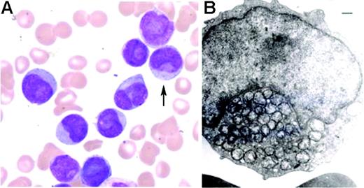
As illustrated in Figure 1, blasts were often medium-sized with scant cytoplasm containing a few azurophilic granules and infrequent Auer rods. Electron microscopy (n = 4) demonstrated condensed collections of mitochondriae within the invaginated nuclear pockets. Compared with the control group, AML with cup-like nuclei had a higher frequency of NPM1 gene mutations detected by PCR amplification of exon 12 followed by direct sequencing (64% versus 20%, P = .043). The most common mutation was type A in 90% of cases. Compared with the control group, AML with cup-like nuclei were significantly associated with female sex (male-female ratio, 6:18 vs 13:7, P = .014), lack of CD34 (71% versus 10%, P < .001), and human leukocyte antigen (HLA)-DR positivity (50% versus 10%, P = .009). There was also a higher frequency of normal cytogenetics (70% versus 45%, P = .10) and FLT3 gene mutations of internal tandem duplication type (88% versus 50%, P = .16), although not significant. There were no differences in blast percentage or white blood cell count. With a median follow-up period of 5 months (range, 1-54 months), the complete remission rate was higher in patients with AML with cup-like nuclei (71% versus 40%, P = .032). There was no difference in the overall survival at 1 year between patients with AML with cup-like nuclei and the control group.
Morphologic features of AML with cuplike nuclei. (A) Blasts are medium-sized and have prominent nuclear invaginations, and scant to moderate cytoplasm with a few, fine azurophilic granules (Wright-Giemsa bone marrow aspirate smear; original magnification, × 1000). Image was obtained using an Olympus BX41 microscope (Olympus, Melville, NY) equipped with a 100×/1.25 numeric aperture oil objective; was acquired using a Spot Insight color camera 3.2.0 (Diagnostic Instruments, Sterling Heights, MI) and Image Pro Plus software version 3.2 (Media Cybernetics, Silver Spring, MD); and was processed using Adobe Photoshop CS version 7 (Adobe Systems, San Jose, CA). (B) Condensed collections of mitochondriae are present within the invaginated nuclear pockets (transmission electron microscopic examination; scale bar indicates 500 nm). Image was obtained using a JEOL 1200 EXII electron microscope (JEOL, Tokyo, Japan) at 20 000× magnification, and was processed using Adobe Photoshop CS version 7.
Morphologic features of AML with cuplike nuclei. (A) Blasts are medium-sized and have prominent nuclear invaginations, and scant to moderate cytoplasm with a few, fine azurophilic granules (Wright-Giemsa bone marrow aspirate smear; original magnification, × 1000). Image was obtained using an Olympus BX41 microscope (Olympus, Melville, NY) equipped with a 100×/1.25 numeric aperture oil objective; was acquired using a Spot Insight color camera 3.2.0 (Diagnostic Instruments, Sterling Heights, MI) and Image Pro Plus software version 3.2 (Media Cybernetics, Silver Spring, MD); and was processed using Adobe Photoshop CS version 7 (Adobe Systems, San Jose, CA). (B) Condensed collections of mitochondriae are present within the invaginated nuclear pockets (transmission electron microscopic examination; scale bar indicates 500 nm). Image was obtained using a JEOL 1200 EXII electron microscope (JEOL, Tokyo, Japan) at 20 000× magnification, and was processed using Adobe Photoshop CS version 7.
Our findings suggest that AML with cup-like nuclei cases are rare, predominantly have AML-M1 morphology, and frequently are associated with NPM1 gene mutations. These neoplasms otherwise resemble other NPM1-mutated cases of AML.
We thank Mannie Steglich for technical assistance.


This feature is available to Subscribers Only
Sign In or Create an Account Close Modal