Abstract
Rac GTPases are key regulators of leukocyte motility. In lymphocytes, chemokine-mediated Rac activation depends on the CDM adaptor DOCK2. The present studies addressed the role of DOCK2 in chemokine-triggered lymphocyte adhesion and motility. Rapid chemokine-triggered activation of both LFA-1 and VLA-4 integrins took place normally in DOCK2–/– T lymphocytes under various shear flow conditions. Consequently, DOCK2–/– T cells arrested normally on TNFα-activated endothelial cells in response to integrin stimulatory chemokine signals, and their resistance to detachment was similar to that of wild-type (wt) T lymphocytes. Nevertheless, DOCK2–/– T lymphocytes exhibited reduced microvillar collapse and lamellipodium extension in response to chemokine signals, ruling out a role for these events in integrin-mediated adhesion strengthening. Strikingly, arrested DOCK2–/– lymphocytes transmigrated through a CCL21-presenting endothelial barrier with similar efficiency and rate as wt lymphocytes but, unlike wt lymphocytes, could not locomote away from the transmigration site of the basal endothelial side. DOCK2–/– lymphocytes also failed to laterally migrate over multiple integrin ligands coimmobilized with chemokines. This is a first indication that T lymphocytes use 2 different chemokine-triggered actin remodeling programs: the first, DOCK2 dependent, to locomote laterally along apical and basal endothelial surfaces; the second, DOCK2 independent, to cross through a chemokine-bearing endothelial barrier.
Introduction
Leukocytes circulating in the blood are recruited to lymphoid organs and to peripheral sites of injury, infection, and inflammation by a series of sequential adhesive steps.1,2 The arrest of lymphocytes and myeloid cells on endothelial cells (ECs) within postcapillary venules requires in situ activation of at least 1 of 3 major integrins: very late antigen 4 (VLA-4), α4β7, or leukocyte function-associated antigen 1 (LFA-1).3 The adhesive potential of these integrins is dynamically regulated by endothelial-displayed chemoattractants, mostly chemokines, which transduce rapid signals via specific leukocyte G-protein–coupled receptors (GPCRs). Once stably arrested on their integrin ligands on the vascular endothelial target, leukocytes embark on a series of coordinated cytoskeletal remodeling events to cross the endothelial lining.4 With few exceptions, lymphocyte emigration (diapedesis or transendothelial migration [TEM]) takes place at endothelial junctions and involves a highly regulated squeezing between 2 or 3 opposed endothelial cells.5 Thus, a lymphocyte temporarily arrested on a given endothelial site away from junctional sites of diapedesis must locomote to one of these sites to successfully transmigrate, while resisting detachment from the vessel wall by the disruptive blood flow.
Chemokine-stimulated GPCRs and their downstream effectors of actin polymerization and cell contraction must allow the arrested leukocyte to sustain integrin-mediated resistance to detachment while the cell polarizes and locomotes from its site of arrest to the TEM site.3 Indeed, interference with actin remodeling on arrested lymphocytes severely impairs lymphocyte locomotion and abrogates subsequent TEM.6 Although chemokine signals to lymphocyte GPCRs and Gi proteins have been shown to remodel the actin polymerization in these cells by stimulating the RhoA, Rac, Cdc42, and Rap GTPases,7-9 the precise involvement of these GTPases in specific adhesive and migratory steps employed by lymphocytes at endothelial contacts is only starting to unfold.10 Rac1 is the predominant Rac GTPase family member in lymphoid cells and is a master regulator of lamellipodium extension in chemoattractant-stimulated cells.11 In addition to Rac1, the rapid activation of the Rap1 GTPase by chemokines has been recently implicated in integrin activation and polarization of lymphocytes at endothelial compartments.8 The dynamic complexity of lymphocyte locomotion on apical endothelial surfaces under shear stress, and of TEM occurring under disruptive shear forces, makes the functional dissection of these processes difficult. Notably, both Rac1, Rap1, as well as other Rho family GTPases are not only implicated in lymphocyte TEM but also contribute to early arrest and adhesion strengthening events8,12,13 ; thus, the outcome of their suppression on lymphocyte diapedesis per se has been difficult to discern.
DOCK2 is a novel member of the Caenorhabditis elegans Ced-5, mammalian DOCK180, and Drosophila melanogaster myoblast city (CDM) family of scaffold proteins. Mice lacking DOCK2 exhibit a striking deficit in T-lymphocyte migration in response to the prototypic lymphoid chemokines CXCL12, CCL19, and CCL2114 and impaired Rac1 and Rac2 GTP loading in response to these chemokines but normal RhoA and Cdc42 activation and function.14 DOCK2–/– lymphocytes are consequently impaired in their exit from the thymus as well as in their homing to and migration within both the spleen and the lymph nodes.14,15 To dissect whether and how Rac activation by chemokines contributes to specific steps of adhesion strengthening, locomotion, and TEM across endothelial cells under defined conditions of shear flow, we chose T lymphocytes derived from the spleens of DOCK2–/– mice as a model for lymphocytes unable to undergo either Rac1 or Rac2 activation by prototypic chemokine signals.14
In this study we dissected the specific migratory steps most affected by the loss of this key Rac regulator using ex vivo analysis of T cells derived from DOCK2 null spleen interacting with an inflamed endothelial barrier. We show here that chemokine triggering of both VLA-4 and LFA-1 was normal in DOCK2–/– T lymphocytes. These lymphocytes exhibited, however, reduced microvillar collapse, lamelopodia extension, and lateral motility over both endothelial and extracellular matrix ligands in response to signals from the prototypic T-cell chemokine, CCL21. Unexpectedly, DOCK2 was not required for lymphocyte TEM triggered by endothelial-associated CCL21, suggesting that lymphocyte TEM, while chemokine triggered,6 does not require chemokine activation of Rac GTPases.
Materials and methods
Reagents and mAbs
Murine ICAM-1–Fc, VCAM-1–Fc, CCL21, CXCL12, and TNFα were purchased from R&D Systems (Minneapolis, MN). Bovine serum albumin (BSA; fraction V), Ca2+, Mg2+-free Hanks balanced salt solution (HBSS), pertussis toxin, and fibronectin (FN) were purchased from Sigma-Aldrich (St Louis, MO). Human serum albumin (HSA; fraction V), LY294002, and protein A were purchased from Calbiochem (San Diego, CA). The α4 integrin function-blocking PS/2 monoclonal antibody (mAb) was purchased from SouthernBiotech (Birmingham, AL). FITC-conjugated anti-CD3 (145-2C11) or anti-CD4 (RM4-5), PE-conjugated anti-CD8 (53-6.7) anti–αL integrin (M17/4, function blocking), anti–αM integrin (M1/170), and anti–α4 (R1-2) mAbs were all purchased from PharMingen (Philadelphia, PA). The αL integrin-specific mAb, M18/2, was purchased from Cymbus Biotechnology (Hants, United Kingdom). CCL19-Fc and CXCL12-Fc were kind gifts from Dr J. Cyster (University of California, San Francisco).
Cells
All animal procedures were approved by the Animal Research Committee at the Weizmann Institute of Science. Splenocyte suspensions were obtained from spleens of either wild-type (wt) or DOCK2–/– C57BL/6 mice at 6 to 9 weeks of age. Splenocytes were cultured for 16 hours before CD3+ lymphocyte purification without stimulation. CD3+, CD4+, and CD8+ murine T lymphocytes were isolated by negative selection with negative cell isolation kits (magnetic-activated cell sorter [MACS]; Miltenyi Biotec, Bergisch Gladbach, Germany) according to manufacturer's instructions 2 hours before functional experiments. Purity of CD3+ lymphocytes was verified by flow cytometry to be more than 90%. The murine brain–derived endothelial-cell line (bEnd.3)16 was a gift from Dr D. Westweber (Munster, Germany). BEnd.3 cells were grown to confluence in RPMI 1640 (Gibco, Carlsbad, CA) containing 1 mM l-glutamine, 10% FCS, and antibiotics all purchased from Biological Industries (Beit Haemek, Israel).
Flow cytometry
Cells were washed once with PBS containing 5 mM EDTA, resuspended in binding medium (HBSS containing 2 mg/mL BSA and 10 mM HEPES [pH 7.4] supplemented with 1 mM CaCl2 and 1 mM MgCl2), and incubated with either fluorescently labeled primary antibodies or with CCL19-Fc or CXCL12-Fc for 20 minutes at 4°C. For Fc staining, cells were washed and incubated with secondary antibodies for 30 minutes at 4°C. All cells were analyzed immediately in a fluorescence-activated cell sorter (FACS) flow cytometer (FACScan; Becton Dickinson, Erembodegem, Belgium).
Scanning electron microscopy
Cells were stimulated with CCL21 (300 ng/mL) for 1 minute in suspension at 37°C and immediately fixed (3% glutaraldehyde in 0.1 M cacodylate buffer supplemented with 7.5% sucrose and 5 mM CaCl2, pH 7.4). Cells were washed with 0.1 M cacodylate buffer, plated on polylysine-coated silicon chips, and postfixed with 1% OsO4 for 1 hour at 22°C. The samples were washed twice prior to treatment with 1% tannic acid for 5 minutes followed by another wash with 0.1 M cacodylate buffer and treatment with 1% uranyl acetate. The samples were dehydrated in graded ethanol, dried, and coated with palladium gold. Samples were photographed with an environmental scanning electron microscope (FEG ESEM XL 30, FEI, Eindhoven, the Netherlands) operated at 10 kV.
Motility and polarization analysis by videomicroscopy
Small motility chamber microslides (Ibidi, Munchen, Germany) were coated overnight at 4°C with 2 μg/mL CXCL12 or 1 μg/mL CCL21 followed, where indicated, by 1-hour coating with FN (5 μg/mL). The slides were extensively washed and blocked with 2 mg/mL HSA. Chambers injected with lymphocytes were immediately mounted on the stage of an inverted microscope (Delta Vision Spectris RT; Applied Precision, Issaquah, WA) and recorded with Softmorx 3.5 (Applied Precisions) for 10 minutes at 6 frames per minutes using a 40 ×/0.95 NA differential interference contrast (DIC) objective. All experiments were performed at 37°C. Cells that acquired and sustained leading and trailing edges for at least 1 minute were considered polarized and were subdivided into polarized stationary or polarized motile categories depending on their ability to locomote for at least 3 cell diameters from their original position during 10 minutes of tracking (Figure 6A; Video S3 [available on the Blood website; see the Supplemental Videos link at the top of the online article]). To block PI3K signaling, cells were pretreated with LY294002 (20 μM) or DMSO for 30 minutes.
Laminar flow adhesion assays
VCAM-1–Fc or ICAM-1–Fc were coated on polystyrene plates precoated with protein A (1 μg/mL), as described.17 The plates were each assembled in a flow chamber (260 μm gap) as previously described.18 Lymphocytes were washed with cation-free H/H medium (cation-free HBSS containing 10 mM HEPES [pH 7.4] and 2 mg/mL BSA), resuspended in binding medium (H/H medium supplemented with 1 mM CaCl2 and 1 mM MgCl2), and perfused through the flow chamber at the desired shear stress. All flow experiments were conducted at 37°C and were recorded with a long integration CCD camera (Applitech, Holon, Israel).17 Tethers were defined as transient if cells attached briefly (less than 2 seconds) to the substrate and as arrests if immediately arrested and remaining stationary for at least 3 seconds of continuous flow. Frequencies of adhesive categories or rates of cell accumulation were determined as a percentage of cells flowing immediately over the substrates, as previously described.17 To assess resistance to detachment over time, cells were settled for 1 minute on integrin ligands alone or coimmobilized with chemokine and were then subjected to a shear stress of 2 dyne/cm2 for 8 minutes. At the indicated time points, the number of cells that remained bound was expressed relative to originally settled cells. Locomotion analysis on purified ligands coimmobilized with chemokines was conducted during the same period.
Analysis of lymphocyte adhesion to, and migration across, endothelial monolayers under shear flow
BEnd.3 cells were plated at confluence on tissue culture dishes spotted with FN (20 μg/mL in PBS) and were stimulated for 24 to 30 hours with TNFα (500 U/mL). CCL21 (2 μg/mL in binding medium) was overlaid for 5 minutes on the bEnd.3 cell monolayer and washed extensively. Frequency of adhesion to the cytokine-stimulated bEnd.3 was measured in a flow chamber as described in this section. For migration analysis, lymphocytes were perfused for 2 minutes over the monolayer at 0.25 dyne/cm2 to allow accumulation. The flow rate was then increased to 2 dyne/cm2 and was kept constant for an additional 15-minute period during which images were recorded at 1 frame per second on a time-lapse SVHS video recorder using a 20 ×/0.4 NA phase-contrast objective as previously described.6 Motion analysis was performed manually on played back video segments. Lymphocytes arrested during the accumulation phase and resisting immediate detachment from the endothelial surface when subjected to the 2 dyne/cm2 shear stress were subdivided into 4 distinct categories: (1) Lymphocytes that either rolled away or detached from the substrate during the 15-minute shear application phase were considered detaching cells. (2) Lymphocytes that remained stationary throughout the assay were considered arrested regardless of their polarization state. (3) Lymphocytes that polarized and locomoted at least 3 cell diameters without detaching or transmigrating through the EC barrier were considered locomoting without TEM. (4) Lymphocytes that underwent stepwise darkening and remained dark underneath the EC with or without subsequent locomotion were considered transmigrating cells.
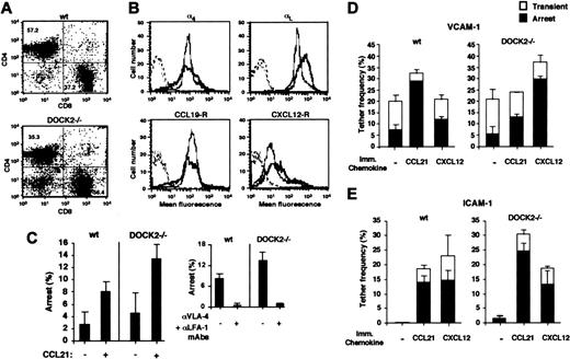
Wt and DOCK2–/–T lymphocytes arrest normally on TNFα-activated murine EC and integrin ligands under shear flow in response to different chemokine signals. (A) CD3+ splenocytes from wt and DOCK2–/– mice were double stained for CD4 and CD8, and the fractions of single-positive subsets are indicated. (B) FACS assay showing expression of α4 integrins (probed with an anti–α4 subunit mAb) and of LFA-1 (probed with an anti–αL subunit mAb) on wt (gray lines) and DOCK2–/– (black lines) CD3+ purified T splenocytes. Functional CCR7 expression was assayed by CCL19-Fc binding probed with antihuman Fc. CXCL12 receptors were assayed by similar staining using CXCL12-Fc. Background staining is depicted by dotted lines. (C) Arrest of CD3+ splenocytes (T lymphocytes) perfused for 1 minute over TNFα-activated bEnd.3 EC monolayers in the absence or presence of overlaid CCL21. The percentages of rapidly arrested cells are expressed as the fractions of the lymphocyte flux in close contact with the endothelial monolayer. Results presented are mean values ± range of 4 independent fields. (Inset) Effect of pretreatment with combined anti-α4 (VLA-4; PS/2) and anti-αL (LFA-1; M17/4) mAbs on CCL21-triggered arrest of either wt or DOCK2–/– T lymphocytes. The experiments are representative of 3 independent runs with different animals and batches of endothelial cells. (D-E) Frequency and strength of tethering of wt and DOCK2–/– T lymphocytes to VCAM-1–Fc coated at 0.5 μg/mL alone or with the indicated immobilized (imm.) chemokines at 2 μg/mL (D) or to ICAM-1–Fc coated alone at 2 μg/mL or with the indicated immobilized chemokines at 2 μg/mL (E). Tethers were determined at a shear stress of 0.5 dyne/cm2. More than 90% of tethers to either VCAM-1 or ICAM-1 were blocked, respectively, by treating lymphocytes with either 20 μg/mL α4-or αL-blocking mAbs as in the inset of panel C (data not shown). Mean values ± range determined in 2 fields are presented.
Wt and DOCK2–/–T lymphocytes arrest normally on TNFα-activated murine EC and integrin ligands under shear flow in response to different chemokine signals. (A) CD3+ splenocytes from wt and DOCK2–/– mice were double stained for CD4 and CD8, and the fractions of single-positive subsets are indicated. (B) FACS assay showing expression of α4 integrins (probed with an anti–α4 subunit mAb) and of LFA-1 (probed with an anti–αL subunit mAb) on wt (gray lines) and DOCK2–/– (black lines) CD3+ purified T splenocytes. Functional CCR7 expression was assayed by CCL19-Fc binding probed with antihuman Fc. CXCL12 receptors were assayed by similar staining using CXCL12-Fc. Background staining is depicted by dotted lines. (C) Arrest of CD3+ splenocytes (T lymphocytes) perfused for 1 minute over TNFα-activated bEnd.3 EC monolayers in the absence or presence of overlaid CCL21. The percentages of rapidly arrested cells are expressed as the fractions of the lymphocyte flux in close contact with the endothelial monolayer. Results presented are mean values ± range of 4 independent fields. (Inset) Effect of pretreatment with combined anti-α4 (VLA-4; PS/2) and anti-αL (LFA-1; M17/4) mAbs on CCL21-triggered arrest of either wt or DOCK2–/– T lymphocytes. The experiments are representative of 3 independent runs with different animals and batches of endothelial cells. (D-E) Frequency and strength of tethering of wt and DOCK2–/– T lymphocytes to VCAM-1–Fc coated at 0.5 μg/mL alone or with the indicated immobilized (imm.) chemokines at 2 μg/mL (D) or to ICAM-1–Fc coated alone at 2 μg/mL or with the indicated immobilized chemokines at 2 μg/mL (E). Tethers were determined at a shear stress of 0.5 dyne/cm2. More than 90% of tethers to either VCAM-1 or ICAM-1 were blocked, respectively, by treating lymphocytes with either 20 μg/mL α4-or αL-blocking mAbs as in the inset of panel C (data not shown). Mean values ± range determined in 2 fields are presented.
Results
T-lymphocyte DOCK2 is not required for subsecond activation of integrins by surface-presented chemokines
To assess the role of DOCK2 in integrin activation by chemokines, spleen-derived T cells were isolated from wt or DOCK2 null C57BL/6 mice. DOCK2–/– spleens contained 2-fold fewer CD3+ splenocytes, which consisted of 35% CD4+ T cells and 56% CD8+ T cells (Figure 1A). Wt CD3+ T cells consisted of a higher proportion of CD4+ cells (57% CD4+ versus 37% CD8+ cells; Figure 1A). Notably, CD3+ T lymphocytes from both mice expressed identical levels of CCR7 based on their equal capacity to bind the high-affinity CCR7 ligand, CCL19 (Figure 1B). In contrast, DOCK2–/– T cells expressed significantly higher levels of CXCL12 receptors (Figure 1B), and therefore most subsequent analysis was conducted on CCR7 signaling in wt and DOCK2–/– CD3+ T cells triggered by the prototypic endothelial-expressed CCR7 ligand, CCL21.19
DOCK2–/– T lymphocytes were recently shown to arrest normally in vivo on high endothelial venules within peripheral lymph nodes.15 Consistent with this finding, CCL21, presented on the apical surface of the murine TNFα-activated EC line, bEnd.3, which expresses VCAM-1, ICAM-1, and MadCAM-1 (data not shown), triggered significant shear-resistant adhesion of both DOCK2–/– and wt T lymphocytes (Figure 1C). Without apical CCL21, T cells, which tethered to the TNFα-activated stimulated endothelial surface, briefly rolled on it but readily detached when subjected to shear stresses above 2 dyne/cm2 (data not shown). T cells settled on the TNFα-activated ECs under shear free conditions and, without exogenously added chemokines, failed to polarize, further ruling out the display of endogenous chemokines in this endothelial model. Firm CCL21-triggered adhesion was Gi dependent (data not shown) and required both intact α4 integrins and LFA-1 on tethered CD3+ T cells (Figure 1C, inset).
We next analyzed the direct triggering of DOCK2–/– T-lymphocyte arrest on either VCAM-1 or ICAM-1 by immobilized chemokines to dissect the inherent capacity of each of the 2 integrin types to undergo subsecond activation by immobilized CCL21 signals under shear stress independent of a prior selectin-mediated capture. CCL21 robustly triggered both VLA-4– and LFA-1–mediated lymphocyte arrest on VCAM-1 or ICAM-1, respectively (Figure 1D-E). Both integrins of DOCK2–/– T lymphocytes also underwent normal subsecond activation by in situ signals from a second prototypic chemokine, CXCL12 (Figure 1D-E). In particular settings, rapid integrin activation in DOCK2–/– T lymphocytes was more efficient than in wt lymphocytes due to either higher levels of CXCL12 receptors or LFA-1 (Figure 1B, right panels). As observed with CCL21-triggered adhesion to the endothelial-cell monolayer, all chemokine-triggered integrin activation events were inhibited by pertussis toxin, an inhibitor of Gi signaling (data not shown). Taken together, these results show that the 2 major integrins that mediate T-lymphocyte arrest on inflamed ECs, VLA-4 and LFA-1, do not require DOCK2 to undergo subsecond activation by the 2 prototypic T-cell–specific chemokines, CCL21 and CXCL12.
Lymphocyte DOCK2 is necessary for microvillar collapse and lamellipodium formation in response to CCL21 signals
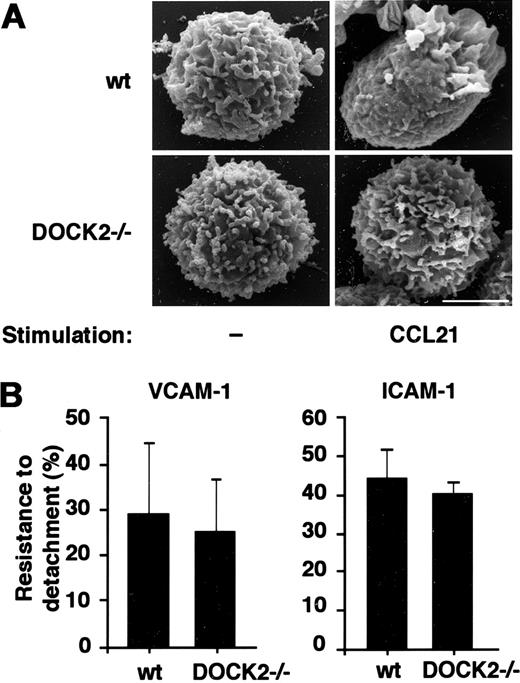
Chemokine signals have been recently shown to trigger a Rac1-mediated collapse of lymphocyte microvilli,20 a process suggested to contribute to lymphocyte adhesion strengthening and spreading on chemokine-presenting endothelial surfaces. Because chemokine-triggered Rac activation is severely impaired in DOCK2–/– lymphocytes, we considered that this microvillar collapse may depend on the presence of DOCK2. Indeed, whereas CCL21 induced both total collapse of microvilli and outgrowth of lamellipodia in more than 95% of treated wt T lymphocytes as assessed by scanning electron microcopy (SEM) (Figure 2A), this chemokine induced microvillar collapse in less than 25% of DOCK2–/– T lymphocytes. Notably, even in this small cell fraction, no lamellipodia extension could be detected (Figure 2A), indicating that DOCK2 is required for optimal CCL21-triggered microvillar collapse and lamellipodia formation in T lymphocytes. Nevertheless, a normal fraction of T cells arrested on either VCAM-1 or ICAM-1 in response to this chemokine (Figure 1D-E), and the strength of adhesion developed by T cells arrested on CCL21 and exposed to an additional 1 minute of continuous shear stress was fully retained in DOCK2–/– T cells (Figure 2B). Thus, deficient microvillar collapse is not associated with reduced ability of lymphocytes to arrest on integrin ligands or to develop adhesion strengthening and resistance to detachment by continuously applied shear stresses.
DOCK2 is critical for lateral chemokine-triggered T-lymphocyte locomotion on integrin ligands under shear stress but not for sustained resistance to detachment
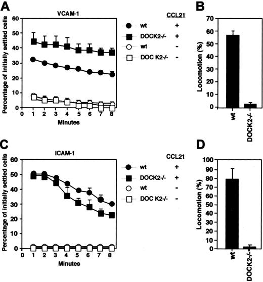
While DOCK2–/– lymphocytes did not exhibit any defect in their integrin-mediated adhesion strengthening, the reduced degree of chemokine-induced microvillar collapse and lamellipodium formation of DOCK2–/– T lymphocytes may result in impaired resistance to detachment at later time points. We therefore exposed wt or DOCK2–/– T lymphocytes, arrested on either VCAM-1 or ICAM-1 coimmobilized with CCL21, with an 8-minute period of continuous shear stress and compared the rate of detachment of these lymphocytes. Surprisingly, DOCK2–/– T lymphocytes exhibited a similar detachment rate to wt T lymphocytes (Figure 3A,C), suggesting that DOCK2-mediated microvillar collapse does not influence the ability of T lymphocytes to generate sustained adhesion strengthening in response to chemokine signals.
CCL21 induces microvillar collapse in wt but not in DOCK2–/–Tlymphocytes. (A) Micrographs from field emission scanning electron microscopy (SEM) of wt and DOCK2–/– T lymphocytes, intact or stimulated for 1 minute with CCL21 (300 ng/mL). Bar = 2 μm. Micrographs are representative of 40 wt cells and 38 DOCK2–/– cells analyzed. (B) Resistance to detachment developed by T cells arrested on either VCAM-1 (left) or ICAM-1 (right) coimmobilized with CCL21 as in Figure 1D-E subjected to continuous application of low shear (0.5 dyne/cm2) up to 1 minute and then subjected to abrupt detachment by 10-fold higher shear stress for 5 seconds. Mean values ± range determined in 2 representative fields are presented.
CCL21 induces microvillar collapse in wt but not in DOCK2–/–Tlymphocytes. (A) Micrographs from field emission scanning electron microscopy (SEM) of wt and DOCK2–/– T lymphocytes, intact or stimulated for 1 minute with CCL21 (300 ng/mL). Bar = 2 μm. Micrographs are representative of 40 wt cells and 38 DOCK2–/– cells analyzed. (B) Resistance to detachment developed by T cells arrested on either VCAM-1 (left) or ICAM-1 (right) coimmobilized with CCL21 as in Figure 1D-E subjected to continuous application of low shear (0.5 dyne/cm2) up to 1 minute and then subjected to abrupt detachment by 10-fold higher shear stress for 5 seconds. Mean values ± range determined in 2 representative fields are presented.
Arrested lymphocytes spread and locomote considerable distances over apical endothelial surfaces presenting chemokines under persistent shear stress.6,21 Leukocytes can also locomote on isolated integrin ligands coimmobilized with chemokines under shear stress conditions.21 In agreement with these earlier findings, shortly after their arrest, firmly adherent wt T lymphocytes established persistent locomotion on both VCAM-1 and ICAM-1 coimmobilized with CCL21 under continuous shear stress (Figure 3B,D). Despite their normal ability to resist detachment from these integrin ligands over prolonged application of shear stress, DOCK2–/– T lymphocytes previously arrested on either VCAM-1 or ICAM-1 failed to locomote on these integrin ligands (Figure 3B,D). DOCK2–/– T lymphocytes also failed to polarize and spread on these ligands (data not shown), consistent with their inability to extend any lamellipodia as assessed by scanning electron microscopy (Figure 2A). Taken together, these results indicate that CCL21-triggered integrin-mediated adhesion of both wt and DOCK2–/– T cells to endothelial surfaces (Figure 1C) and to integrin ligands (Figures 1D-E, 2B, and 3A,C) does not require T-cell polarization on these ligands. These results thus implicate DOCK2 in chemokine-triggered postarrest lamellipodia formation by T lymphocytes and lateral locomotion on integrin ligands rather than in chemokine-triggered arrest and adhesion strengthening on these ligands.
Resistance to detachment and lateral locomotion of CCL21-stimulated wt or DOCK2–/–T lymphocytes on integrin ligands under prolonged application of shear stress. (A) Wt and DOCK2–/– T lymphocytes were settled for 1 minute on VCAM-1 alone or coimmobilized with CCL21 as described in Figure 1D. Cells were then subjected to a shear stress of 2 dyne/cm2 for the indicated periods, and the percentage of initially settled cells remaining adherent was determined as a function of shear application time. (B) Locomotion of adherent wt and DOCK2–/– T lymphocytes (which resisted detachment for at least 1 minute of shear flow application) on VCAM-1 coimmobilized with CCL21, determined as described in “Materials and methods.” (C) Resistance to detachment with time of wt and DOCK2–/– T cells from ICAM-1 coated alone or with CCL21, determined as in panel A. (D) Locomotion on ICAM-1 coimmobilized with CCL21, determined as in panel B. Mean values ± range determined in 2 representative fields are presented. Experiments are each representative of 3 independent experiments.
Resistance to detachment and lateral locomotion of CCL21-stimulated wt or DOCK2–/–T lymphocytes on integrin ligands under prolonged application of shear stress. (A) Wt and DOCK2–/– T lymphocytes were settled for 1 minute on VCAM-1 alone or coimmobilized with CCL21 as described in Figure 1D. Cells were then subjected to a shear stress of 2 dyne/cm2 for the indicated periods, and the percentage of initially settled cells remaining adherent was determined as a function of shear application time. (B) Locomotion of adherent wt and DOCK2–/– T lymphocytes (which resisted detachment for at least 1 minute of shear flow application) on VCAM-1 coimmobilized with CCL21, determined as described in “Materials and methods.” (C) Resistance to detachment with time of wt and DOCK2–/– T cells from ICAM-1 coated alone or with CCL21, determined as in panel A. (D) Locomotion on ICAM-1 coimmobilized with CCL21, determined as in panel B. Mean values ± range determined in 2 representative fields are presented. Experiments are each representative of 3 independent experiments.
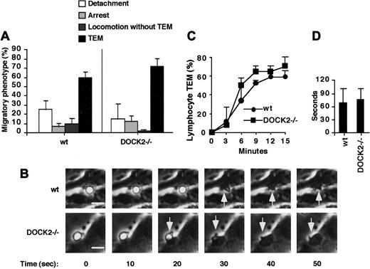
Apical endothelial CCL21 promotes comparable TEM of wt and DOCK2–/–T lymphocytes under physiologic shear flow. (A) Wt and DOCK2–/– T lymphocytes accumulated at low shear flow (0.25 dyne/cm2) for 2 minutes on TNFα-stimulated bEnd.3 overlaid with CCL21 (2 μg/mL) were subjected to physiologic shear stress (2 dyne/cm2) for 15 minutes. The migratory phenotype of each population was determined as described in “Materials and methods” and was expressed as a fraction of T cells accumulated on the endothelial monolayer at the first 2 minutes. Data are mean ± range values determined in 2 fields. (B) Phase-contrast images taken from time-lapse recordings of wt and DOCK2–/– T cells in the process of TEM. Time zero was set at the beginning of each TEM. Micrographs are representative of about 100 transmigrating T cells. Bar = 10 μm. Arrows depict the lymphocyte-cell region that undergoes TEM. (C) Time course of TEM determined for wt and DOCK2–/– lymphocyte populations. Time zero was set at the end of the accumulation phase. (D) Mean duration of the passage time required for individual apically adherent T cells to complete their TEM. At least 20 cells were analyzed for each experimental group. The experiments described in all panels are representative of 5 independent runs with different mice and bEnd.3 batches.
Apical endothelial CCL21 promotes comparable TEM of wt and DOCK2–/–T lymphocytes under physiologic shear flow. (A) Wt and DOCK2–/– T lymphocytes accumulated at low shear flow (0.25 dyne/cm2) for 2 minutes on TNFα-stimulated bEnd.3 overlaid with CCL21 (2 μg/mL) were subjected to physiologic shear stress (2 dyne/cm2) for 15 minutes. The migratory phenotype of each population was determined as described in “Materials and methods” and was expressed as a fraction of T cells accumulated on the endothelial monolayer at the first 2 minutes. Data are mean ± range values determined in 2 fields. (B) Phase-contrast images taken from time-lapse recordings of wt and DOCK2–/– T cells in the process of TEM. Time zero was set at the beginning of each TEM. Micrographs are representative of about 100 transmigrating T cells. Bar = 10 μm. Arrows depict the lymphocyte-cell region that undergoes TEM. (C) Time course of TEM determined for wt and DOCK2–/– lymphocyte populations. Time zero was set at the end of the accumulation phase. (D) Mean duration of the passage time required for individual apically adherent T cells to complete their TEM. At least 20 cells were analyzed for each experimental group. The experiments described in all panels are representative of 5 independent runs with different mice and bEnd.3 batches.
DOCK2 is not required for chemokine-triggered TEM of T lymphocytes under shear stress
We next analyzed the migratory patterns of wt and DOCK2–/– T lymphocytes arrested on CCL21-bearing TNFα-activated bEnd.3 endothelium under physiologic shear flow. Within 2 to 3 minutes following arrest, both wt and DOCK2–/– T lymphocytes started to cross the endothelial monolayer and remained confined to the basal endothelial surface (Figure 4A [fourth category] and 4B). Under optimal conditions of stimulation by endothelial-immobilized CCL21, as many as 60% and 70% of originally arrested wt and DOCK2–/– T lymphocytes, respectively, transmigrated through the inflamed endothelial barrier during the 15-minute period of the assay (Figure 4A,C). Both wt and DOCK2–/– T lymphocytes required intact LFA-1 for successful TEM, as the LFA-1 mAb M18/2 abolished all TEM, even under conditions in which this mAb did not interfere with CCL21-triggered LFA-1 adhesiveness to the endothelial surface (data not shown). In most cases, and in contrast to human peripheral-blood lymphocytes (PBLs) responding to chemokines presented on TNFα-activated human umbilical vein ECs (HUVECs),6 transmigrating murine T lymphocytes crossed the bEnd.3 endothelial barrier within a single-cell diameter of their original arrest point (Figure 4B). The average time required for DOCK2–/– T lymphocytes to cross the endothelial barrier was also similar to the passage time of wt T lymphocytes (Figure 4D). Some recruited wt T lymphocytes locomoted away from their initial site of arrest but failed to transmigrate through the endothelial monolayer (Figure 4A [third category]); this category of lymphocytes was essentially absent in DOCK2–/– T lymphocytes (Figure 4A [third category] and data not shown), consistent with the failure of these lymphocytes to locomote over isolated VCAM-1 or ICAM-1 (Figure 3B,D). Together, these results suggest that despite the inability of DOCK2–/– T lymphocytes to locomote laterally in response to CCL21 signals on integrin ligands and on the apical surface of an endothelial monolayer expressing these ligands, DOCK2–/– T lymphocytes successfully transmigrate through the endothelial barrier in response to these chemokine signals.
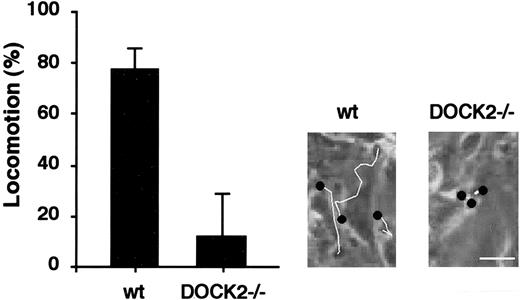
Locomotion of transmigrating wt and DOCK2–/–T cells underneath the endothelial layer. Lymphocyte motion underneath the endothelial monolayer was tracked immediately after completing TEM. The fraction of transmigrating wt or DOCK2–/– T cells that successfully locomoted for at least 3 cell diameters on the basal substrate away from the TEM site is depicted for each population. (Inset) Tracks of 3 individual wt and DOCK2–/– T cells taken immediately after TEM completion. Black dots denote the sites of the lymphocyte TEM, and white lines denote the lymphocyte locomotion tracks after TEM underneath the endothelium. Bar = 30 μm. Results are representative of 5 independent experiments.
Locomotion of transmigrating wt and DOCK2–/–T cells underneath the endothelial layer. Lymphocyte motion underneath the endothelial monolayer was tracked immediately after completing TEM. The fraction of transmigrating wt or DOCK2–/– T cells that successfully locomoted for at least 3 cell diameters on the basal substrate away from the TEM site is depicted for each population. (Inset) Tracks of 3 individual wt and DOCK2–/– T cells taken immediately after TEM completion. Black dots denote the sites of the lymphocyte TEM, and white lines denote the lymphocyte locomotion tracks after TEM underneath the endothelium. Bar = 30 μm. Results are representative of 5 independent experiments.
The endothelial monolayers studied here were grown on an extracellular matrix enriched with the key integrin ligand, fibronectin. Subsequent to transendothelial migration, both lymphocytes and neutrophils locomote away from their site of TEM and across the fibronectin matrix.6,21 Accordingly, nearly 80% of the wt T lymphocytes that completed their transmigration route through the CCL21-bearing, TNFα-activated endothelium rapidly locomoted away from their TEM site at the basal endothelial compartment (Figure 5; Video S1). In contrast, less than 20% of successfully transmigrating DOCK2–/– T lymphocytes locomoted away from their site of TEM at the same basal endothelial compartment (Figure 5; Video S2), suggesting a major defect in the lateral locomotion of these lymphocytes across subendothelial fibronectin matrix. Because this locomotion takes place in a compartment inaccessible to shear stress signals, DOCK2 appears essential for lateral T-lymphocyte locomotion in both apical and basal endothelial compartments, both in the presence of directly applied shear stress and in its absence (Figures 3B-D, 4A [third category], and 5).
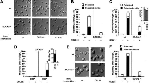
Chemokine-induced polarization and locomotion of wt and DOCK2–/–T cells. (A) DIC images of wt and DOCK2–/– T lymphocytes settled on FN (5 μg/mL) or coimmobilized with saturating levels of CXCL12 (2 μg/mL) or CCL21 (1 μg/mL). Bar = 10 μm. (B-C) Cell shape and motility of wt or DOCK2–/– T lymphocytes monitored for 10 minutes on FN coated alone (-) or with CXCL12 (B) or with CCL21 (C). At least 60 cells were monitored in each group. Results represent the mean ± range determined in 2 fields. (Inset) Mean velocity of locomoting lymphocytes within wt or DOCK2–/– populations. (D) Shape and motility of DOCK2–/– CD4+ or CD8+ T cells on FN coimmobilized with CCL21 as in panel C. (Inset) Effect of PI3K inhibition by LY294002 (LY) on the fraction of CD8+ T cells locomoting on FN coimmobilized with CCL21. (E) Defective GPCR-triggered locomotion of DOCK2–/– T lymphocytes on a surface devoid of integrin ligands. DIC images of wt and DOCK2–/– T lymphocytes settled on CCL21 (1 μg/mL) alone, monitored as in panel A. Intact lymphocytes (-) were monitored for the same time periods. Bar = 5 μm. (F) Cell shape and motility of wt and DOCK2–/– T lymphocytes compared under the experimental conditions described in panel E. Results are the mean ± range of 2 fields with at least 30 cells in each field. Experiment is a representative of 3.
Chemokine-induced polarization and locomotion of wt and DOCK2–/–T cells. (A) DIC images of wt and DOCK2–/– T lymphocytes settled on FN (5 μg/mL) or coimmobilized with saturating levels of CXCL12 (2 μg/mL) or CCL21 (1 μg/mL). Bar = 10 μm. (B-C) Cell shape and motility of wt or DOCK2–/– T lymphocytes monitored for 10 minutes on FN coated alone (-) or with CXCL12 (B) or with CCL21 (C). At least 60 cells were monitored in each group. Results represent the mean ± range determined in 2 fields. (Inset) Mean velocity of locomoting lymphocytes within wt or DOCK2–/– populations. (D) Shape and motility of DOCK2–/– CD4+ or CD8+ T cells on FN coimmobilized with CCL21 as in panel C. (Inset) Effect of PI3K inhibition by LY294002 (LY) on the fraction of CD8+ T cells locomoting on FN coimmobilized with CCL21. (E) Defective GPCR-triggered locomotion of DOCK2–/– T lymphocytes on a surface devoid of integrin ligands. DIC images of wt and DOCK2–/– T lymphocytes settled on CCL21 (1 μg/mL) alone, monitored as in panel A. Intact lymphocytes (-) were monitored for the same time periods. Bar = 5 μm. (F) Cell shape and motility of wt and DOCK2–/– T lymphocytes compared under the experimental conditions described in panel E. Results are the mean ± range of 2 fields with at least 30 cells in each field. Experiment is a representative of 3.
DOCK2 is critical for lateral T-cell motility triggered by immobilized chemokines regardless of integrin occupancy
The subendothelial lymphocyte motility defective in DOCK2–/– T lymphocytes could reflect defects in signaling from chemokines, inflammatory cytokines, or integrin occupancy events (outside-in signaling). We therefore wished to differentiate the involvement of DOCK2 in integrin-mediated versus integrin-independent motility triggered by prototypic chemokines. Although in some settings T lymphoblasts can polarize spontaneously on integrin-binding ligands such as fibronectin, VCAM-1, or ICAM-1,22 neither wt nor DOCK2–/– T lymphocytes polarized on any of these ligands in the absence of chemokine stimuli (Figure 6A [left panels] and 6B-C and data not shown). However, when settled on fibronectin and encountering coimmobilized CCL21 or CXCL12 signals, nearly all wt T lymphocytes polarized, and a significant fraction could also locomote at moderate speeds on the integrin ligand (Figure 6A-C; Video S3). In contrast, CXCL12 failed to trigger any DOCK2–/– T-lymphocyte locomotion despite the higher expression of CXCL12 receptors on these cells as compared with wt (Figures 1B and 6B). Interestingly, a small but significant fraction of DOCK2–/– T lymphocytes could polarize and locomote on fibronectin coimmobilized with CCL21 (Figure 6C; Video S4), suggesting that CCR7 can still trigger polarization and motility in a small subset of DOCK2–/– T cells occupied by fibronectin. Further characterization revealed that this subset of DOCK2–/– contained CD8+ but not CD4+ T cells (Figure 6D). Notably, the locomotion of this subset was completely eliminated by inhibition of PI3K activity (Figure 6D, inset).
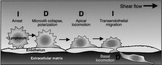
A summary of adhesive and migratory steps triggered by chemokine signals at lymphocyte-endothelial and lymphocyte matrix interfaces and the contribution of DOCK2 to each step. The dependence on DOCK2 for a particular step is denoted by the symbol “D.” DOCK2 independence of a particular step is denoted by the symbol “I.”
A summary of adhesive and migratory steps triggered by chemokine signals at lymphocyte-endothelial and lymphocyte matrix interfaces and the contribution of DOCK2 to each step. The dependence on DOCK2 for a particular step is denoted by the symbol “D.” DOCK2 independence of a particular step is denoted by the symbol “I.”
Although without chemokine stimulation T cells failed to spread on fibronectin, chemokine-triggered T-cell motility on the integrin ligand could involve an outside-in signaling component from chemokine-activated fibronectin-binding integrins such as VLA-4. We therefore next compared the ability of wt and DOCK2–/– T lymphocytes to locomote on a chemokine-bearing albumin-coated substrate devoid of any integrin ligand. Notably, almost all wt T lymphocytes could polarize and locomote on the albumin-coated substrate, in response to immobilized CCL21 signals, even without fibronectin occupancy signals (Figure 6E-F). Similar results were obtained on CXCL12-bearing albumin-coated substrate (data not shown). By contrast, none of the DOCK2–/– T lymphocytes could locomote on this substrate in response to CCL21 (Figure 6E-F) or CXCL12 (data not shown). Although immobilized albumin is recognized by the β2 integrin, Mac-1, this integrin was absent on most (more than 95%) wt T lymphocytes and thus could not have participated in the robust locomotion of these cells on immobilized CCL21 (data not shown). These results collectively suggest that spleen T lymphocytes require DOCK2 to optimally locomote in response to CCL21 signals regardless of their state of integrin occupancy on the chemokine-bearing surface.
Discussion
For lymphocytes to reach their target tissues, these cells must locomote from their arrest site on the endothelial target to sites of diapedesis while resisting detachment from the endothelial surface by continuously applied shear forces. Upon successful crossing of the endothelial barrier, they must navigate considerable distances within the extracellular space.4,9 The Rac family GTPases are key regulators of cell motility,11 but their functions in leukocyte adhesion to and migration on endothelial surfaces under physiologic shear flow have just begun to unfold.13 Similar to polarization processes in shearfree environments, endothelium-adherent lymphocytes extend actin-enriched lamellipodia, which constitute their leading edge.9 Chemoattractant signals induce morphologic asymmetry in a variety of leukocytes via the compartmentalized activation of Rac and RhoA GTPases and their effectors.11,23,24 In the present study we dissected the specialized roles for the hematopoietic Rac regulator, DOCK2, in chemokine-triggered lateral motility of primary T lymphocytes under both shear stress and shearfree conditions. Despite the pleotropic cytoskeletal remodeling activities of chemokine-activated Rac and, potentially, of its upstream regulator, DOCK2, integrin-mediated lymphocyte arrest, subsequent adhesion strengthening, and transendothelial migration were found to be DOCK2 independent (Figure 7). Our results suggest that chemokine signaling to T-lymphocyte Rac, while obligatory for microvillar collapse, lamellipodia extension, and lateral motility on both endothelial and extracellular matrices,14 is not involved in shear-resistant adhesion to endothelium or in TEM under shear stress (Figure 7), 2 processes tightly regulated by proper stimulation of lymphocyte integrins by apical endothelial chemokines.10,25
Integrin activation by endothelial chemokines is tightly controlled by GTPases distinct of Rac such as RhoA and Rap1.10 Although chemokine-triggered lymphocyte TEM has been postulated to involve Rap1 pathways,8,26 this GTPase regulates earlier adhesion strengthening and polarization of lymphocytes on endothelial surfaces under shear flow and, thus, its direct contribution to TEM has been difficult to discern. The critical role of RhoA in earliest activation of LFA-1 by endothelial chemokines underlying initial leukocyte arrest12,27 complicates any direct assessment of its roles in TEM initiation and termination.28 In contrast to these GTPases, early arrest and subsequent adhesion strengthening of T cells triggered by apical endothelial chemokines were found independent of DOCK2. Thus, our in vitro studies assign a unique contribution of this key Rac activator to chemokine-triggered lymphocyte motility rather than TEM (Figure 7).
Endothelial-displayed chemokines can induce localized cytoskeletal changes, a striking example of which is the collapse of leukocyte microvilli, thought to facilitate initial adhesion to endothelial ligands under shear flow.29 The actin-binding linker proteins ezrin/radixin/moesin (ERM), when properly phosphorylated, link the actin cytoskeleton to many plasma-membrane proteins and thereby control microvillus rigidity.20 Chemokine-stimulated Rac was recently suggested to induce rapid dephosphorylation and inactivation of ERMs, resulting in the disassembly of microvilli and ERM dephosphorylation.20,30 The collapse of microvilli enlarges the leukocyte-endothelium contact and therefore was assumed to provide increased resistance of adherent leukocytes to detachment by shear forces.20 Our results argue against this postulated role of microvillus collapse, because DOCK2–/– T cells, which failed to undergo collapse of their microvilli, exhibited normal arrest and postarrest adhesion strengthening and resistance to detachment by prolonged shear flow. Thus, integrin affinity and microclustering, as well as integrin anchorage states, are 3 microscopic properties that control both rapid arrest and subsequent adhesion strengthening of integrin bonds31-34 ; none of these 3 parameters appears to involve the Rac regulator, DOCK2.
In accordance with defective Rac activation by chemokines in DOCK2–/– T cells, both ultrastructural analysis of chemokine-stimulated lymphocytes (Figure 2A) and real-time imaging of lymphocytes overlaid on surface-bound chemokines (Videos S3 and S4) revealed dramatically impaired lamellipodia formation in DOCK2–/– T cells. This impaired polarization also resulted in defective lateral motility of these lymphocytes on multiple substrates studied both under shear stress and under shearfree conditions. DOCK2-facilitated Rac activation by chemokines is therefore essential for lateral motility triggered by local chemokine signals regardless of the adhesive information involved and independently of integrin outside-in signaling. One of the most unexpected results of this study is that chemokine signaling to Rac, although blocked in DOCK2–/– T cells, is nonessential for lymphocyte TEM, a process that involves local signaling primarily in the presence of shear flow to the LFA-1 integrin on adherent lymphocytes by apically presented endothelial chemokines.6,35 Because lymphocyte TEM involves extensive remodeling of the lymphocyte plasma membrane triggered at TEM sites, predominantly at interendothelial junctions,36 we conclude that integrin activation by chemokine signals coordinates lymphocyte crossing through the endothelial junctions via a DOCK2-independent (and therefore most probably Rac activation-independent) process. Rab GTPases, for instance, can induce lamellipodia in some cell types37 and thus could participate in leukocyte migration through endothelial barriers. Because lymphocyte TEM involves extensive protrusions into the endothelial cells, the CDC42 GTPase, which regulates polarized filopodia formation,9 is also a candidate.
DOCK2 is a member of the CDM adaptor family that regulates Rac activation and function38 in both T and B lymphocytes by acting as a docking scaffold protein and is thought to function also as a Rac GEF.39,40 DOCK2 functions downstream of T-cell and B-cell GPCRs as well as of the T-cell receptor.14,40 We could not study the role of DOCK2 in the lateral migration of B lymphocytes over endothelium due to the B-lymphocyte requirement for intact DOCK2 in earliest chemokine stimulation of integrins.15 DOCK2 has 59% identity to DOCK180, which forms a complex with the adaptor protein ELMO1,41 which promotes integrin-induced Rac activation and migration on extracellular matrix in fibroblasts.38,42,43 PBLs show little or no expression of this DOCK member44-46 and do not spread spontaneously on integrin ligands (data not shown), although upon prolonged activation they do.22 It is not known whether lymphocyte activation and acquisition of spreading ability is associated with DOCK180 up-regulation. Interestingly, while in vitro lymphoid-cell chemotaxis toward homeostatic chemokines and in vivo migration to lymph nodes and spleen are severely impaired by DOCK2 deficiency, these processes are only mildly sensitive to PI3K inhibition.15 In contrast, chemoattractant-stimulated macrophage polarization and chemotaxis do not require DOCK2 14 but instead use PI3K products for actin polymerization, polarization, and motility.47 Notably, chemokine-triggered Rac activation in T cells is DOCK2 dependent and PI3K independent.14 Nevertheless, lymphocyte adhesion and motility may be coregulated by PI3K products of chemokine and integrin signaling. Indeed, PI3K blocking markedly reduced LFA-1–mediated TEM triggered by endothelial-bound CCL21 (data not shown), in accordance with previous reports,48,49 whereas DOCK2 deficiency did not alter this key property. Furthermore, the small fraction of DOCK2–/– T lymphocytes capable of locomoting on fibronectin in response to CCL21 signals (Figure 6C) did so via a DOCK2-independent pathway that was entirely eliminated by PI3K inhibition (Figure 6D, inset). Thus, to successfully complete their apical and transendothelial migration, lymphocytes must translate endothelial-displayed chemokine signals as well as integrin outside-in signals to coordinate activation of both their PI3K and DOCK2-dependent Rac machineries. Because the contribution of these 2 Rac-activating machineries is likely to vary between distinct endothelial beds, combinations of pharmacologic reagents targeting these 2 machineries may therefore prove selective in attenuating the trafficking of specific subsets of lymphocytes to distinct target organs.
Prepublished online as Blood First Edition Paper, June 13, 2006; DOI 10.1182/blood-2006-04-017608.
Supported by MAIN, the EU6 Program for Migration and Inflammation, and by G.I.F., the German-Israeli Foundation for Scientific Research and Development. R. Alon is the Incumbent of The Tauro Career Development Chair in Biomedical Research.
Z.S. designed the models, designed and performed the research, and wrote the paper; R.P. and E.W. performed part of the research; V.G. assisted in flow chamber experiments; S.W.F. performed part of the research and provided editorial help; N.E. assisted in live imaging; Y.F. contributed the DOCK2–/– mice; and R.A. supervised the research and wrote the paper.
The online version of this article contains a data supplement.
The publication costs of this article were defrayed in part by page charge payment. Therefore, and solely to indicate this fact, this article is hereby marked “advertisement” in accordance with 18 U.S.C. section 1734.
We thank Drs E. Klein and O. Yeger for assistance in SEM and Dr S. Schwarzbaum for editorial aid. We also thank Dr S. Reich-Zeliger for helpful comments.

This feature is available to Subscribers Only
Sign In or Create an Account Close Modal