Abstract
Platelet dense granules are lysosome-related organelles which contain high concentrations of several biologically important low-molecular-weight molecules. These include calcium, serotonin, adenine nucleotides, pyrophosphate, and polyphosphate, which are necessary for normal blood hemostasis. The synthesis of dense granules and other lysosome-related organelles is defective in inherited diseases such as Hermansky-Pudlak syndrome (HPS) and Chediak-Higashi syndrome (CHS). HPS and CHS mutations in 8 human and at least 16 murine genes have been identified. Previous studies produced contradictory findings for the function of the murine ashen (Rab27a) gene in platelet-dense granules. We have used a positional cloning approach with one line of ashen mutants to establish that a new mutation in a second gene, Slc35d3, on mouse chromosome 10 is the basis of this discrepancy. The platelet-dense granule defect is rescued in BAC transgenic mice containing the normal Slc35d3 gene. Thus, Slc35d3, an orphan member of a nucleotide sugar transporter family, specifically regulates the contents of platelet-dense granules. Unlike HPS or CHS genes, it has no apparent effect on other lysosome-related organelles such as melanosomes or lysosomes. The ash-Roswell mouse mutant is an appropriate model for human congenital-isolated delta-storage pool deficiency.
Introduction
Platelet activation induces secretion of the contents of several subcellular organelles, including α granules, lysosomes, and dense granules.1,2 α Granules contain multiple proteins critical to adhesion and repair such as von Willebrand factor, thrombospondin, and fibrinogen. Lysosomes hold a battery of hydrolytic enzymes postulated to function in elimination of circulating platelet aggregates. Dense granules like lysosomes are acidic organelles but, unlike the aforementioned organelles, contain extremely high concentrations of small nonprotein components, including calcium, serotonin, adenine nucleotides, and pyrophosphate, which are thought to exist as an insoluble complex within platelets.2 They are among the first components released from activated platelets and participate in clotting in numerous ways. For example, serotonin causes vasoconstriction. ADP activates and recruits nearby platelets. More recently, high concentrations of polyphosphate were found within platelet-dense granules.3 Platelet-derived polyphosphate is a potent modulator of blood coagulation and fibrinolysis.4
The biosynthesis of platelet dense and α granules is thought to proceed through a common multivesicular body intermediate.5 A wide variety of genes causative for the inherited diseases, Hermansky-Pudlak syndrome (HPS) and Chediak-Higashi syndrome (CHS), regulate the biosynthesis of dense granules, probably through control of vesicle trafficking. Platelet dense granules are absent or greatly reduced in a number in these diseases, and/or dense granule contents are significantly lower, leaving “empty” organelles. HPS is a genetically heterogeneous disease affecting the biosynthesis and/or intracellular transport of lysosome-related organelles (LROs) and their precursor vesicles.6-10 LROs commonly affected include melanosomes, platelet-dense granules, and lamellar bodies of lung type II cells. Clinical presentation includes oculocutaneous albinism and lowered visual acuity, prolonged bleeding and, in many cases, early death from fibrotic lung disease. There is presently no permanent treatment for HPS, although bone marrow transplantation has restored normal platelet function and bleeding times in mouse HPS models.11,12
Mutations in 15 genes have been identified as causal for HPS in murine models.12-15 Eight of the corresponding genes are mutated in human patients with HPS.10,16,17 The majority of murine HPS genes10,14 fall into 2 classes: (1) 5 genes of known function in vesicle trafficking and (2) 10 novel genes found only in higher eukaryotes whose precise functions are unknown. All novel genes encode protein subunits of 1 of 3 heteromeric protein complexes termed BLOCs (Biogenesis of Lysosome-related Organelles Complexes) whose precise functions are unknown.12,18 Platelet dense granules are likewise deficient in CHS, a rare inherited disease caused by mutations in the lysosomal trafficking regulator (LYST) gene.19
The assignment of the ashen or Rab27a gene as a murine HPS gene has been controversial.20-22 An ashen mutant line originally obtained from The Jackson Laboratory and subsequently maintained at Roswell Park Cancer Institute (hereafter referred to as ash-Roswell) displays the typical features of HPS, including a prolonged bleeding time accompanied by substantial platelet-dense granule deficiency in whole-mount electron microscopy.22 However, the original ashen mutant line independently maintained at The Jackson Laboratory (hereafter referred to as ash-Jackson or ash-Jax) has normal bleeding times and normal concentrations of platelet-dense granule components.20 In this study we determine that a mutation in a second gene, Slc35d3,23,24 which encodes an orphan transporter with significant sequence homology to sugar nucleotide transporters,23 has occurred in the ash-Roswell mutant line and is the cause of its platelet dysfunction. Slc35d3 causes platelet dysfunction by regulating the contents of platelet-dense granules. It differs from well-established HPS and CHS genes in that its effects on LROs are specific to platelet-dense granules with no effect on pigmentation (melanosomes) or lysosomes.
Materials and methods
Mice and genetic crosses
Mutant ash mice (ash-Roswell) were obtained from the colony of Drs Nancy Jenkins and Neal Copeland of the Frederick Cancer Center (Frederick, MD) who in turn had obtained them from The Jackson Laboratory (Bar Harbor, ME). They were bred for 7 years at Roswell Park Cancer Institute (Buffalo, NY). Control C3H/HeSnJ and ash-Jackson mice were purchased from The Jackson Laboratory. All procedures (mouse protocol 125M) were approved by the Roswell Park Institutional Animal Care and Use Committee and adhered to the principles of the National Institutes of Health Guide for the Care and Use of Laboratory Animals.
A line of mice (Slc35d3−/−, Rab27a+/+) homozygous for the Chr10 IAP insertion mutation (see “Mice and genetic crosses”) in the Slc35d3 gene and containing the normal (nonmutated or +) Rab27a gene was produced by crossing the parental lines ash-Roswell and C3H/HeSnJ followed by molecular identification of (Slc35d3−/−, Rab27+/+) F2 offspring. The Rab27a mutation was monitored as described.22 To identify offspring containing the Slc35d3 IAP mutation, genomic DNA was amplified with a forward primer (5′-ATACAAAAGCTGGCCCACAT-3′) within the intergenic region just upstream of Slc35d3 and a reverse primer (5′-GGGGCTGAACAAGAAAGACC-3′) within intron 1 of Slc35d3. This produced a large 8-kb (kilobase) product from ash-Roswell compared with a 2.4-kb product from wild-type controls, and heterozygotes exhibited both bands.
Another line of mice (Slc35d3−/−, Rab27a+/+, a/a) with the homozygous Chr 10 IAP insertion mutation in the Slc35d3 gene on the C57BL/6J (nonagouti or a) background was produced by crossing parental C57BL/6J and homozygous Slc35d3 mutants followed by selection of Slc35d3−/−, Rab27a+/+, a/a F2 offspring.
Positional gene identification
To obtain high-resolution genetic and physical maps of the critical chromosomal subregion containing the dense granule mutation homozygous ash-Roswell mutants were backcrossed with inbred PWK mice (subspecies Mus musculus musculus).25 Platelet serotonin levels were typed in 393 backcross progeny at 6 weeks of age. Mutant platelet serotonin concentrations were less than 0.2 μg/109 platelets. Offspring with platelet serotonin levels greater than 2.0 μg/109 platelets were considered the high serotonin group. Also, offspring were typed for coat color (controlled by Rab27a). To narrow the chromosomal location of the low serotonin trait, progeny serotonin concentrations were compared with the genetic segregation of 3 to 4 polymorphic microsatellites from each of the 20 mouse chromosomes.26
Cells and tissues
For serotonin analyses in platelets of BAC transgenic offspring, blood was drawn into 0.38% citrate by eye bleed under isofluorane anesthesia followed by differential centrifugation. For other platelet analyses, platelets were isolated from mice under anoxia with CO2. Blood was immediately drawn by heart puncture into 0.38% citrate, and platelets were isolated by differential centrifugation.27 Mouse tissues were isolated from 6- to 10-week-old animals, washed with phosphate-buffered saline, and stored at −70°C for genomic DNA or protein preparation or immediately used for RNA isolation.
Antibodies
Antibodies to Rabs 1b, 3b, 4, 6, and 8 were obtained from Santa Cruz Biotechnology (Santa Cruz, CA). Dr Richard Haslam (McMaster University, Hamilton, Canada) supplied the antibody to Rab11A; Dr Kazuhiro Osanai (Kanazawa Medical University, Kahokugun, Japan) to Rab 38, and Dr Miguel Seabra (Imperial College, London, United Kingdom) to Rabs 27a and 27b. Actin antibody was rabbit polyclonal (catalog no. AANO1; Cytoskeleton, Denver, CO).
Immunoblotting
Platelets and tissues were homogenized in proteinase inhibitor cocktail (Roche Molecular Biochemicals, Indianapolis, IN), denatured in SDS, electrophoresed (30 μg protein/lane) on polyacrylamide gels, and blotted to polyvinylidene difluoride transfer membranes (Hybond-P; Amersham Biosciences, Buckinghamshire, United Kingdom).28 Bound antibody was detected using the enhanced chemiluminescence Plus system (Amersham Biosciences, Piscataway, NJ). Equivalent loading and transfer were verified using actin as internal standard.
Expression and transcript analyses
Multiple tissue Northern blots were from Clontech, Mountain View, CA. For Northern blot analysis of Slc35d3 transcripts, poly(A)–mRNA was isolated via the Qiagen (Valencia, CA) Oligotrex direct mRNA kit. Total RNA was reverse transcribed as described.25 A transcript-specific 303–base pair (bp) 32P-radiolabeled probe (exon 2) was generated by reverse transcriptase–polymerase chain reaction (RT-PCR) amplification of total RNA from C3H/HeSnJ brain (primers 5′-TCTATTCATTGCTGGCGTTG-3′ and 5′-TGCCCTTCTGCTCTTGAACT-3′).
Generation of 5′-RACE fragments from brain Poly-A mRNA was performed with the SMART RACE cDNA Amplification Kit (Clontech) using 2 nested antisense primers in exon 1 of Slc35d3. PCR product sizes were verified by agarose gel electrophoresis and sequenced.
For quantitative real-time PCR analyses, total RNAs from tissues were reverse transcribed (Applied Biosystems, Foster City, CA; TaqMan protocol). PCR primers bridged the end of exon 1 and the beginning of exon 2 of Slc35d3. PCR products were analyzed on an Applied Biosystems Real-Time Cycler HT-7900 and by agarose gel electrophoresis after 40 cycles to verify correct product size. Expression levels were standardized to GAPDH mRNA levels by calculating cycle thresholds (Ct).
BAC rescue of normal platelet-dense granule phenotype in transgenic mice
Purified bacterial artificial chromosome (BAC) DNA (RP23-344M6) was injected into pronuclei derived from hybrid (C3H/HeRos × C57BL/10 Rospd) F2 females as described.15 Transgenic BAC-positive mice were identified by PCR analyses of genomic DNA isolated from tail biopsies. BAC presence was verified using BAC vector primers (5′-AAGTGACATCGTCCTTTTCCCCAAG-3′ and 5′-TTCTTCTTTGCTTCCTCGCCAGTTC-3′). BAC-positive pups were mated with ash-Roswell mutants to produce F1 progeny. BAC-positive pups were backcrossed to ash-Roswell mutants to produce F2 progeny. Each F2 pup was typed for the BAC transgene and the IAP insertion in the Slc35d3 gene as described earlier in this section and for coat color.
Platelet serotonin analyses
Platelets were enumerated in a Coulter counter, lysed in 1 mL distilled water, and assayed fluorometrically for serotonin.29
Screening of patients with HPS
Genomic DNA was isolated from peripheral blood of 12 patients with HPS and 2 patients with mild storage pool deficiency enrolled in a protocol approved by the National Human Genome Research Institute institutional review board to study the molecular and clinical aspects of HPS. Mutation analysis of both coding exons of human SLC35D3 was performed on each patient's DNA using standard PCR and sequencing protocols.
PCR products spanning the 2 exons of SLC35D3 plus the adjacent intron and noncoding sequences were screened in genomic DNA of 10 other patients with HPS and 3 patients with undefined storage pool deficiency who likewise lacked mutations in the known human HPS genes.
Results
Further characterization of the ash-Roswell mutant
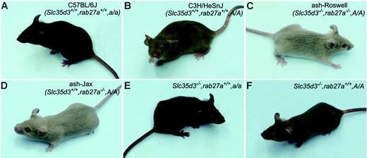
Because of conflicting data20-22 on platelet dysfunction in ash-Roswell and ash-Jax inbred lines, a series of experiments was performed to characterize the basis of this difference. Side-by-side comparison of the ash-Roswell and ash-Jackson mutants revealed no discernible difference in coat color (Figure 1C-D). Both exhibited the hypopigmentation expected from homozygosity for the ashen mutation at the Rab27a gene. Also, repeat tests confirmed the reported21,22 depressed platelet serotonin levels (0.24 μg/109 platelets) in ash-Roswell and the reported20 normal serotonin levels (2.3 μg/109 platelets) in ash-Jackson (P < .001). The dense granule defect in ash-Roswell is genetically recessive as heterozygotes produced by breeding ash-Roswell to C3H/HeSnJ had normal platelet serotonin levels (not shown).
Effects of the Slc35d3, Rab27a, and agouti (A) genes on pigmentation. Control C57BL/6J (A) and C3H/HeSnJ (B) mice display typical nonagouti (a) and agouti (A) pigmentation, respectively. The ash-Roswell (C) and ash-Jax (D) mutants, which arose on the C3H/HeSnJ inbred line, are hypopigmented because of the homozygous Rab27a (ashen) mutation. The Slc35d3 mutation does not affect coat or eye color when transferred in homozygous form to the C57BL/6J (E) or C3H/HeSnJ (F) backgrounds.
Effects of the Slc35d3, Rab27a, and agouti (A) genes on pigmentation. Control C57BL/6J (A) and C3H/HeSnJ (B) mice display typical nonagouti (a) and agouti (A) pigmentation, respectively. The ash-Roswell (C) and ash-Jax (D) mutants, which arose on the C3H/HeSnJ inbred line, are hypopigmented because of the homozygous Rab27a (ashen) mutation. The Slc35d3 mutation does not affect coat or eye color when transferred in homozygous form to the C57BL/6J (E) or C3H/HeSnJ (F) backgrounds.
To determine whether a secondary mutation in the Rab27a gene was responsible for the platelet dysfunction in the ash-Roswell line, the complete sequences of the open reading frames of the Rab27a mRNA were compared in ash-Roswell and ash-Jax. Both mutants contained the previously described22 (A→T) transversion in the splice donor site located downstream of exon 4 of the Rab27a gene. However, neither contained any additional sequence alterations in the cDNA of Rab27a, indicating that secondary mutations in Rab27a do not explain the ash-Roswell phenotype. Also, the open reading frame sequence of the cDNA of the closely related Rab27b gene, which is highly expressed in platelets20 and can in some instances substitute for the function of Rab27a,30 was normal. This indicates that a secondary alteration in Rab27b is not responsible for the ash-Roswell phenotype. This conclusion was confirmed by the finding of normal levels of Rab27b protein in immunoblots of platelets from ash-Roswell mutants together with the expected absence of expression of Rab27a protein (Figure 2). Therefore, the abnormal expression of either Rab is not responsible for the ash-Roswell platelet phenotype. Also, a series of Rab proteins, including Rabs 1b, 3b, 4, 6, 8, 11a, and 38, were present at normal levels and normal apparent sizes (not shown) in immunoblots of ash-Roswell platelets, suggesting that abnormalities in these important regulators of vesicle trafficking are likewise not responsible for the dense granule defect. Finally, the possibility that the gene responsible for the dense granule defect originated from contamination of either the ash-Roswell or ash-Jackson colonies maintained at Roswell Park is highly unlikely because PCR amplification of a series of 47 microsatellites randomly scattered across the mouse genome uniformly produced sizes expected for the parental C3H/HeSnJ inbred line.
Expression of Rab27a and Rab27b proteins in platelets of ash-Roswell and ash-Jax mutants. Platelet protein (30 μg) was electrophoresed and immunoblotted with the indicated antibodies. β-Actin served as loading control. The vertical dotted lines indicate that empty spacer gel lanes were removed during the digital composition of this figure.
Expression of Rab27a and Rab27b proteins in platelets of ash-Roswell and ash-Jax mutants. Platelet protein (30 μg) was electrophoresed and immunoblotted with the indicated antibodies. β-Actin served as loading control. The vertical dotted lines indicate that empty spacer gel lanes were removed during the digital composition of this figure.
Positional identification of the ash-Roswell mutant gene
Together, the data presented in “Further characterization of the ash-Roswell mutant” suggested that a second mutation, in addition to the original Rab27a mutation, had occurred within another gene in the ash-Roswell mutant line to produce its abnormal dense granule phenotype. To test this hypothesis, ash-Roswell mutants were backcrossed with the wild-type PWK inbred line to generate 393 progeny (see “Materials and methods”), and segregation of the low platelet serotonin phenotype was compared with the segregation of 53 polymorphic microsatellite loci uniformly distributed among the 20 mouse chromosomes. The segregation among progeny of the serotonin phenotype followed the classical 1:1 ratio expected for a single Mendelian gene. Further, genetic linkage of the platelet phenotype with microsatellites D10MIT168 and D10MIT17 of chromosome 10 was apparent. In contrast, coat color segregated, as expected,22 with polymorphic microsatellites located near the Rab27a gene on chromosome 9. This indicated that a new gene on mouse chromosome 10, distinct from the Rab27a gene, controls platelet-dense granules in ash-Roswell mice.
Further analyses of the segregation in the backcross of a battery of additional polymorphic microsatellites on chromosome 10 narrowed the genetic interval containing the platelet regulatory gene to 2.0 centiMorgans. This interval contains 11 genes within 2.1 megabases of DNA (Figure 3). Genes within the critical interval were systematically amplified by RT-PCR procedures using cDNA prepared from brain tissues. PCR products were analyzed on agarose gels to detect possible abnormalities in size, expression, or both. Also, the coding region sequences of the cDNAs of all 11 genes were determined in their entirety in ash-Roswell. Ten contained completely wild-type sequences. However, one gene, Slc35d3, produced no RT-PCR product with several sets of primers designed within exon 1. Long-range genomic PCR amplification produced a large 8-kb product from ash-Roswell compared with a 2.4-kb product from C3H/HeSnJ controls (not shown). Sequencing revealed an approximate 5-kb intracisternal A particle (IAP) within the 8-kb DNA product derived from ash-Roswell (Figure 3C).
Identification of the Slc35d3 gene by positional cloning. (A) High-resolution genetic map of the region surrounding the Slc35d3 gene on mouse chromosome 10. Numbered markers are polymorphic MIT microsatellites. ARDR26 (amplified with primers 5′-GGGTGGAGCGGTACTATTCA-3′ and 5′-TGTCTTCAGAGACAACAATCCAG-3′) and ARDR22 (amplified with primers 5′-TTTTCTTTAGGTGGTTTTCTACAGC-3′ and 5′-AGGACAGACAGAGCCCTGAG-3′) are polymorphic microsatellites identified by genome scanning. (B) High-resolution physical map [based on the National Center for Biotechnology Information map viewer (build 35.1)]. The positions of 11 known genes in the critical genetic interval are indicated with their transcription orientations indicated by arrows. (C) The region of the genome from the beginning of exon 1 to the end of exon 2 of Slc35d3. A 5.5-kb IAP element is inserted between nucleotides 12296009 and 12296010 (Contig NT_039 492.5 of Mus musculus build 35.1) in exon 1 of Slc35d3 in the ash-Roswell mutant (Slc35d3−/−). (D) Transcripts of Slc35d3 in ash-Roswell (Slc35d3−/−) and control C3H/HeSnJ brains. Slc35d3 m-RNA of ash-Roswell mutants contains a new and in-frame ATG start site derived from the IAP element. More detailed views of normal and mutant Slc35d3 transcripts are given in Figure 4. (E) Predicted N-terminal sequences of mutant and control Slc35d3 proteins. The substitution of 7 new N-terminal mutant amino acids for the original 10 N-terminal wild-type amino acids is indicated.
Identification of the Slc35d3 gene by positional cloning. (A) High-resolution genetic map of the region surrounding the Slc35d3 gene on mouse chromosome 10. Numbered markers are polymorphic MIT microsatellites. ARDR26 (amplified with primers 5′-GGGTGGAGCGGTACTATTCA-3′ and 5′-TGTCTTCAGAGACAACAATCCAG-3′) and ARDR22 (amplified with primers 5′-TTTTCTTTAGGTGGTTTTCTACAGC-3′ and 5′-AGGACAGACAGAGCCCTGAG-3′) are polymorphic microsatellites identified by genome scanning. (B) High-resolution physical map [based on the National Center for Biotechnology Information map viewer (build 35.1)]. The positions of 11 known genes in the critical genetic interval are indicated with their transcription orientations indicated by arrows. (C) The region of the genome from the beginning of exon 1 to the end of exon 2 of Slc35d3. A 5.5-kb IAP element is inserted between nucleotides 12296009 and 12296010 (Contig NT_039 492.5 of Mus musculus build 35.1) in exon 1 of Slc35d3 in the ash-Roswell mutant (Slc35d3−/−). (D) Transcripts of Slc35d3 in ash-Roswell (Slc35d3−/−) and control C3H/HeSnJ brains. Slc35d3 m-RNA of ash-Roswell mutants contains a new and in-frame ATG start site derived from the IAP element. More detailed views of normal and mutant Slc35d3 transcripts are given in Figure 4. (E) Predicted N-terminal sequences of mutant and control Slc35d3 proteins. The substitution of 7 new N-terminal mutant amino acids for the original 10 N-terminal wild-type amino acids is indicated.
Additional analysis of the upstream end of the ash-Roswell Slc35d3 mRNA by 5′ RACE revealed that 468 bp of control mRNA was substituted by 39 bp in ash-Roswell (Figure 4C). The mutant sequence derived from the IAP contains a new ATG start site together with 18 IAP-derived nucleotides in-frame with the control mRNA sequence (Figure 4C), resulting in a predicted substitution of 7 new mutant amino acids for 10 wild-type amino acids at the N-terminus of the Slc35d3 protein (Figure 3E).
Genomic and transcript structures of Slc35d3 in ash-Roswell and C3H/HeSnJ. Insertion of an IAP element (A) into exon 1 of the Slc35d3 gene (B) alters the 5′ terminal sequence of the Slc35d3 cDNA (C) to introduce a new IAP-derived ATG start site (bold and underlined in panel C) in the ash-Roswell mutant. This results in substitution of 21 new in-frame coding nucleotides (parentheses in panel C) for the 30 coding nucleotides (parentheses in D) found in control C3H DNA. The predicted result is the substitution of 7 new N-terminal amino acids in mutant Slc35d3 (see Figure 3E). IAP-derived sequences are in bold. A 7-bp duplication of endogenous gene sequences (GGCATCT), which is typical of IAP transpositions, is underlined. ATG start signals derived from the IAP and from the Slc35d3 gene are underlined. An additional 420 nucleotides at the 5′ end of the C3H/HeSnJ wild-type Slc35d3 cDNA are not listed.
Genomic and transcript structures of Slc35d3 in ash-Roswell and C3H/HeSnJ. Insertion of an IAP element (A) into exon 1 of the Slc35d3 gene (B) alters the 5′ terminal sequence of the Slc35d3 cDNA (C) to introduce a new IAP-derived ATG start site (bold and underlined in panel C) in the ash-Roswell mutant. This results in substitution of 21 new in-frame coding nucleotides (parentheses in panel C) for the 30 coding nucleotides (parentheses in D) found in control C3H DNA. The predicted result is the substitution of 7 new N-terminal amino acids in mutant Slc35d3 (see Figure 3E). IAP-derived sequences are in bold. A 7-bp duplication of endogenous gene sequences (GGCATCT), which is typical of IAP transpositions, is underlined. ATG start signals derived from the IAP and from the Slc35d3 gene are underlined. An additional 420 nucleotides at the 5′ end of the C3H/HeSnJ wild-type Slc35d3 cDNA are not listed.
The information that a second gene influences the low serotonin phenotype was used to breed a line of mice (Slc35d3−/−, Rab27a+/+) which were homozygous for the ash-Roswell mutation, but which contained the wild-type Rab27a gene (see “Materials and methods”). Platelets of these mice contained low (0.18 μg serotonin/109 platelets) levels of serotonin. This demonstrates that the mutant platelet-dense granule phenotype is due solely to the mutation in the ash-Roswell chromosome 10 gene rather than to genetic interaction between the mutant chromosome 10 and Rab27a genes and/or their gene products.
Expression of Slc35d3 in tissues of control and ash-Roswell mice
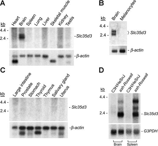
A multiple tissue Northern blot (Figure 5A-B) indicated expression of a 2.6-kb doublet Slc35d3 mRNA in brain tissues with no significant expression in 14 other tissues, including melanocytes (Figure 5C), of wild-type mice. Slc35d3 is expressed at quite low levels, even in the brain. It was necessary to prepare poly (A)–RNA (4 μg brain RNA) and expose for several days to obtain a satisfactory Northern blot signal. Northern blot analyses of poly(A)–RNA from brain tissues revealed lack of expression of the normal 2.6-kb Slc35d3 mRNA in mutant brain (Figure 5D). However, this tissue and other mutant tissues such as spleen exhibited greatly amplified expression of an abnormal 2.2-kb transcript (Figure 5D) with additional transcripts at 3.1 and 3.8 kb. Confirmation of amplification of Slc35d3 transcripts in most other mutant tissues was obtained by the sensitive and quantitative real-time PCR (QPCR) technique (Table 1) We observed greatly enhanced (8- to 2000-fold compared with levels in corresponding control C3H tissues) expression of Slc35d3 in multiple tissues derived from 3 ash-Roswell mutants. This indicates that stable, highly expressed mutant Slc35d3 transcripts exist in multiple tissues in ash-Roswell mice, although their functional relevance is uncertain. Significant expression of Slc35d3 was observed in bone marrow and platelets of control C3H/HeSnJ (Table 1), consistent with a role for this gene in platelets.
Relative expression of Slc35d3 in control and mutant tissues. (A-B) Multiple tissue Northern blots (Clontech) were treated with a 303-bp cDNA probe derived from nucleotides 1281 to 1583 of Slc35d3 cDNA together with a β-actin probe (below) as a loading control. Poly(A)–RNA was isolated from Melan-A melanocytes and C3H/HeSnJ brain for Northern blotting (C). Brain and spleen poly(A)-RNA blots (4 μg/lane) (D) were probed with the above Slc35d3 probe and a G3PDH probe (below). The apparent decreased expression of the 2.6-kb mRNA in control C3H brain tissue in this experiment compared with that of control brain tissue in panel C is due to the greatly decreased time (8 hours compared with 4 days) of exposure of this blot to film.
Relative expression of Slc35d3 in control and mutant tissues. (A-B) Multiple tissue Northern blots (Clontech) were treated with a 303-bp cDNA probe derived from nucleotides 1281 to 1583 of Slc35d3 cDNA together with a β-actin probe (below) as a loading control. Poly(A)–RNA was isolated from Melan-A melanocytes and C3H/HeSnJ brain for Northern blotting (C). Brain and spleen poly(A)-RNA blots (4 μg/lane) (D) were probed with the above Slc35d3 probe and a G3PDH probe (below). The apparent decreased expression of the 2.6-kb mRNA in control C3H brain tissue in this experiment compared with that of control brain tissue in panel C is due to the greatly decreased time (8 hours compared with 4 days) of exposure of this blot to film.
Relative concentrations of Slc35d3 mRNA in tissues of C3H and ash-Roswell mutants
| Tissues . | ΔCt values . | Relative Slc35d3 mRNA level (ash-Roswell/C3H) . | |
|---|---|---|---|
| C3H . | Ash/R . | ||
| Brain | 15.8 ± 0.17 | 12.7 ± 0.40* | 8 |
| Spleen | 15.9 ± 0.76 | 11.1 ± 0.34† | 32 |
| Bone marrow | 15.9 ± 0.37 | 11.8 ± 0.37* | 16 |
| Adult liver | 24.1 ± 0.24 | 13.3 ± 0.31‡ | 2048 |
| Kidney | 25.6 ± 0.51 | 14.1 ± 0.34‡ | 2432 |
| Lungs | 18.7 ± 0.56 | 10.6 ± 0.17‡ | 256 |
| Platelets | 12.1 | 3.0 | 512 |
Quantitative, real-time PCR analyses of mRNA concentrations were determined using TaqMan probes. Expression levels were standardized to levels of GAPDH mRNA by calculating cycle thresholds (Ct). ΔCt = Slc35D3 Ct − GAPDH Ct. Values are mean ± SEM of 3 separate mouse samples. RNA was combined from the platelets isolated from 3 mice.
P < .01 compared with same tissue of C3H mice.
P < .02 compared with same tissue of C3H mice.
P < .001 compared with same tissue of C3H mice.
Effect of Slc35d3 mutation on pigmentation
Because mutations in HPS genes typically (although not universally) cause hypopigmentation in patients with HPS and HPS mutant mice, the effect of the ash-Roswell Slc35d3 mutation on coat and eye pigmentation was investigated on 2 pigment backgrounds, agouti (A) and nonagouti (a). The mutant form of the Slc35d3 gene, which arose in the agouti C3H background, was transferred to a nonagouti C57BL/6J background by mating ash-Roswell mutants with C57BL/6J controls and identifying Slc35d3−/− homozygotes on the nonagouti background in the F2 generation. No dilution of coat or eye pigmentation occurred in Slc35d3 homozygotes on either the agouti (Figure 1F) or nonagouti (Figure 1E) backgrounds compared with controls (Figure 1B and 1A, respectively). Also, pigmentation segregated independently of platelet serotonin phenotype in the above 393 progeny backcross. Taken together, these data demonstrate that Slc35d3 does not control pigmentation.
Transgenic confirmation of the importance of Slc35d3 in the regulation of platelet-dense granules
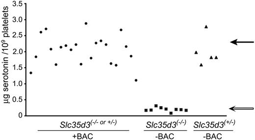
The effect of mutation and “rescue” of the Slc35d3 mutation on platelet phenotype was investigated in transgenic mice. A BAC (RP23-344M6) containing the wild-type copy of the Slc35d3 gene was inserted into fertilized eggs and transferred to the Slc35d3−/− genetic background and to other backgrounds by appropriate matings (see “Materials and methods”). BAC-positive mice in the F2 generation are either Slc35d3−/− or Slc35d3+/− at the Slc35d3 genomic locus. We were unable to devise a molecular test to directly distinguish these 2 genotypes in BAC-positive F2 offspring because differentiation between the Slc35d3 (+) allele contributed by genomic DNA and the Slc35d3 (+) gene contributed by the inserted BAC was not possible. We therefore used an indirect approach to determine whether BAC344M6 rescued the deficient platelet-dense granule phenotype in Slc35d3−/− progeny. Among 36 total F2 progeny, all 22 BAC-positive offspring contained high platelet serotonin concentrations (> 1.1 μg serotonin/109 platelets) (Figure 6). The laws of Mendelian segregation predict equal numbers of Slc35d3−/− and Slc35d3+/− progeny among these BAC-positive mice. Therefore, the probability that there is at least one true “rescued” F2 offspring [ie, (−/−) at the Slc35d3 genomic locus] among these 22 offspring is greater than 99.999 98% by the binomial distribution test, strongly indicating that rescue has indeed occurred and that BAC344M6 contains the platelet gene. BAC RP23-344M6 contains the Slc35d3 gene and the Pex7 gene, which is adjacent to Slc35d3 on chromosome 10. However, sequencing of the entire open reading frame of the cDNA of Pex7 together with 37 bp of 5′ and 309 bp of 3′ sequences revealed a completely wild-type sequence. This fact, together with the observed IAP insertion mutation in and accompanying abnormal expression of Slc35d3, indicates that Slc35d3 is the gene responsible for regulation of platelet-dense granules. It was possible to determine the Slc35d3 genotypes of the 14 F2 progeny that were BAC negative by molecular procedures. All 5 Slc35d3+/− offspring were high (> 1.6 μg serotoninin/109 platelets) and all 9 Slc35d3−/− offspring were low (< 0.3 μg serotonin/109 platelets) in platelet serotonin (Figure 6), further emphasizing the critical role of Slc35d3 in platelet-dense granule production. Finally, although BAC344M6 rescued the platelet deficiency, it had no effect on coat color, thus further proving that Slc35d3 does not affect pigmentation.
Platelet serotonin concentrations in BAC-positive and BAC-negative F2 progeny. Platelet serotonin levels were determined in each of 22 BAC-positive, Slc35d3(−/− or +/−) (•), 9 BAC-negative, Slc35d3−/− (▪), and 5 BAC-negative Slc35d3+/− (▴) F2 progeny. Each symbol represents the serotonin concentration in platelets from a single mouse. The average serotonin levels in wild-type C3H/HeSnJ (solid arrow) and ash-Roswell mutants (open arrow) are indicated.
Platelet serotonin concentrations in BAC-positive and BAC-negative F2 progeny. Platelet serotonin levels were determined in each of 22 BAC-positive, Slc35d3(−/− or +/−) (•), 9 BAC-negative, Slc35d3−/− (▪), and 5 BAC-negative Slc35d3+/− (▴) F2 progeny. Each symbol represents the serotonin concentration in platelets from a single mouse. The average serotonin levels in wild-type C3H/HeSnJ (solid arrow) and ash-Roswell mutants (open arrow) are indicated.
Screening for SLC35D3 mutations in patients with HPS
Screening of 22 patients with symptoms of Hermansky-Pudlak syndrome (albinism and prolonged bleeding) in which no known HPS-causing gene was found defective did not yield any SLC35D3 mutations, nor did screening of 3 patients with unclassified storage pool deficiency. (The latter patients have low levels of important low-molecular-weight components within platelet-dense granules, exhibit prolonged bleeding and have no other obvious clinical symptoms.) One variation (Q317H) was identified in 2 patients with HPS in the heterozygous state, which is likely not disease causing because no mutations on the other allele were identified, the Q at position 317 in SLC35D3 is not conserved in lower organisms, and one of the patients was recently identified to carry a homozygous mutation in the HPS3 gene, which explains her HPS phenotype.
Discussion
These studies establish that the Slc35d3 gene plays an important role in hemostasis as a regulator of the biosynthesis of platelet-dense granules. Platelet serotonin levels in the ash-Roswell mutant are less than 10% of normal.22 Likewise, platelet adenine nucleotides are significantly reduced, and analyses with mepacrine indicate that empty vesicles are present but lack normal dense granule contents.21,22 That Slc35d3 regulates platelet-dense granules was verified by rescue of normal platelet serotonin levels in Slc35d3−/− mutants by transgenic insertion of normal copies of the Slc35d3 gene (these studies).
We originally ascribed the platelet dysfunctions of the ash-Roswell mutant to a mutation in the ashen or Rab27a gene.21,22 However, normal platelet serotonin levels were observed20 in ostensibly identical ashen mutant mice obtained from The Jackson Laboratory. The present studies show that the explanation for these conflicting and confounding results derives from the occurrence of a mutation in another gene (Slc35d3) in the ash-Roswell mutant line maintained at Roswell Park Cancer Institute. That is, the ash-Roswell line contains mutations, not only in Rab27a, but also in Slc35d3. Mutations in the former gene result in hypopigmentation, as expected for a model of human Griscelli syndrome (Rab27a mutations), and mutations in the latter gene cause platelet storage pool deficiency. These analyses demonstrate that the Slc35d3 gene, not the Rab27a gene, regulates platelet-dense granules, a result consistent with the fact that platelet functional defects are not observed in patients with Griscelli syndrome.31 Thus, Griscelli syndrome is distinct from platelet storage pool deficiency syndromes in both human and mouse models.
The abnormal platelets of ash-Roswell arise from insertion of an IAP element within exon 1 of Slc35d3. IAPs are common (∼ 1000 copies per genome in mice) and mobile endogenous retroviral elements capable of inserting at new genomic sites causing both gene activation and inactivation.32 For example, IAP insertions cause ubiquitous ectopic expression of at least 4 separate agouti (A) alleles resulting from expression of the agouti gene from new promoters within the IAP element.33 Subsequently, novel dominant phenotypes such as obesity, diabetes, and increased susceptibility to tumors appear. It is likewise possible that the increased ectopic expression of Slc35d3 in most tissues of ash-Roswell derives from utilization of new IAP promoters. Loss of most of the upstream region of wild-type Slc35d3 mRNA in ash-Roswell mutants is consistent with this possibility. However, the Slc35d3 IAP insertion causes no apparent novel phenotypes in these tissues. Also, the enhanced Slc35d3 expression does not cause gain of function because Slc35d3+/− heterozygotes display normal platelet-dense granule numbers and serotonin levels. Rather, the mutation behaves as a typical recessive loss-of-function genetic trait in platelets, implying that the mutant protein (assuming it is stable) is nonfunctional.
The Slc35d3 IAP insertion predicts substitution of 7 new amino acids at the N-terminus for 10 wild-type N-terminal amino acids. It is possible this causes altered mutant membrane topology leading in turn to loss of function. Certain (although not all) protein topology prediction programs suggest that the mutant ash-Roswell N-terminal protein sequence changes the number of transmembrane domains of Slc35d3 from the wild-type 10 to 9 in ash-Roswell mutants. If true, the orientation of the mutant N-terminus would change in Golgi membranes from cytosolic to Golgi lumenal (assuming that Slc35d3 protein, like other Slc35 family members, resides in Golgi membranes and normally exposes both N- and C-termini to the cytosol23 ). Alternatively, the ash-Roswell mutation may directly alter a transporter function, folding, or the degradation rate of mutant Slc3d3 protein. Clearly, additional experiments are required to discriminate among these and other possibilities.
Previous studies established that the granule defect in ash-Roswell mutants is specific to platelet-dense granules.21 The quantity and intracellular trafficking of other organelles are apparently unaffected. Tissue and platelet lysosomal enzyme levels and rates of secretion of lysosomal enzymes from thrombin-stimulated platelets are normal. Likewise levels of platelet α granules and their rates of secretion from thrombin-treated platelets are normal. Platelet ultrastructure, except for the apparent deficiency of dense granules, is normal. Thus, Slc35d3 specifically regulates platelet-dense granules.
Current evidence for the steps in platelet-dense granule synthesis is limited. Dense granules are synthesized in immature megakaryocytes simultaneously with the appearance of α granules, and both granules derive from multivesicular bodies.5 Dense granules are unlike other intracellular granules in that their contents apparently exclusively consist of low-molecular-weight components such as serotonin, adenine nucleotides, pyrophosphate, calcium, and polyphosphate.2,3 Dense granule membrane proteins include P-selectin, CD63 (granulophysin), LAMP-2, GP1b, and αIIbβ3 though none of these are found exclusively in dense granules.34 Our studies indicate that valuable insight into the mechanisms of dense granule synthesis will derive from analyses of Slc35d3 gene products and interactions.
The Slc35d3 gene encodes an orphan receptor of unknown function and is a member, by sequence homology, of the SLC35 nucleotide sugar transporter family.23,24 The members of this family thus far characterized are antiporters, transporting nucleotide sugars from the cytosol into the lumen of the Golgi apparatus and/or the endoplasmic reticulum in exchange for the corresponding nucleoside monophosphates.23 The transported sugars are in turn used by glycosyltransferases to synthesize sugar chains of glycoproteins, glycolipids, and polysaccharides. There are at least 17 members of the SLC35 family, all encoding hydrophobic membrane proteins. The sugar nucleotide transported has been identified for 5 members of this family. However, the substrates or other function(s) of the Slc35d3 protein are unknown. Similarly, the mechanism by which Slc35d3 regulates platelet-dense granules and their contents is unknown. Nevertheless, the presence of empty dense granules22 in platelets of the ash-Roswell mutant (together with a putative antiporter function of Slc35d3) suggest it may act by regulating transport into or retention of dense granule interior components rather than by affecting the synthesis or trafficking of the dense granule membrane per se. A putative sugar nucleotide transporter function is consistent with the likely derivation of dense granules from the Golgi apparatus5,34 and the presence of membrane glycoproteins within dense granules.34 In this regard it is intriguing that substantial amounts of carbohydrate (probably sugar diphosphates35 ) occur in acidocalcisomes, subcellular organelles similar in numerous respects to platelet-dense granules.3 Acidocalcisomes were first described in trypanosomatids but are now known to be conserved from bacteria to human.36 The enhanced expression of Slc35d3 in brain tissues suggests a possible role in behavior. We have observed no overt behavioral abnormalities of ash-Roswell mutants up to 8 months of age. Nevertheless, specific behavioral tests should be performed to resolve this issue.
The ash-Roswell mutant is both similar to and distinct from the typical HPS mouse in phenotype. Platelet dense granules are highly deficient by whole-mount electron microscopy, the gold standard for HPS classification.22 However, the Slc35d3 mouse mutant differs from other mouse HPS mutants14 and most patients with HPS8 in its lack of effect on pigmentation. The latter fact is consistent with the lack of detectable expression of the Slc35d3 gene in melanocytes. It is also uncertain if the Slc35d3 gene directly regulates vesicle trafficking, a hallmark of all other HPS genes. Altogether, the ash-Roswell mutant appears to be a more appropriate model for congenital isolated δ-storage pool deficiency37 (which presents with deficiencies of dense granule constituents and normal pigmentation) than for HPS. The absence of deleterious SLC35D3 mutations in 22 patients with HPS analyzed here is consistent with this conclusion. The Slc35d3 gene is to our knowledge unique in that it regulates the contents of dense granules with no effects on other lysosome-related organelles such as melanosomes or lysosomes.
Authorship
Contribution: S.C., J.T., R.G., M.E.R., X.G., M.H., S.H., and E.K.N. designed and performed the research; W.L., W.A.G., and R.A.S. designed the research; R.T.S. designed the research and wrote the paper.
Conflict-of-interest disclosure: The authors declare no competing financial interests.
S.C. and J.T. contributed equally to this study.
Correspondence: Richard T. Swank, Roswell Park Cancer Institute, Elm & Carlton Sts, Buffalo, NY 14263; e-mail: richard.swank@roswellpark.org.
An Inside Blood analysis of this article appears at the front of this issue.
The publication costs of this article were defrayed in part by page charge payment. Therefore, and solely to indicate this fact, this article is hereby marked “advertisement” in accordance with 18 USC section 1734.
Acknowledgments
We thank Dr Richard Haslam for the antibody to Rab11A; Dr Kazuhiro Osanai for the antibody to Rab38; Dr Miguel Seabra for the antibodies to Rab27a and Rab27b; Dr David Bellnier for aid in serotonin analyses; and Madonna Reddington, Mary Kay Ellsworth, and Debra Tabaczynski for excellent technical assistance.
This work was supported by the National Institute of Health (grants HL-51480, HL-31698, and EY-12104) (R.T.S.), (grants AR39892 and EY015626) (R.A.S.) and by the National Natural Science Foundation of China (30525007) (W.L.). This research used core facilities supported in part by Roswell Park Cancer Institute's National Cancer Institute–funded Cancer Center Support Grant CA-16056. This study was partly supported by the Intramural Research Program of the National Human Genome Research Institute, National Institutes of Health (M.H. and W.A.G.).


![Figure 3. Identification of the Slc35d3 gene by positional cloning. (A) High-resolution genetic map of the region surrounding the Slc35d3 gene on mouse chromosome 10. Numbered markers are polymorphic MIT microsatellites. ARDR26 (amplified with primers 5′-GGGTGGAGCGGTACTATTCA-3′ and 5′-TGTCTTCAGAGACAACAATCCAG-3′) and ARDR22 (amplified with primers 5′-TTTTCTTTAGGTGGTTTTCTACAGC-3′ and 5′-AGGACAGACAGAGCCCTGAG-3′) are polymorphic microsatellites identified by genome scanning. (B) High-resolution physical map [based on the National Center for Biotechnology Information map viewer (build 35.1)]. The positions of 11 known genes in the critical genetic interval are indicated with their transcription orientations indicated by arrows. (C) The region of the genome from the beginning of exon 1 to the end of exon 2 of Slc35d3. A 5.5-kb IAP element is inserted between nucleotides 12296009 and 12296010 (Contig NT_039 492.5 of Mus musculus build 35.1) in exon 1 of Slc35d3 in the ash-Roswell mutant (Slc35d3−/−). (D) Transcripts of Slc35d3 in ash-Roswell (Slc35d3−/−) and control C3H/HeSnJ brains. Slc35d3 m-RNA of ash-Roswell mutants contains a new and in-frame ATG start site derived from the IAP element. More detailed views of normal and mutant Slc35d3 transcripts are given in Figure 4. (E) Predicted N-terminal sequences of mutant and control Slc35d3 proteins. The substitution of 7 new N-terminal mutant amino acids for the original 10 N-terminal wild-type amino acids is indicated.](https://ash.silverchair-cdn.com/ash/content_public/journal/blood/109/4/10.1182_blood-2006-08-040196/4/m_zh80040708270003.jpeg?Expires=1765901955&Signature=OnagmnQ4xY~YJSVPKnznVrtRzsR53da8NzOP3YYhrz4BtlEgBsITkVS6n0Bz4lyv6RHxfRSnOeIZmT8zM2lYu1BC6stBFPIcL~YKOmHbOPiZntUdAbnw6i5gm03o9fdJF8jKe-6NtkbkiV-ymfpKm1BG5fjLeLSg~egddmXHFLEILdb11UikwwPMKtOfNrzt2Y946Fa~J3Ogl3AF3yje4MDaBPnRu8KSGwP9bengMjAGL4geSpA4vHl4Q0NVKx6arXTPVU6pbx3TlRtuB33Ip5RB4jf1Barg1ZpvCFQSN0AE1bstrHWSrBxo~cDxQXxMpJTFzvNwtQ2MGeEAzoeM5g__&Key-Pair-Id=APKAIE5G5CRDK6RD3PGA)


This feature is available to Subscribers Only
Sign In or Create an Account Close Modal