Abstract
Using deconvolution microscopy, we visualized in real time fibrin network formation in the hydrated state. Individual mobile fibers were observed before the gel point determined by eye. After gelation, an initial fibrin network was seen, which evolved over time by addition of new fibers and elongation and branching of others. Furthermore, some fibers in the network moved for a time. We quantified network formation by number of branch points, and longitudinal and lateral growth of fibers. Eighty percent of branch points were formed, and 70% of all fibers reached their maximum length at the gel point. In contrast, at the gel point, fiber diameter, measured as fluorescence intensity, was less than 25% and turbidity was less than 15% of the maximum values of the fully formed clot. The cumulative percentage of fibers reaching their final length and the number of branch points attained maximum values at 60% of maximum turbidity. Lateral fiber growth reached a plateau at the same time as turbidity. Measurements of clot mechanical properties revealed that the clots achieved maximum stiffness and minimum plasticity after the structural parameters reached their maxima. These results provide new information on the relative time sequence of events during fibrin network formation.
Introduction
A wide variety of studies of fibrin polymerization have been carried out using different techniques, including light scattering, x-ray diffraction, electron microscopy, and laser tweezers,1 leading to much knowledge on the molecular mechanisms of fibrin polymerization. The polymerization of fibrin takes place upon cleavage of the fibrinopeptides A and B from fibrinogen, exposing the knobs “A” and “B,” respectively, in the center of the molecule, that interact with complementary holes “a” and “b” at the ends of the molecules. Specific A:a interactions cause longitudinal polymerization of fibrin monomers into structures called double-stranded protofibrils,2-4 which aggregate laterally to form fibers. The fibrin band pattern and repeat of 22.5 nm, or half the molecular length, shown through x-ray diffraction and electron microscopy, indicate that the interactions involved in the molecular packing, including lateral aggregation, are highly specific.5-7 The process of lateral growth of fibers is limited due to the twisting of protofibrils in fibers, since the increase of diameter requires more stretching of protofibrils to accommodate the increase of path length, so that lateral aggregation stops when the energy required to stretch an added protofibril equals the energy of binding.8 Fibers form branches that lead to a 3-dimensional network or gel. Knowledge of the transformation of soluble polymers to an insoluble elastic gel is a key for understanding and controlling blood clotting, fibrinolysis, wound healing, and thrombosis. The mechanical properties and permeation of fluid through different types of clots have been studied and correlated with clot structure, visualized by confocal microscopy, for different concentrations of thrombin and other conditions.9-13 Correlations between the fibrin gel architecture and rate and nature of fibrinogen activation have been demonstrated.14-16
However, there are still significant gaps in our knowledge, particularly quantitative analyses of structural features (branch points, and fiber length and thickness changes) in real time during clot formation, partly due to limitations of the methods available. In studies using electron microscopy, the fibrin clot must be fixed and dehydrated, which is unnatural because clots normally consist of approximately 98% water, and fixation and staining could produce some artifacts. Furthermore, the necessity of fixation means that only snapshots can be obtained at different times, rather than a more continuous record of the same clot.
The light microscopy techniques, confocal and deconvolution microscopy, are among the most effective methods for studying biologic systems. On one hand, in comparison with electron microscopy, these techniques allow the investigation of unfixed, hydrated samples; 3-dimensional reconstruction of samples; and also monitoring of changes occurring in a sample in a time-dependent manner. On the other hand, these techniques provide better quality of images compared with those from epifluorescence microscopy.
Confocal and deconvolution microscopic techniques use different approaches to increase the quality of images. The confocal imaging system is based on the principle of rejecting out-of-focus fluorescent light by having a pinhole placed before the collection aperture that blocks all light from the sample except the light coming from the focal plane, which greatly increases the ratio of signal to noise. The practical effect of this is that images coming from thin sections of sample have a small depth of field, and by scanning many thin sections through a sample it is possible to build a very clean 3-dimensional image of a sample. To go deep into a sample, the confocal imaging system requires a very bright laser, which can be a problem because of fading of fluorescence. Compared with the confocal imaging system, the deconvolution imaging system uses a computational process to eliminate light coming from above and below the focal plane. The real advantage of the deconvolution imaging system is that the same image quality can be accomplished at very low light levels using a mercury arc lamp, which allows imaging light-sensitive specimens over long time periods. We used deconvolution microscopy to avoid fading of fluorescence, allowing us to make quantitative comparisons.
In the present work, the dynamics of fibrin network formation of plasma clots have been investigated, and early stages of network formation were visualized for the first time. Direct measurements of fiber network features (fiber length, branch points, and fiber thicknesses) were made in real time during plasma clot formation, which will be useful for understanding thrombus formation, blood clotting, fibrinolysis, and wound healing. These studies were also synchronized with clot turbidity and viscoelastic measurements.
Methods
Approval was obtained from the University of Pennsylvania institutional review board for these studies. Informed consent was obtained in accordance with the Declaration of Helsinki.
Polymerization mixture and gel point measurement
Pooled platelet-poor plasma from 6 healthy volunteers was collected with 12 mM sodium citrate (Na3C6H5O7) added to prevent coagulation. Collected plasma was stored at −80°C in 150-μL aliquots. The fibrin polymerization reactions were performed by mixing 150 μL pooled platelet-poor plasma from 6 healthy volunteers with 10% of total volume of labeled fibrinogen with 26 mM CaCl2 and clotted by 0.12 U/mL tissue factor from BIO/DATA (Horsham, PA) in 50 mM Tris-HCl, 140 mM NaCl, pH 7.4, which corresponded approximately to 0.4 U/mL thrombin based on clotting time. The stock concentration of labeled fibrinogen was 0.9 mg/mL, and the sample was prepared by mixing CaCl2 with tissue factor and adding it to the plasma together with labeled fibrinogen. After all components were mixed, the sample was inserted into a Sigmacoat-treated (Sigma-Aldrich, St Louis, MO) glass chamber made from a microscope slide and a coverslip separated by a spacer made of 2 strips of double-stick tape. Sigmacoat produced a very thin hydrophobic film on the glass surface, which eliminates surface effects. The thickness of the chamber was 350 μm.
The gel point is the stage at which the liquid became a gel. To measure the gel point, the polymerization mixture as described in the previous paragraph was prepared in transparent Sigmacoat-treated glass tubes. After all components were mixed, a timer was started, and the gel point was observed by eye, with 5 replicates for each sample.
Clot turbidity studies
The whole clot turbidity measurements were done at a wavelength of 350 nm using a DU 640 spectrophotometer (Beckman Coulter, Fullerton, CA). The polymerization mixture was prepared as described in “Polymerization mixture and gel point measurement.” Polymerization was followed by monitoring changes in turbidity in the slide chamber of the same design as used for microscopy studies. Turbidity curves were averaged from 3 experiments.
Labeling of fibrinogen with Alexa 488
Purified human fibrinogen (plasminogen free) from HYPHEN BioMed (Neuville-Sur-Oise, France) was labeled using an Alexa Fluor 488 Protein labeling kit from Molecular Probes (Eugene, OR). The unreacted dye was removed by gel filtration on a column with a molecular weight cutoff of more than 15 000 equilibrated with 10 mM potassium phosphate, 150 mM NaCl (pH 7.4), with 0.2 mM sodium azide. The degree of labeling was 3 to 4 molecules of the dye per 1 molecule of fibrinogen. The preparation was frozen in aliquots that were thawed only once before each experiment. A whole variety of control experiments, such as the measurement of turbidity and electron microscopy of clots, demonstrated that the introduction of labeled fibrinogen had no effect on the polymerization of plasma (data not shown).
Microscopy studies
Polymerization reactions were carried out as described in “Polymerization mixture and gel point measurement.” To monitor morphologic changes during plasma clot formation, images were taken every 10 seconds for 45 minutes using a Delta Vision Spectris Restoration microscopy system equipped with Olympus IX-70 inverted microscope with a 63×/1.4 NA Plan Apo and 20× objective lens (Olympus, Melville, NY). Epifluorescence illumination was provided by a HBO 100-W mercury arc lamp (USHIO, Tokyo, Japan), and the excitation wavelength was selected with an electronic filterwheel containing a filter for Alexa 488, a dichromatic mirror, and an emission filter for Alexa 488. SoftWorx software (Applied Precision, Issaquah, WA), which is a part of DeltaVision Spectris Restoration microscopy system, was used for processing the images. Deconvolution procedures that are based on a constrained-iterative method were used.17 More than 10 deconvolution microscopy experiments were completed to do quantitative analysis.
Image analysis
Before image analysis, all stacks of images were deconvolved using SoftWorx software. Lispix software (National Institute of Standards and Technology, Gaithersburg, MD) was used for single-fiber fluorescence intensity measurements. Fluorescence intensity from a selected area 5 × 5 pixels that corresponded to 0.5 × 0.5 μm2 of a single-fiber was measured and the background was subtracted. Fluorescence intensity was measured for each single-fiber at the same coordinates and the same size area at each time point. Changes of fluorescence intensity at each time point corresponded to the progressive aggregation of new fluorescently labeled fibrin monomers. The intensity of more than 100 single-fibers from 10 different experiments was measured. The same experiments were used to do fiber length measurements and branch point calculations. For fiber length measurements, all fibers for each single optical section were mapped. Fiber length was measured along each fiber as observed in the images. The parameters of single-fibers at each time point were used for statistical analysis. The number of fibers and branch points (the juncture of 3 or more fibers) were counted for each time point.
Measurements of viscoelastic properties of plasma clots
The polymerization mixture was prepared as described in “Polymerization mixture and gel point measurement.” Plasma mixture (100 μL) was polymerized between 2 coverslips in a custom-built Plazek torsion pendulum.18 A short impulse was applied to the pendulum, causing oscillations. The dynamic storage modulus (G′), loss modulus (G″), and loss tangent (tan; δ = G″/G′) were calculated from recordings of the oscillation. Three replicates were done for each sample, and measurements were taken every 10 minutes after the time of the gel point for each experiment.
Results
Deconvolution microscopy was used to visualize the dynamics of fibrin network assembly during plasma clot formation, correlated with measurement of turbidity and viscoelastic properties.
Initial events before the scaffold formation
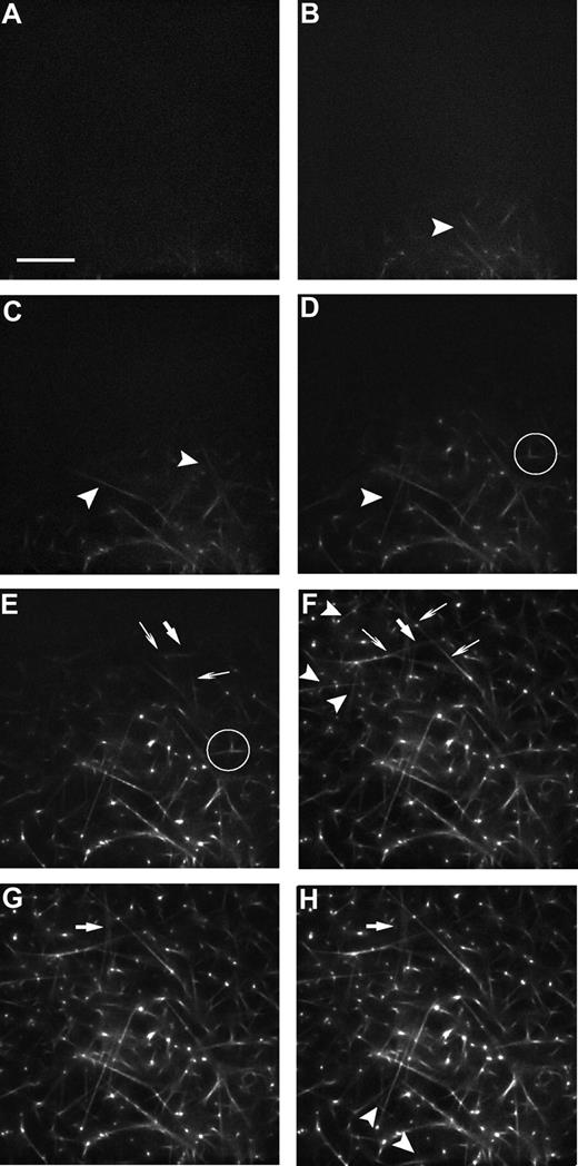
Examination of the time series of images during plasma clot formation revealed morphologic changes of these clots both at the single-fiber and network levels (Figure 1). It was observed for the first time that during the earliest stages of assembly individual fibers were mobile when they first became visible. It was difficult to identify the same fibers from images at one time point to the next, since they moved in 3 dimensions and grew in length.
Series of micrographs from the deconvolution microscope showing the dynamics of fibrin network formation. Fibrin was labeled with Alexa 488 as described in “Labeling of fibrinogen with Alexa 488.” Images were taken every 10 seconds (Videos S1,S2, available on the Blood website; see the Supplemental Materials link at the top of the online article). Selected images at the following times are shown: (A) 3 minutes and 50 seconds, (B) 4 minutes, (C) 4 minutes and 10 seconds, (D) 4 minutes and 20 seconds, (E) 4 minutes and 40 seconds, (F) 5 minutes, (G) 6 minutes, and (H) 20 minutes. Arrowheads ( ) indicate new fibers. Thick arrows (↙) indicate fibers that change position. Thin arrows (↑) indicate fibers that change length. The circled area shows a single-fiber with a branch point that is not connected to the scaffold in panel D, but it has connected to the scaffold later, as in panel E. Bar represents 15 μm.
Series of micrographs from the deconvolution microscope showing the dynamics of fibrin network formation. Fibrin was labeled with Alexa 488 as described in “Labeling of fibrinogen with Alexa 488.” Images were taken every 10 seconds (Videos S1,S2, available on the Blood website; see the Supplemental Materials link at the top of the online article). Selected images at the following times are shown: (A) 3 minutes and 50 seconds, (B) 4 minutes, (C) 4 minutes and 10 seconds, (D) 4 minutes and 20 seconds, (E) 4 minutes and 40 seconds, (F) 5 minutes, (G) 6 minutes, and (H) 20 minutes. Arrowheads ( ) indicate new fibers. Thick arrows (↙) indicate fibers that change position. Thin arrows (↑) indicate fibers that change length. The circled area shows a single-fiber with a branch point that is not connected to the scaffold in panel D, but it has connected to the scaffold later, as in panel E. Bar represents 15 μm.
An important aspect of these studies was to determine the time courses for 2 processes, branch point formation and longitudinal growth. These are 2 aspects of fibrin polymerization about which we know least, and characterizing the sequence of events is necessary to understand the mechanisms involved. We found that longitudinal growth and branch point formation of fibers were proceeding simultaneously. Branch points were observed on mobile fibers before they attached to other fibers. Mobile fibers grew in length and branched until connecting with other fibers, thereby establishing a scaffold.
Establishment of the scaffold and network formation
A scaffold or skeleton, consisting of the initial 3-dimensional network of fibers that first appeared, was visualized by deconvolution microscopy in our conditions at approximately 4 minutes (± 10 seconds) after polymerization was initiated. The gel point, or the time of the change from liquid to solid, was determined 5 times by examination of clots in transparent tubes by eye. The average value was 4 minutes and 50 seconds (± 16 seconds), approximately 40 seconds after fibers first appeared. After the appearance of the scaffold, we observed that there were no more individual fibers moving entirely independently of each other. At this point, it was possible to measure the length and the fluorescence intensity of single-fibers during polymerization (“Quantitative analysis of fibrin network formation”). Sometimes the addition of new fibers caused rearrangements of existing fibers relative to each other. For example, growth of a fiber can push other fibers that are part of the network. Usually, there was slow movement of a large group of interconnected fibers within the image (Videos S1,S2).
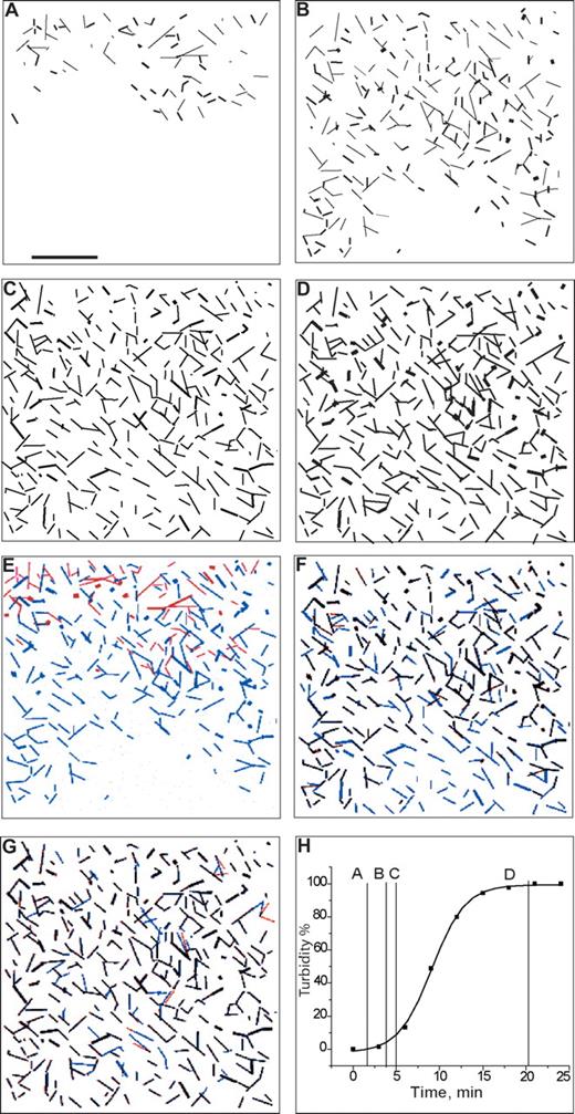
To do morphologic analysis of the networks as a function of time, we made high-contrast images by thresholding to show all fibers as they appeared during plasma clot formation, using a real-time image series (Figure 2). These images were made from low-magnification (20× objective lens) micrographs that showed more extensive arrays of fibers to visualize network formation, in comparison with more detail visible at higher magnification (63× objective lens; Figure 1). During the lag period of the turbidity curve, fibers started to appear: some single-fibers and some connected to another single-fiber or fiber with a branch point (Figure 2A). At the time when turbidity started increasing, the scaffold appeared (Figure 2B). Thresholded images at the time corresponding to the gel point measured by eye (Figure 2C) may be compared with those at the time when the turbidity curve reaches a plateau (Figure 2D). It is easy to see that the network becomes denser when the turbidity curve reaches the plateau.
Images of real-time series of fibrin network formation. The images of a low-magnification time series of micrographs were thresholded and colorized as described in “Establishment of the scaffold and network formation.” Original images from a similar time course of polymerization are shown in Videos S1,S2. (A) First fibers appear, corresponding to the lag period of the turbidity curve. (B) The scaffold appears, corresponding to the time when the turbidity curve starts increasing. (C) Corresponds to the gelation time measured by eye. (D) Network is fully formed, corresponding to the time when the turbidity curve reached a plateau. (E) Represents merged panels A and B, where the panel A was colored red, and panel B blue. (F) Represents merged panels B and C, where panel B was colored red, and panel C blue. Fibers that have purple color (mixture of red and blue) represent the same fibers at the 2 time points. Fibers that have blue color represent newly formed fibers. Fibers that have red color are the fibers that changed position or were rearranged. (G) Represents merged panels C and D, where panel C was colored red, and panel D was blue. Purple fibers are the same fibers at the same positions, red fibers are rearranged fibers, and blue fibers are newly formed fibers. (H) Turbidity curve with marked time points A, B, C, D, each corresponding to the time point represented in panels A through D, respectively. Bar represents 40μm.
Images of real-time series of fibrin network formation. The images of a low-magnification time series of micrographs were thresholded and colorized as described in “Establishment of the scaffold and network formation.” Original images from a similar time course of polymerization are shown in Videos S1,S2. (A) First fibers appear, corresponding to the lag period of the turbidity curve. (B) The scaffold appears, corresponding to the time when the turbidity curve starts increasing. (C) Corresponds to the gelation time measured by eye. (D) Network is fully formed, corresponding to the time when the turbidity curve reached a plateau. (E) Represents merged panels A and B, where the panel A was colored red, and panel B blue. (F) Represents merged panels B and C, where panel B was colored red, and panel C blue. Fibers that have purple color (mixture of red and blue) represent the same fibers at the 2 time points. Fibers that have blue color represent newly formed fibers. Fibers that have red color are the fibers that changed position or were rearranged. (G) Represents merged panels C and D, where panel C was colored red, and panel D was blue. Purple fibers are the same fibers at the same positions, red fibers are rearranged fibers, and blue fibers are newly formed fibers. (H) Turbidity curve with marked time points A, B, C, D, each corresponding to the time point represented in panels A through D, respectively. Bar represents 40μm.
To visualize the changes during network formation, images at 2 different time points were colored red and blue and then merged to observe changes between the 2 time points. Structures that do not change between the 2 time points will appear to be purple (mixture of blue and red), while fibers that are either red or blue retain their original colors because they moved or grew between the 2 time points. The first comparison was between an image at the time of the first appearance of fibers (Figure 2A red) and another at the first appearance of the network (Figure 2B blue) that were merged (Figure 2E). We see that no one fiber in Figure 2A matched any fiber in Figure 2B. In other words, fibers appearing at early stages of network formation corresponding to the lag period of the turbidity curve are mobile. Mobile fibers are red (Figure 2E). The second comparison was between an image at the first appearance of the network (Figure 2B red) and another at the time of the gel point measured by eye (Figure 2C blue) that were merged (Figure 2F). Since there are many purple fibers, that part of network, formed early, remains at later stages of network formation. However, fibers that we see as blue represent newly formed fibers, while fibers that have moved between the 2 time points are red. It is likely that these fibers change location as a result of adding new fibers to the existing network, with the consequence of forces being exerted. Fibers that are blue at one end and purple at the other end are fibers that grew in length between the 2 time points.
We consider the appearance of the scaffold as a very important moment in network formation because, after that, fibers stopped moving as individual fibers and started the process of adding new fibers to the scaffold and longitudinal growth and branching of existing fibers. The third comparison was between an image at the time of the gel point measured by eye (Figure 2C red) and another at the time that the turbidity reached a plateau and the network was fully formed (Figure 2D blue) that were merged (Figure 2G). Blue fibers represent newly formed fibers, while red ones correspond to fibers that changed position. This comparison showed a lot of similarity, but also new fibers appeared and there was some rearrangement of previously existing fibers.
Quantitative analyses of fibrin network formation
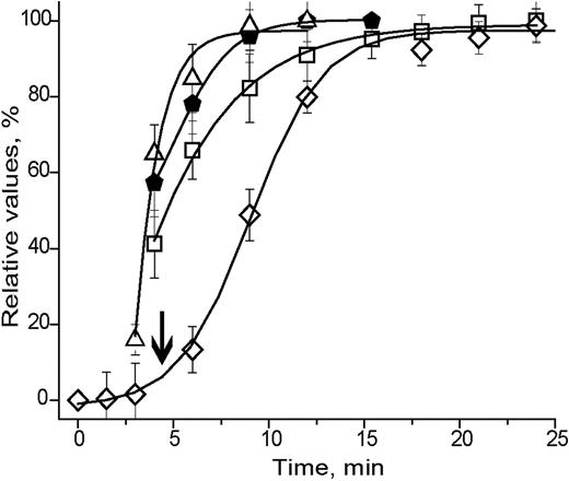
At the appearance of a scaffold, we observed that there were no more individual fibers moving entirely independently of each other. At this point, it was possible to measure the length and the fluorescence intensity (reflecting fiber cross-sectional area or mass) of a single-fiber during polymerization. To investigate the dynamics of plasma clot formation quantitatively at the single-fiber level, we quantified the deconvolution microscope images for comparison with whole clot turbidity as a function of time. We measured the fluorescence intensity changes of 121 fibers, and the fiber lengths and number of branch points of all fibers from the images of 10 experiments. Mean turbidity curves were obtained by averaging curves from 3 turbidity experiments under the same conditions as described in “Clot turbidity studies.” The changes of turbidity, fluorescence intensity, fiber length, and branch point number as a function of time illustrate the dynamics in a manner not apparent from the images alone (Figure 3). At the gel point, the longitudinal growth, branch point formation, and the average fluorescence intensity of many fibers, which corresponds to fiber mass (cross-sectional area), were not completed. Longitudinal growth of fibers and branch point formation were completed by the time turbidity reached approximately 60% of maximum. At that same time, fluorescence intensity was approximately 80% of maximum. After longitudinal growth and branch point formation were completed, the fluorescence intensity of fibers continued increasing and reached a maximum at almost the same time as the turbidity curve reached its maximum.
Dynamics of fibrin polymerization in terms of turbidity (◇), fluorescence intensity (□), number of branch points (▵), and fiber length (⬟). The error bars represent 1 standard deviation. Curves that represent branch point formation and length changes reach a plateau before lateral growth, which is represented by the fluorescent intensity curve. Lateral growth of fibrin fibers reached a plateau at the same time as the turbidity curve.  shows the gel point measured by eye.
Dynamics of fibrin polymerization in terms of turbidity (◇), fluorescence intensity (□), number of branch points (▵), and fiber length (⬟). The error bars represent 1 standard deviation. Curves that represent branch point formation and length changes reach a plateau before lateral growth, which is represented by the fluorescent intensity curve. Lateral growth of fibrin fibers reached a plateau at the same time as the turbidity curve.  shows the gel point measured by eye.
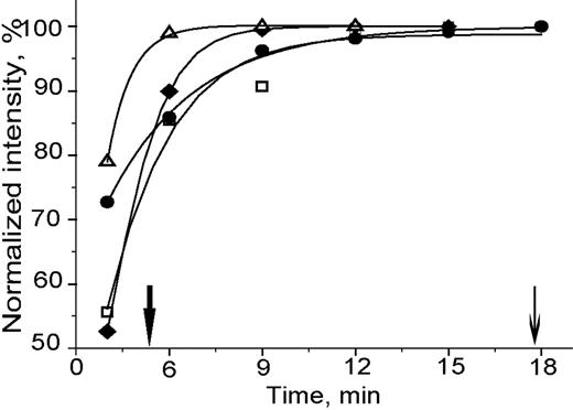
The increasing fluorescence intensity of a single-fiber indicates changes of fiber thickness due to adding of fluorescently labeled fibrin molecules. The increasing of fluorescence until it reached a plateau was a constant feature of all individual fibers examined. However, the time that it took for the fluorescence intensity to reach a plateau was not the same for all single-fibers as for the clot turbidity (Figure 4). Some fibers reached a maximum, constant intensity at an early stage of clot formation, close to the gel point, while other fibers reached a maximum intensity at a much later stage of clot formation, when the turbidity curve reached a plateau.
Dynamics of normalized fluorescence intensity of single-fibers. Each curve (▵, ●, ◆, □) represents a single-fiber. The kinetics of single-fiber assembly was different. The graph shows that polymerization of single-fibers reach a maximum intensity at different stages of network formation. The fluorescence intensity of single-fibers was measured and normalized by the maximum of intensity of each fiber. The first arrow ( ) shows the gel point measured by eye. The second arrow (↓) shows the time when turbidity curve reached a plateau.
Dynamics of normalized fluorescence intensity of single-fibers. Each curve (▵, ●, ◆, □) represents a single-fiber. The kinetics of single-fiber assembly was different. The graph shows that polymerization of single-fibers reach a maximum intensity at different stages of network formation. The fluorescence intensity of single-fibers was measured and normalized by the maximum of intensity of each fiber. The first arrow ( ) shows the gel point measured by eye. The second arrow (↓) shows the time when turbidity curve reached a plateau.
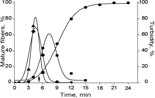
The distribution of mature fibers as a function of time by fluorescence intensity, fiber growth, and numbers of branch points, or, in other words, the rate of change of these parameters over time, were compared (Figure 5). We see that the number of mature fibers in terms of longitudinal growth and branch point formation have a maximum at the time of the gel point and represent approximately 75% of all fibers. However, at this time, 22% of fibers reached a maximum of intensity, while the turbidity increase was almost 13%.
Distribution of mature fibers. The rates of changes as a function of time of fluorescence intensity, longitudinal growth, and branch points. Turbidity (●), fluorescence intensity (■), number of branch points (▴), and fiber length (◆) were calculated for each time point. ↑ shows the gel point measured by eye. Distribution of mature fibers in term of longitudinal growth and branch point formation have a maximum at the same time that corresponds to the gel point measured by eye. The distribution of mature fibers in terms of fluorescence intensity has a maximum after longitudinal growth and branch point formation were completed. The software package Origin version 6 (OriginLab, Northampton, MA) was used to do the Gaussian fitting.
Distribution of mature fibers. The rates of changes as a function of time of fluorescence intensity, longitudinal growth, and branch points. Turbidity (●), fluorescence intensity (■), number of branch points (▴), and fiber length (◆) were calculated for each time point. ↑ shows the gel point measured by eye. Distribution of mature fibers in term of longitudinal growth and branch point formation have a maximum at the same time that corresponds to the gel point measured by eye. The distribution of mature fibers in terms of fluorescence intensity has a maximum after longitudinal growth and branch point formation were completed. The software package Origin version 6 (OriginLab, Northampton, MA) was used to do the Gaussian fitting.
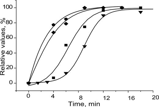
The cumulative number of mature fibers for each time point calculated from the distribution graphs reveals the times at which most of these events occur (Figure 6). The clot turbidity curve and the fibers' intensity curve, which represented the total percentage of fibers reaching a maximum of intensity, have similar slopes, 0.261 and 0.254, respectively. This result indicates that the rates of these 2 phenomena are similar, which is consistent with the turbidity arising from the average fiber cross-sectional area.19 Curves that represent branch points and length have slopes, 0.152 and 0.184, respectively, suggesting that branching and fiber extension are directly related. The parameters of the fitting curves were used to calculate the slopes.
The cumulative fraction of mature fibers that have reached maximum (■) intensity, (◆) length, and (⬟) number of branch points compared with turbidity (▾) as a function of time. Curves that represent the cumulative fraction of mature fibers in terms of length and branch point formation have similar slopes and reached a plateau before turbidity reached a plateau. Curves that represent the cumulative fraction of mature fibers in terms of intensity have similar slopes with turbidity and reached a plateau at the same time. The software package Origin version 6 was used to do the sigmoid fitting.
The cumulative fraction of mature fibers that have reached maximum (■) intensity, (◆) length, and (⬟) number of branch points compared with turbidity (▾) as a function of time. Curves that represent the cumulative fraction of mature fibers in terms of length and branch point formation have similar slopes and reached a plateau before turbidity reached a plateau. Curves that represent the cumulative fraction of mature fibers in terms of intensity have similar slopes with turbidity and reached a plateau at the same time. The software package Origin version 6 was used to do the sigmoid fitting.
Dynamics of mechanical properties during fibrin network formation
To see how changes in the mechanical properties of plasma clots as a function of time are related to the dynamics of plasma clot formation, we measured the mechanical properties during clot formation under the same conditions as described in “Measurement of viscoelastic properties of plasma clots.” We were not able to measure the mechanical properties at early times when the scaffold appeared or at the gel point measured by eye, because at this time clots were not mechanically stable, so that any disturbance to them could cause artifacts and alter network formation. We found that the storage modulus or stiffness (G′) and loss modulus or inelastic component (G″) reached 50% to 60% of the total value at the time when branch point formation and length changes were completed (Figure 7A,B). The loss tangent δ (tan δ = G″/G′), which is a measure of the energy dissipated by viscous processes relative to that stored by elastic processes, was still high when the turbidity reached 80% of its maximum, indicating that by this time, the storage modulus was low compared with the loss modulus (or inelastic component) or, in other words, the clot has low stiffness. Only after the turbidity curve reached a plateau, and branch points, and length and thicknesses of fibers were completed, did the proportion between loss and storage modulus reach an equilibrium (Figure 7C).
Dynamics of the mechanical properties of a plasma clot was measured using a torsion pendulum. Samples for measurements were prepared as described in “Polymerization mixture and gel point measurement.” (A) Storage modulus (G′) or stiffness (■). (B) Loss modulus (G″) or inelastic component (■). (C) Tangent (δ = G″/G′) (■). (D) Turbidity (■). The error bars represent 1 standard deviation. G′ and G″, which represent stiffness and inelastic component, respectively, reached a plateau after polymerization was completed, as represented by turbidity curve. Tan δ, which is the ratio of G″ and G′, reached a plateau also after polymerization was completed.
Dynamics of the mechanical properties of a plasma clot was measured using a torsion pendulum. Samples for measurements were prepared as described in “Polymerization mixture and gel point measurement.” (A) Storage modulus (G′) or stiffness (■). (B) Loss modulus (G″) or inelastic component (■). (C) Tangent (δ = G″/G′) (■). (D) Turbidity (■). The error bars represent 1 standard deviation. G′ and G″, which represent stiffness and inelastic component, respectively, reached a plateau after polymerization was completed, as represented by turbidity curve. Tan δ, which is the ratio of G″ and G′, reached a plateau also after polymerization was completed.
Discussion
We have followed the physical process of fibrin polymerization correlated with the whole clot turbidity. The shape and parameters of the turbidity curves reflect the steps involved in polymerization. Coordinated studies of light scattering and electron microscopy established that the lag period is a result of the time necessary for cleavage of fibrinopeptide A and assembly of the resulting fibrin monomer into oligomers, followed by their elongation to make protofibrils.20 Lateral aggregation of protofibrils to produce fibers does not occur until protofibrils reach sufficient length. The rapid rise in turbidity is caused by the lateral aggregation of protofibrils, since turbidity is directly related to the average fiber cross-sectional area.19 The turbidity reaches a maximum when the remaining fibrin is incorporated into the fibers.
Heretofore, there have been some direct observations of the early stages of fibrin polymerization.20,21 Using confocal light microscopy, some movement of fibers prior to the gel point was observed.21 In the present study, these observations were extended and the mobility of the prefibers has been characterized (Videos S1,S2). Extensive movement of elongated structures was observed prior to the gel point. It is not yet known whether single protofibrils can be visualized by this technique, but it does not seem unreasonable to presume that they can,22 since individual actin filaments or microtubules have been imaged. In fact, one study demonstrated that individual microtubules in Toxoplasma gondii could be visualized by our deconvolution microscope but not by a confocal microscope.23
Although the gel point or clotting time was one of the earliest measures used to characterize coagulation, there has been little information on its physical meaning. Hematologists first determined the clotting time by visual observation of the transition from liquid to solid. Newer automated instruments use other characteristics to determine the clotting time, including changes in mechanical properties that indicate gelation. One model for gelation was developed from observations of the structures of plasma clots formed under different conditions.12,14,15 For example, it was observed that the structure of plasma clots was determined primarily by the concentration of thrombin added, with clots made at higher thrombin concentrations having thinner fibers. Since more thrombin will be generated in the plasma by activation of prothrombin to thrombin, it was proposed that the final clot structure was determined at the gel point by the thrombin added and was independent of thrombin later produced in the plasma. The model proposed was that the entire framework of the clot was produced at the gel point and that subsequent fibrin was used to thicken the fibers but no new fibers were generated.12,14,15 Although this model is attractive because it explains some experimental observations, our results are not entirely consistent with it. On the other hand, our results support aspects of models that propose the establishment of a network of thin fibers at the gel point with subsequent thickening of these fibers.12,24,25 Our results differ in that we have observed new fibers being added to the network and rearrangement of existing fibers after the gel point measured by eye. Therefore, it is apparent that a new, quantitative model is necessary to account for all of the observations. It is possible that a kinetic model for fibrin polymerization20,26 could be extended.
Although branching is necessary for making a gel, there has been little information on branch point formation during clotting. Electron microscopy of clots made of very thin fibers suggested that branch points could be of 2 types, tetramolecular or trimolecular.27 Tetramolecular branch points form by divergence of 2 protofibrils. Trimolecular branch points form when one fibrin molecule associates with 2 others via knob-hole interactions. There is considerable evidence that branching is a process opposite to that of lateral aggregation.9 Clots made of thicker fibers tend to have fewer branch points than those made of thinner fibers. In other words, conditions that favor lateral aggregation produce clots in which protofibrils are associated over long distances, whereas conditions that inhibit lateral aggregation, allow protofibrils to diverge, yielding clots with many branch points and thin fibers. Our results show the time sequence of branch point formation, and longitudinal and lateral growth during clot formation. We have shown that lateral aggregation continues after longitudinal growth and branch point formation has been completed, but it is important to note that branch points and longitudinal growth continued after the gel point measured by eye, that the framework of the clot network is not fully formed at that time. The number of branch points reached a maximum after the gel point but considerably before the maximum clot turbidity, although lateral aggregation reached a maximum at the same time as turbidity. The branch point maximum was reached at the same time as the maximum of longitudinal growth, which makes sense since fiber elongation will cease when all branch points are established.
A hypothesis has been developed to relate turbidity of the clot to clot structure, according to which the turbidity is directly related to the average cross-sectional area of the fibers making up the clot.19 Our results provide the first direct structural evidence for this relationship, since the turbidity curve and our curve that represents the summarized percentage of fibers that reach a maximum of intensity as a function of time have similar slopes and time course (Figure 6). The measurement of fluorescence intensity of individual fibers is directly proportional to protein content or cross-sectional area per unit length. However, plotting of single-fiber intensity changes as a function of time allows the observation of microscopic changes in clot structure, rather than overall, averaged properties. It is apparent from these results that some fibers reach maximum size early, while others grow much more slowly. This is another indication that network formation is a very dynamic process and is not completed by the gel point.
The measurement of the mechanical properties of the clots in parallel with structural parameters allows the correlation of macroscopic stability with structure. The stiffness or elastic modulus is first measurable after the gel point and then increases continuously as a function of time. At low stiffness, movement of the network was observed, contrary to expectations that a stable network is established at the gel point. There is movement of individual fibers or whole sections of the clot at early times after gelation. These movements are likely to be caused by forces generated during the assembly of new fibers and branch points, although the nature of these forces remains to be determined. Some random movements of the relatively weak early network could result from Brownian motion, but much of the motion observed is not random.
In this study, we demonstrate that fibers are mobile at early times of polymerization. The time when the fibers are mobile corresponds to the lag period of the turbidity curve. The scaffold forms by the time that the turbidity starts rising. Our observations show that the appearance of the initial scaffold on the microscopic level corresponds to the gel point at the macroscopic level, because of the juxtaposition of these events at the same time. After that time, little independent movement of single-fibers was observed. In contrast to existing theories12,20 of polymerization, branch point formation and longitudinal growth of fibers continued after the gel point and were completed when the turbidity reached 60%. After branch point formation and longitudinal growth are completed, lateral growth of fibers continued and reached a plateau at the same time as turbidity. The mechanical properties of plasma clots developed much more slowly, somewhat after branch point formation, longitudinal and lateral growth, as well as turbidity reached a plateau.
In summary, our results require modification of existing models12,20 of fibrin network formation. It appears that network formation begins as protofibrils move around and contact each other. Some of these events result in initiation points, such that the network is further extended by both branching and contact with other fibers. These initial structures also grow in thickness by adding new protofibrils and fibers. This time of scaffold formation corresponds to the lag period of the turbidity curve. Branch points can move as a result of forces from the growth of adjoining fibers, depending on how big they are. At the time when the turbidity curve starts to rise, the network stops moving and a more stable scaffold forms. After the scaffold is formed, branching, longitudinal growth, and lateral growth of fibers continue until the network is fully completed.
An Inside Blood analysis of this article appears at the front of this issue.
The online version of this article contains a data supplement.
The publication costs of this article were defrayed in part by page charge payment. Therefore, and solely to indicate this fact, this article is hereby marked “advertisement” in accordance with 18 USC section 1734.
Acknowledgments
We thank Dr Rustem Litvinov and Dr Alina Hategan for their comments and valuable discussions in preparing this paper.
This work was supported by National Institutes of Health grant HL30954.
National Institutes of Health
Authorship
Contribution: I.N.C. designed and performed research, analyzed data, and wrote the paper; J.W.W. designed research, analyzed data, and wrote the paper.
Conflict-of-interest disclosure: The authors declare no competing financial interests.
Correspondence: John W. Weisel, Department of Cell and Developmental Biology, University of Pennsylvania School of Medicine, 421 Curie Blvd, 1054 BRB II/III, Philadelphia, PA 19104-6058; e-mail: weisel@mail.med.upenn.edu.


This feature is available to Subscribers Only
Sign In or Create an Account Close Modal