Abstract
Despite the great importance of nonhematopoietic cells constituting the microenvironment for normal hematopoiesis, the cellular interactions between nonhematopoietic cells themselves are largely unknown. Using the Cre-loxP system in mice to inactivate Mind bomb-1 (Mib1), an essential component for Notch ligand endocytosis, here we show that the development of an MPD is dependent on defective Notch activation in the microenvironment. Our 2 independent Mib1 conditional knockout (CKO) mouse lines each developed a myeloproliferative disease (MPD), with gradual accumulations of immature granulocytes. The mutant mice showed hepatosplenomegaly, anemia, granulocytosis, and leukocyte infiltration in multiple organs and finally died at approximately 20 weeks of age. We were surprised to find that the transplantation of wild-type bone marrow cells into the Mib1-null microenvironment resulted in a de novo MPD. Moreover, by introducing the constitutively active intracellular domain of Notch1 in the Mib1-null background, we show that active Notch1 expression in the Mib1-null microenvironment significantly suppressed the disease progression, suggesting that the MPD development in the Mib1 CKO mice is due to defective Notch activation in the nonhematopoietic cells. These findings demonstrate that normal hematopoiesis absolutely requires Notch activation through the Notch ligand-receptor interaction between microenvironmental cells themselves and shed light on the microenvironment that fosters hematopoietic disorders.
Introduction
Maintenance of hematopoietic stem cells (HSCs) and regulation of their self-renewal and differentiation in vivo is thought to depend on their specific microenvironment, known as the HSC niche.1-3 The bone marrow (BM) microenvironment mostly consists of mesenchymally originated cells, including osteoblasts, fibroblasts, adipocytes, and endothelial cells.2,4 A subset of osteoblasts, sinusoid endothelial cells, and CXCL12-secreting reticular cells have been identified as the HSC niches, maintaining the quiescence of HSCs and regulating the proliferation, migration, and differentiation of HSCs.1,2 The stem cell niches maintain stem cells during lifetime by preventing their depletion and overexuberant proliferation.3 A variety of interactions between the BM microenvironment and hematopoietic cells in paracrine and juxtacrine manners, including stem cell factor/c-Kit,5 Tie2/Angiopoietin-1,6 CXCR4/CXCL12,7 and Notch signaling,8-12 are believed to play a role in the maintenance of HSCs in the BM. In addition, cell extrinsic mediators, such as Wnt, Shh, and BMP, secreted by the niches can affect, at least in part, the cell cycle of HSCs through cell cycle regulators, including Bmi1,13 p16Ink4a/p19Arf,14,15 p21cip1,16 and p18Ink4c.17 Thus, it is possible that the deregulation of HSCs by their niches could cause hematopoietic disorders
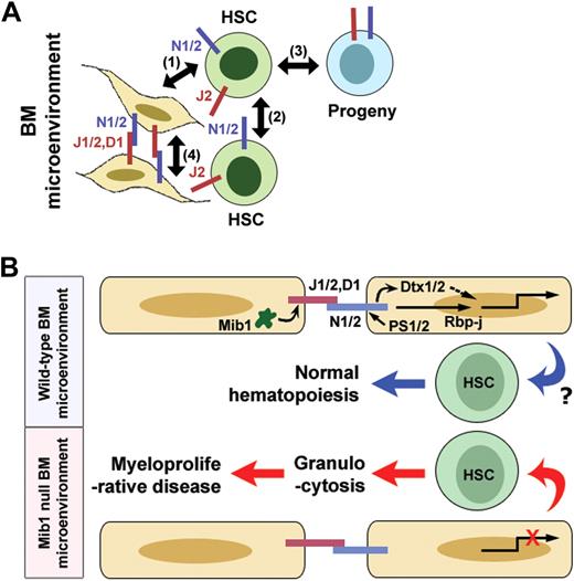
Notch signaling is thought to have a role in the maintenance of HSCs12 and in lineage decisions at multiple stages of lymphopoiesis.18 The activation of Notch signaling results in an increase of HSCs or progenitors,10 whereas the inhibition of Notch signaling leads to the accelerated differentiation of HSCs and the depletion of HSCs.9 Although the up-regulation of Jagged-1 (Jag1) on osteoblasts may expand HSCs, potentially through Notch activation,8 the conditional inactivation of Jag1 does not affect HSC maintenance or hematopoiesis.19 Because the BM microenvironment expresses multiple Notch ligands, such as Jag1, Jag2, and Deltalike-1 (Dll1), redundancy may exist between Notch ligands.20 The BM microenvironment also expresses Notch receptors Notch1 and Notch2, and hematopoietic cells express Notch ligands Dll1, Dll4, Jag1, and Jag2 besides the Notch receptors, Notch1 and Notch2.18,20 Therefore, multiple Notch interactions between hematopoietic cells, between BM microenvironmental cells, and between hematopoietic cells and BM microenvironmental cells could exist through the various Notch ligand-receptor pairs (Figure 7A). Although it is well known that HSCs require Notch activation to maintain their stemness, the cellular source of Notch signals remains to be clarified.
Notch signaling is initiated by the interaction of the Notch receptors with their ligands, which leads to sequential proteolytic cleavages that result in the release of the Notch intracellular domain (NICD) and the Notch extra-cellular domain (NECD).21 The NICD acts in the nucleus as a transcriptional regulator,22 and the NECD seems to undergo transendocytosis along with Delta in the signal-sending cell.23 A surprising, but poorly understood finding is that the internalization of Delta in the signal-sending cell is required to activate the Notch signaling in the receiving cells.24 To date, 2 structurally distinct E3 ubiquitin ligases, Neuralized (Neur)–1/2 (Neur in Drosophila) and Mind bomb (Mib)–1/2, have been shown to regulate the endocytosis of Notch ligands in vertebrates and invertebrates.24-29 Mib1-null mice exhibited pan-Notch defects in somitogenesis, neurogenesis, vasculogenesis, and cardiogenesis,27 and Mib1 regulates all known canonical Notch ligands, Dll1, Dll4, Jag1, and Jag2, in the Notch signal-sending cells. In contrast, Neur1 and Neur2 double mutant and Mib2 mutant mice exhibited no abnormality in a Notch signaling-dependent developmental process. Our extensive genetic mutant analyses of these 4 E3 ligases revealed that Mib1 has an obligatory role in the regulation of Notch ligands in mammalian development.30 Therefore, the genetic inactivation of Mib1 can provide an excellent model to elucidate Notch ligand-receptor interactions between hematopoietic cells and the microenvironment, between hematopoietic cells themselves, or between microenvironmental cells themselves, regulate hematopoiesis.
In this study, we have generated conditional Mib1 knockout mice under the control of 2 independent promoters, MMTV and Mx1. Unexpectedly, both mouse lines developed a myeloproliferative disease (MPD). Reciprocal BM transplantation (BMT) experiments revealed that the MPD in these mice was not intrinsic to the hematopoietic cells, but was caused by the Mib1-null microenvironment. Furthermore, intriguingly, the conditional activation of the active form of Notch1 (N1ICD) in the Mib1-null microenvironment significantly suppressed the MPD in the MMTV-Cre;Mib1f/f mice. These findings demonstrate that defective Notch activation between microenvironmental cells is responsible for the MPD development in the Mib1-null mice.
Methods
Mice
Floxed Mib1 (Mib1f/f) mice were generated.30 C57BL/6 (CD45.2+), congenic CD45.1, transgenic MMTV-Cre, and Mx1-Cre mice were purchased from The Jackson Laboratory (Bar Harbor, ME). Rosa-Notch1 and TNR mice were a kind gift from Drs D. A. Melton and N. Gaiano, respectively. All mouse lines were maintained in specific pathogen-free conditions at the POSTECH animal facility under institutional guidelines.
Flow cytometry
For cell staining, the following allophycocyanin (APC)-, fluorescein isothiocyanate (FITC)-, phycoerythrin (PE)-, or biotin-conjugated monoclonal antibodies (mAbs) were purchased from BD Biosciences (San Jose, CA) unless otherwise indicated: CD45.1 (A20), CD11b (M1/70), Gr-1 (8C5), B220 (6B2), CD71 (C2), IgE (R35-72), Sca-1 (E13-161.7), c-Kit (2B8), CD3 (2C11), CD4 (GK1.5), CD8 (53-6.7), CD19 (1D3), Ter119, and CCR-3 (83 101; R&D Systems, Minneapolis, MN). Biotin-conjugated mAbs were detected with streptavidin–peridinin chlorophyll protein (PerCP; BD Biosciences). For blood analysis, erythrocytes were first removed by suspending in red blood cell (RBC) lysis buffer, and nonspecific binding was reduced by preincubation with unconjugated anti-FcγRII/III (2.4G2). Single-cell suspensions were stained with the respective antibodies (Abs) and were analyzed using FACSCalibur or sorted by FACSVantage-SE (BD Biosciences). For Lin−Sca-1+c-Kit+ (LSK) sorting, whole BM cells were stained with biotinylated Abs specific for the following lineage markers (Lin): CD3, CD4, CD8, B220, CD19, Gr-1, CD11b, and Ter119. Lin+ cells were partially removed with streptavidin-magnetic beads (Dynabeads M-280; Invitrogen, Carlsbad, CA), and the remaining cells were stained with streptavidin-PerCP, anti–Sca-1–FITC, and anti–c-Kit–PE. Dead cells were excluded by 7-amino-actinomycin D (7-AAD) staining. For DNA content analysis, CD11b-stained splenocytes were fixed overnight in the dark in 2% paraformaldehyde in phosphate-buffered saline (PBS), washed twice, and resuspended in 500 μL PBS/0.05% Tween-20 containing 1 mg/mL propidium iodide before flow cytometric analysis.
Cell transplantation
Donor BM cells from CD45.1 or CD45.2 mice, or sorted CD45.1+ LSK or sorted CD45.1+ myeloid progenitor-enriched population (MP) cells along with CD45.2+ whole BM cells, were injected into the tail vein of lethally irradiated (9.6 Gy) recipient mice.
Colony-forming assay and cell proliferation
Filtered whole splenocytes (105 cells) and blood cells (105 cells) were incubated in RBC lysis buffer and plated into methylcellulose medium (StemCell Technologies, Vancouver, BC) supplemented with 2 ng/mL granulocyte macrophage–colony-stimulating factor (GM-CSF; Peprotech, Rocky Hill, NJ). Colony formation was scored after 10 days of culture. For cell proliferation, 5 × 10⁵ unfractionated BM cells were seeded per well of a 96-well plate, containing Dulbecco modified Eagle medium supplemented with 10% fetal bovine serum and macrophage–colony-stimulating factor (M-CSF), granulocyte-colony-stimulating factor (G-CSF), and GM-CSF in the described concentrations and were grown for 48 hours. Twelve hours before harvest, [3H]thymidine was added and analyzed for [3H]thymidine incorporation by standard procedures.
Cell proliferation and cell–cycle analysis
Bromodeoxyuridine (BrdU; 1 mg) was injected into the tail vein of mice, and the BM and blood cells were analyzed by FACSCalibur 6 hours after BrdU injection.
Histology and immunohistochemistry
Tissues were fixed in 4% paraformaldehyde (PFA)/PBS, paraffin-embedded, sectioned, and stained with hematoxylin-eosin. Four-micrometer sections were incubated with Abs for MPO (Dako Denmark A/S, Glostrup, Denmark), TER119 (BD Biosciences), Ki67 (Dako Denmark A/S), and PCNA (Santa Cruz Biotechnology, Santa Cruz, CA), and then were visualized with Alexa Fluor 488– or Alexa Fluor 594–conjugated secondary Abs (Invitrogen). Blood and BM smears were fixed with methanol and stained with Wright-Giemsa staining solutions (Sigma). For cytospin preparations, 104 sorted LSKs were cytocentrifuged onto glass slides, fixed with 4% PFA, stained with rabbit anti-cleaved Notch1 Abs (1:200; Cell Signaling Technology, Danvers, MA), and visualized with an Alexa Fluor 594–conjugated secondary Ab. Slides were viewed with an Axioskop2 Plus upright microscope for brightfield and fluorescence applications (Carl Zeiss, Jena, Germany). Images were acquired using the DP70 digital microscope camera (Olympus, Tokyo, Japan), and were processed with DPController software (Olympus).
BM stromal cell culture
Total BM cells were flushed from long bones (tibias and femurs) preincubated with 1% collagenase at 37°C, and were cultured in α-minimal essential medium supplemented with 20% fetal bovine serum. After 2 days of culture, nonadherent cells were removed, and adherent cells were further grown to confluence. For culture of BM stromal cells which were depleted for CD45+ and CD11b+ cells, adherent cells with confluence were detached and stained by Biotin-conjugated CD11b and CD45 Abs and negatively isolated by conjugation of streptavidin-magnetic beads.
RT-PCR and Western blot analysis
Total RNAs from whole BM cells and cultured BM stromal cells were extracted using the RNeasy Micro Kit (QIAGEN, Valencia, CA), according to the manufacturer's instructions. The RNA was converted into cDNA using the Omniscript Kit (QIAGEN). Primer information for reverse transcription-polymerase chain reaction (RT-PCR) is available upon request. Protein extraction and Western blot analyses were performed as described previously.27 The anti-Mib1 (DIP-1) Ab was a generous gift from Dr P. J. Gallagher (Indiana University School of Medicine, Indianapolis, IN), and the anti-Bclxl Ab was obtained from BD Transduction Laboratories (Lexington, KY).
Results
Mib1-null mice develop an MPD
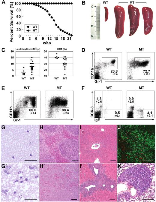
MMTV-Cre;Mib1f/f mice were generated by crossing Mib1f/f mice30 with MMTV-Cre–transgenic mice. Mib1 was almost completely inactivated in the whole BM cells and BM stromal cells in MMTV-Cre;Mib1f/f mice (Figure S1, available on the Blood website; see the Supplemental Materials link at the top of the online article). They were born in the expected Mendelian frequency, but most of the mutant mice died by 20 weeks of age (Figure 1A). The mutant mice developed severe splenomegaly and hepatomegaly (Figure 1B and Table S1). The splenomegaly was associated with extensive extramedullary hematopoiesis: a greatly increased myeloid progenitor-enriched population (Lin−Sca-1−c-Kit+; MP), an HSC-enriched population (Lin−Sca-1+c-Kit+; LSK), and enhanced erythropoiesis (CD71+Ter119+) (Figure S2A,B). The enhanced erythropoiesis was further confirmed by TER119 immunostaining; an increased number of erythroblasts, which were TER119-positive cells with nuclei, were found in the mutant spleens (Figure S2C).
Mib1 conditional knockout mice develop an MPD. (A) Cumulative survival of MMTV-Cre;Mib1+/f (WT; n = 277) and MMTV-Cre;Mib1f/f (MT; n = 292) mice. (B) Splenomegaly in the 14-week-old MT mice (right) compared with an age-matched wild-type (WT) control (left). (C) Hemograms of 14- to 16-week-old WT (n = 6) and MT (n = 16) mice. Bars indicate the mean. (D-F) Flow cytometric analysis of peripheral blood leukocytes (PBLs) (D,F) and BM cells (E) from 12- to 16-week-old WT (left) and MT (right) mice. Numbers around rectangles indicate the mean plus or minus SD (n = 10). Note that marked eosinophilia with CCR-3+ and basophilia with IgE+ (F) are evident in the mutant mice. (G and G′) Giemsa staining of blood smears from the WT (G) and MT (G′) peripheral blood. (H,I′) Hematoxylin and eosin staining of the WT (H and I) and MT (H′,I′) spleens (H,H′) and livers (I,I′). (J) Myeloid peroxidase (MPO; in green) and proliferating cell nuclear antigen (PCNA; in red) double immunohistochemistry of the liver (×400) from 15-week-old MMTV-Cre;Mib1f/f mice. (K) Hematoxylin and eosin staining of MT lung (×200). Scale bars: 50 μm (G, G′, and J); 100 μm (H, H′, I, I′, and K).
Mib1 conditional knockout mice develop an MPD. (A) Cumulative survival of MMTV-Cre;Mib1+/f (WT; n = 277) and MMTV-Cre;Mib1f/f (MT; n = 292) mice. (B) Splenomegaly in the 14-week-old MT mice (right) compared with an age-matched wild-type (WT) control (left). (C) Hemograms of 14- to 16-week-old WT (n = 6) and MT (n = 16) mice. Bars indicate the mean. (D-F) Flow cytometric analysis of peripheral blood leukocytes (PBLs) (D,F) and BM cells (E) from 12- to 16-week-old WT (left) and MT (right) mice. Numbers around rectangles indicate the mean plus or minus SD (n = 10). Note that marked eosinophilia with CCR-3+ and basophilia with IgE+ (F) are evident in the mutant mice. (G and G′) Giemsa staining of blood smears from the WT (G) and MT (G′) peripheral blood. (H,I′) Hematoxylin and eosin staining of the WT (H and I) and MT (H′,I′) spleens (H,H′) and livers (I,I′). (J) Myeloid peroxidase (MPO; in green) and proliferating cell nuclear antigen (PCNA; in red) double immunohistochemistry of the liver (×400) from 15-week-old MMTV-Cre;Mib1f/f mice. (K) Hematoxylin and eosin staining of MT lung (×200). Scale bars: 50 μm (G, G′, and J); 100 μm (H, H′, I, I′, and K).
The mutant mice exhibited increased numbers of peripheral blood leukocytes, accompanied by anemia and decreased numbers of platelets, but comparable BM cellularity (Figure 1C, Figure S2D, and Table S1). Flow cytometric analyses revealed elevated numbers of CD11b+Gr-1+ granulocytes in the mutant blood (Figure 1D), BM (Figure 1E), and spleen (Figure S3A). Blood smears also showed elevated numbers of granulocytes (Figure 1G′). Eosinophilia, displayed by CCR-3+ or SSChi, was detected in all of the mutant blood samples (Figure 1F), as well as in the BM, spleen, lymph nodes, and thymus (Figure S3B-F). Basophilia, displayed by IgE+, was also often observed in the blood (Figure 1F) and BM of the mutant mice (Figure S3F). The mutant spleens exhibited a severely disrupted architecture as a result of massive leukocyte infiltration (Figure 1H′) and significant reticulin fibrosis (Figure S4A). Moreover, massive leukocyte infiltration was observed in the liver (Figure 1I′) and lung (Figure 1K). Myeloid peroxidase (MPO) immunostaining revealed that most of the invading cells in the liver and spleen were of granulocytic origins (Figure 1J and Figure S4B). In the mutant spleens, a significant proportion of the leukocytes were proliferating cell nuclear antigen (PCNA)–positive, indicating that the infiltrating cells were actively proliferating (Figure S4B). Collectively, the phenotypes of the MMTV-Cre;Mib1f/f mice correspond best to those of a “myeloproliferative disease” based on Bethesda proposals.31
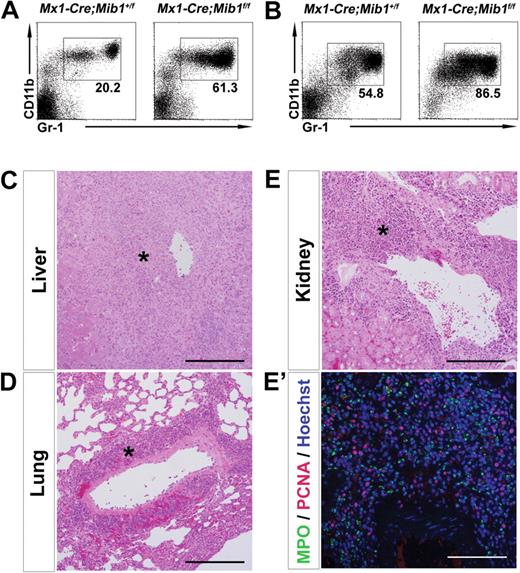
To confirm that the MPD in the MMTV-Cre;Mib1f/f mice depends on the Mib1 deficiency rather than the MMTV promoter effect, we generated Mx1-Cre;Mib1f/f mice. The Mx1 promoter allows for the expression of Cre recombinase in various hematopoietic systems, including the BM microenvironment, in response to interferon or interferon-inducing agents, such as polyinosinic-polycytidylic acid (pIpC).32,33 As expected, after plpC injection, the Mx1-Cre;Mib1f/f mice developed an MPD, leading to death approximately 6 months after the pIpC injections, with the same features as the MMTV-Cre;Mib1f/f mice, such as the marked expansion of CD11b+Gr-1+ granulocytes in the blood and BM (Figure 2A,B), and the massive leukocyte infiltration in multiple organs, such as the liver, lung, and kidney (Figure 2C-E′). MPO/PCNA double immunostaining revealed that more than half of the infiltrated cells were of granulocytic origins, and significant proportions of the infiltrated cells were highly proliferative (Figure 2E′). It is noteworthy that the time period until death of the Mx1-Cre;Mib1f/f mice after plpC injection was similar to the age of death of MMTV-Cre;Mib1f/f mice. Collectively, since the deletion of the Mib1 gene by 2 independent promoters, MMTV and Mx1, led to the exact phenocopy, the MPD should be caused directly by the inactivation of Mib1.
Mx1-Cre;Mib1f/f mice also develop an MPD. (A,B) Flow cytometric analysis of blood (A) and BM cells (B) from Mx1-Cre;Mib1+/f (left) and Mx1-Cre;Mib1f/f (right) mice 5 months after pIpC treatment (200 μg/every other day, 4 times). (C-E) Massive leukocyte infiltration (*) in the liver (C), lung (D), and kidney (E) (hematoxylin and eosin stain, ×200) of the Mx1-Cre;Mib1f/f mice. (E′) MPO/PCNA double immunostaining of the kidney (×200) from Mx1-Cre;Mib1f/f mice, 15 weeks after pIpC injection. Results are representative examples of 3 mutant mice. Scale bars: 100 μm.
Mx1-Cre;Mib1f/f mice also develop an MPD. (A,B) Flow cytometric analysis of blood (A) and BM cells (B) from Mx1-Cre;Mib1+/f (left) and Mx1-Cre;Mib1f/f (right) mice 5 months after pIpC treatment (200 μg/every other day, 4 times). (C-E) Massive leukocyte infiltration (*) in the liver (C), lung (D), and kidney (E) (hematoxylin and eosin stain, ×200) of the Mx1-Cre;Mib1f/f mice. (E′) MPO/PCNA double immunostaining of the kidney (×200) from Mx1-Cre;Mib1f/f mice, 15 weeks after pIpC injection. Results are representative examples of 3 mutant mice. Scale bars: 100 μm.
The gradual accumulation of immature granulocytes and progenitors
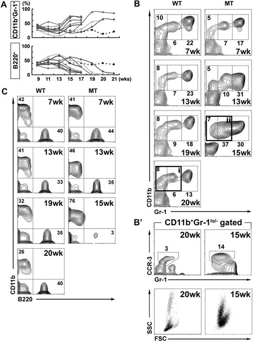
Because most of the MMTV-Cre;Mib1f/f mice died by 20 weeks of age, we monitored the blood cells from the control and mutant mice biweekly to examine the disease progression that eventually leads to death (Figure 3A). In most of the mutant mice, the CD11b+Gr-1hi granulocytes gradually increased at the early stages, whereas the B220+ lymphoid cells increased initially in some of the mutant mice (Figure 3A,B). At the late stages, however, CD11b+Gr-1lo/− immature granulocytes progressively increased, and a significant proportion of CD11b+Gr-1lo/− cells were CCR-3+ or SSChi eosinophils (Figure 3B,B′). In addition, the B220+ cells disappeared progressively with age, in inverse proportion to the increase of CD11b+ cells in the mutant mice (Figure 3C). All of the mutant mice died within 2 weeks after the prominent increase of CD11b+Gr-1lo/− cells in the peripheral blood, suggesting that MMTV-Cre;Mib1f/f mice exhibit a disease progression with the accumulation of immature granulocytes.
The disease progression in the MMTV-Cre;Mib1f/f mice. (A) Biweekly monitoring of the peripheral blood cells from MMTV-Cre;Mib1+/f (dotted line, representative data) and MMTV-Cre;Mib1f/f (solid line, 9 individuals) mice by flow cytometry. (B,C) The representative CD11b/Gr-1 (B) and B220/CD11b (C) expression profiles of the blood cells of MMTV-Cre;Mib1+/f (WT) and MMTV-Cre;Mib1f/f (MT) mice are shown. (B′) Flow cytometric analysis of the CD11b+Gr-1lo/− gated blood cells from WT (i) and MT (ii) at 20 and 15 weeks of age, respectively. Note that the CD11b+Gr-1lo/− cells consisted of a significant proportion of CCR-3+ and SSChi cells in the blood cells of MT.
The disease progression in the MMTV-Cre;Mib1f/f mice. (A) Biweekly monitoring of the peripheral blood cells from MMTV-Cre;Mib1+/f (dotted line, representative data) and MMTV-Cre;Mib1f/f (solid line, 9 individuals) mice by flow cytometry. (B,C) The representative CD11b/Gr-1 (B) and B220/CD11b (C) expression profiles of the blood cells of MMTV-Cre;Mib1+/f (WT) and MMTV-Cre;Mib1f/f (MT) mice are shown. (B′) Flow cytometric analysis of the CD11b+Gr-1lo/− gated blood cells from WT (i) and MT (ii) at 20 and 15 weeks of age, respectively. Note that the CD11b+Gr-1lo/− cells consisted of a significant proportion of CCR-3+ and SSChi cells in the blood cells of MT.
Because the CD11b+Gr-1lo/− cells progressively increased in the mutant mice, we examined the frequencies of stem and progenitor cells. In the 14- to 20-week-old MMTV-Cre;Mib1f/f mice, the HSC-enriched (Lin−Sca-1+c-Kit+) population was increased more than 2-fold compared with that of the control mice (Figure 4A). Further analyses revealed that the numbers of Lin−Sca-1+c-Kit+CD150− cell populations containing multipotent progenitor were exclusively increased in the mutant BM, whereas the numbers of long-term HSCs (LT-HSC, Lin−Sca-1+c-Kit+CD150+) were similar to those of the control mice (Figure 4A,B). Moreover, the mutant mice displayed a massive expansion of the granulocyte-macrophage progenitor (GMP; Lin−IL-7Rα−Sca-1−c-Kit+CD34+FcγRII/IIIhi) population in the BM and spleen (Figure 4C,D).
MMTV-Cre;Mib1f/f mice exhibit the increased numbers of granulocyte progenitors, GM-CSF responsive cells, GMPs, and MPPs. (A,B) Flow cytometric analysis of BM cells from WT and MT mice. Absolute cell numbers of LSK and its subpopulations, LSKCD150+ and LSKCD150− (A), and relative ratios of them (B). Statistical differences (t test) were P = .0015 (*), P = .0006 (**). (C,D) Flow cytometric analysis of BM cells (C) and splenocytes (D) from moribund group I/II MMTV-Cre;Mib1f/f (MT) and littermate control (WT) mice. A distribution of GMP (Lin−IL-7Rα−Sca-1−c-Kit+CD34+FcγRII/IIIhi) in the BM (C) and spleen (D) is shown. Numbers indicate the percentage of described populations in total BM (C) and spleen cells (D). Numbers around rectangles indicate the mean plus or minus SD (n = 3). (E) [3H]Thymidine incorporation by 14- to 15-week-old WT and MT BM cells in response to M-CSF, G-CSF, and GM-CSF. BM cells were cultured with the indicated amounts of cytokines for 48 hours. Pulse labeling was performed for the last 12 hours with [3H]thymidine. Bars indicate mean plus or minus SD. Statistical differences (t test) were P less than .0001 (*). (F,G) Colony numbers in splenocytes (F) and blood cells (G) from 15- to 19-week-old WT and MT mice, which were cultured in semisolid media in the presence of single GM-CSF (2 ng/mL). Bars indicate mean plus or minus SD. Colony numbers were barely (0.5, *) or not detected (**). (H) Flow cytometric analysis of splenocytes stained with propidium iodide and CD11b. (Bottom panels) DNA content of the SSChiCD11b+ granulocytes. (I) Cytospin of sorted CD11b+Gr-1lo and CD11b+Gr-1hi cells from moribund mutant spleens (Wright Giemsa, ×400). Scale bars: 50 μm.
MMTV-Cre;Mib1f/f mice exhibit the increased numbers of granulocyte progenitors, GM-CSF responsive cells, GMPs, and MPPs. (A,B) Flow cytometric analysis of BM cells from WT and MT mice. Absolute cell numbers of LSK and its subpopulations, LSKCD150+ and LSKCD150− (A), and relative ratios of them (B). Statistical differences (t test) were P = .0015 (*), P = .0006 (**). (C,D) Flow cytometric analysis of BM cells (C) and splenocytes (D) from moribund group I/II MMTV-Cre;Mib1f/f (MT) and littermate control (WT) mice. A distribution of GMP (Lin−IL-7Rα−Sca-1−c-Kit+CD34+FcγRII/IIIhi) in the BM (C) and spleen (D) is shown. Numbers indicate the percentage of described populations in total BM (C) and spleen cells (D). Numbers around rectangles indicate the mean plus or minus SD (n = 3). (E) [3H]Thymidine incorporation by 14- to 15-week-old WT and MT BM cells in response to M-CSF, G-CSF, and GM-CSF. BM cells were cultured with the indicated amounts of cytokines for 48 hours. Pulse labeling was performed for the last 12 hours with [3H]thymidine. Bars indicate mean plus or minus SD. Statistical differences (t test) were P less than .0001 (*). (F,G) Colony numbers in splenocytes (F) and blood cells (G) from 15- to 19-week-old WT and MT mice, which were cultured in semisolid media in the presence of single GM-CSF (2 ng/mL). Bars indicate mean plus or minus SD. Colony numbers were barely (0.5, *) or not detected (**). (H) Flow cytometric analysis of splenocytes stained with propidium iodide and CD11b. (Bottom panels) DNA content of the SSChiCD11b+ granulocytes. (I) Cytospin of sorted CD11b+Gr-1lo and CD11b+Gr-1hi cells from moribund mutant spleens (Wright Giemsa, ×400). Scale bars: 50 μm.
To further examine whether the progenitor populations increased in the mutant mice in the late stage of the progression, we tested the proliferating activity of BM cells in response to cytokines, M-CSF, G-CSF, and GM-CSF. The mutant BM cells showed markedly increased proliferation in response to recombinant GM-CSF, but not to G-CSF or M-CSF (Figure 4E). Moreover, the mutant BM cells displayed elevated expression of the specific α-subunit of the GM-CSF receptor (Figure S5). These results suggest that there are significantly increased GM-CSF responsive myeloid progenitors in MMTV-Cre;Mib1f/f mice. Next, the numbers of granulocyte progenitors in the splenocytes and blood cells of MMTV-Cre;Mib1f/f mice were quantified in vitro, using colony-forming assays. As expected, there were marked increases in the GM-CSF responsive colony-forming cells in the mutant spleens, and even in the peripheral blood (Figure 4F,G). Therefore, the myeloproliferative phenotype in MMTV-Cre;Mib1f/f mice is most likely due to the massive expansion of myeloid progenitors responsive to GM-CSF.
Granulocytes are highly proliferative in Mib1-null mice
The increased numbers of GMPs and GM-CSF-responsive progenitors in the Mib1-null mice might be due to either the disrupted lineage commitment or the active expansion of these progenitors. To distinguish between these possibilities, we examined the cell cycle status of whole BM and blood cells by using 7-AAD and BrdU. At 6 hours after BrdU injection, the percentages of BrdU+ cells in both the BM and blood were markedly increased in the MMTV-Cre;Mib1f/f mice, compared with the control mice (Figure S6A,B). It is noteworthy that the percentage of BrdU+ blood cells in the S and G2/M phases was dramatically increased in the MMTV-Cre;Mib1f/f mice, whereas those cells were barely detected in the control mice (Figure S6A). Given that the whole BM cellularity of MMTV-Cre;Mib1f/f mice is comparable with that of control mice, these results suggest that dividing granulocyte progenitors in the mutant BM might be actively released into the blood.
To characterize the accumulated immature granulocytes, the relative numbers of cycling myeloid cells were quantified in vivo. Although the majority of the CD11b+ cells were arrested in the G0/G1 phases of the cell cycle, with less than 7% in the S and G2/M phases in control mice, more than 20% of the CD11b+ cells were in the S and G2/M phases in MMTV-Cre;Mib1f/f mice (Figure 4H). Because only progenitor cells are mitotically active, these results suggest that MMTV-Cre;Mib1f/f mice have increased numbers of cycling granulocyte progenitors. Among the CD11b+ cells, both the Gr-1lo and Gr-1hi splenocytes of the mutant mice included relatively large, blastlike cells (Figure 4I), suggesting that the increased granulocyte progenitors have large cell sizes. In addition, the up-regulation of Bclxl, a known target of the GM-CSF receptor/STAT5 signaling pathway in the myeloid lineage,34 was observed in the mutant CD11b+Gr-1+ (Gr-1hi+lo) splenocytes, which might contribute to the progression of the MPD (Figure S6C).
Mib1-null microenvironment causes an MPD
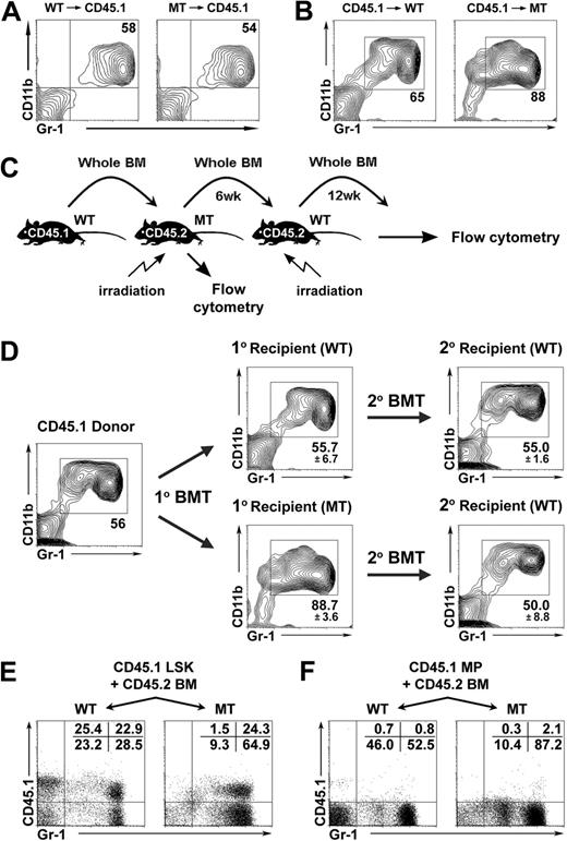
To investigate whether Mib1 plays a role in either the hematopoietic cells or nonhematopoietic cells, we performed reciprocal BMT experiments. When lethally irradiated CD45.1 wild-type mice were reconstituted with BM cells from the CD45.2 moribund mutant and control mice (15-20 weeks old), none of the CD45.1 recipient mice developed the MPD (Figure 5A), suggesting that the MPD development is not an autonomous effect caused by the loss of Mib1 in hematopoietic cells (Figure S1A). In contrast, when lethally irradiated CD45.2 MMTV-Cre;Mib1+/f and MMTV-Cre;Mib1f/f mice (7- to 9-weeks old) were reconstituted with BM cells from CD45.1 congenic wild-type mice, all of the reconstituted mutant mice showed significantly increased CD11b+Gr-1+ granulocytes in the BM, spleen, and blood, whereas the reconstituted wild-type mice were comparable with the untransplanted wild-type mice (Figure 5B and data not shown). To rule out the possibility that the MPD could arise from the remaining mutant recipient BM cells that had survived the lethal irradiation, we investigated the origin of the reconstituted BM cells by CD45.1 staining, and confirmed that more than 99% of them were of donor origins (Figure S10A). Moreover, all of the reconstituted mutant mice developed an MPD, leading to death generally within 8 weeks after BMT (Figure 6E). In addition, interestingly, the increased LSK and GMP populations, which were found in the MMTV-Cre;Mib1f/f mice, were also found in the reconstituted mutant mice, indicating that these phenotypes were dependent on the Mib1-null microenvironment (Figure S7).
MPD caused by the Mib1-null microenvironment. (A,B) Reciprocal BMT. Lethally irradiated CD45.1 mice were injected intravenously with BM cells from 12-week-old CD45.2 MMTV-Cre;Mib1+/f (left, n = 5) and MMTV-Cre;Mib1f/f (right, n = 5) mice. Twelve weeks after transplantation, the reconstituted BM cells were analyzed by flow cytometry (A). A representative of 10 independent experiments is shown. Lethally irradiated 7- to 9-week-old CD45.2 MMTV-Cre;Mib1+/f (left, n = 5) and MMTV-Cre;Mib1f/f (right, n = 5) mice were injected intravenously with CD45.1 congenic BM cells. Six weeks after transplantation, the reconstituted BM cells were analyzed by flow cytometry (B). A representative of 10 independent experiments is shown. (C,D) Secondary BMT. Primary and secondary BMT were performed as schematically depicted (C) and were analyzed by flow cytometry (D). Numbers around rectangles indicate the mean plus or minus SD (n = 3). (E,F) Microenvironment-induced MPD initiation by LSK. Lethally irradiated 7- to 9-week-old CD45.2 MMTV-Cre;Mib1+/f (WT, n = 2) and MMTV-Cre;Mib1f/f (MT, n = 3) mice were injected intravenously with 4.0 × 10³ sorted CD45.1 congenic LSK cells (E) or 8.0 × 10⁴ MP cells (F) along with 5.0 × 10⁶ CD45.2 wild-type whole BM cells. The blood cells from each recipient mouse were analyzed by flow cytometry 3.5 weeks after transplantation.
MPD caused by the Mib1-null microenvironment. (A,B) Reciprocal BMT. Lethally irradiated CD45.1 mice were injected intravenously with BM cells from 12-week-old CD45.2 MMTV-Cre;Mib1+/f (left, n = 5) and MMTV-Cre;Mib1f/f (right, n = 5) mice. Twelve weeks after transplantation, the reconstituted BM cells were analyzed by flow cytometry (A). A representative of 10 independent experiments is shown. Lethally irradiated 7- to 9-week-old CD45.2 MMTV-Cre;Mib1+/f (left, n = 5) and MMTV-Cre;Mib1f/f (right, n = 5) mice were injected intravenously with CD45.1 congenic BM cells. Six weeks after transplantation, the reconstituted BM cells were analyzed by flow cytometry (B). A representative of 10 independent experiments is shown. (C,D) Secondary BMT. Primary and secondary BMT were performed as schematically depicted (C) and were analyzed by flow cytometry (D). Numbers around rectangles indicate the mean plus or minus SD (n = 3). (E,F) Microenvironment-induced MPD initiation by LSK. Lethally irradiated 7- to 9-week-old CD45.2 MMTV-Cre;Mib1+/f (WT, n = 2) and MMTV-Cre;Mib1f/f (MT, n = 3) mice were injected intravenously with 4.0 × 10³ sorted CD45.1 congenic LSK cells (E) or 8.0 × 10⁴ MP cells (F) along with 5.0 × 10⁶ CD45.2 wild-type whole BM cells. The blood cells from each recipient mouse were analyzed by flow cytometry 3.5 weeks after transplantation.
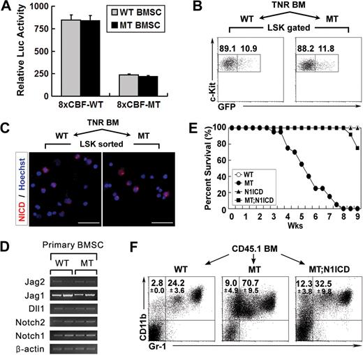
Defective Notch activation in the Mib1-null microenvironment leads to an MPD. (A) Potential of Mib1-inactivated primary BM stromal cells to readily activate Notch signaling. The wild-type and mutant stromal cells were cocultured with C2C12-Notch1 cells transfected with the 8× wild-type and mutant CBF-Luc vectors. Twenty-four hours after coculture, luciferase activity was measured. The 8× mutant CBF-Luc lacks the CBF-binding sites and was used as a control. (B,C) Intact Notch activation in LSKs in the Mib1-null microenvironment. Lethally irradiated 4.5-week-old WT (n = 3) and MT (n = 3) mice were injected intravenously with 5 × 10⁶ BM cells from transgenic notch reporter mice.9 At 6.5 weeks after transplantation (mutant recipient mice suffered from an MPD), LSKs from reconstituted BM cells were analyzed by flow cytometry (B) and immunocytochemistry with cleaved Notch1 antibody (Val1744, in red) (C). Numbers indicate the distribution of GFP-negative (left rectangle) and -positive (right rectangle) LSKs. Scale bar: 10 μm. (D) Semiquantitative RT-PCR analysis of Notch/Notch ligands in cultured primary BM stromal cells. β-Actin was used for normalization. (E,F) Survival rate (E) and flow cytometric analysis of blood cells (F) from the reconstituted mice. Lethally irradiated 7- to 9-week-old CD45.2 MMTV-Cre;Mib1+/f (WT, n = 10), MMTV-Cre;Mib1f/f (MT, n = 20), MMTV-Cre;Rosa-Notch1 (N1ICD, n = 10), and MMTV-Cre;Mib1f/f; Rosa-Notch1 (MT;N1ICD, n = 12) mice were injected intravenously with 5 × 10⁶ CD45.1 BM cells. Flow cytometric analysis of each BMT recipient mouse 7 weeks after transplantation (F). Numbers indicate the mean plus or minus SD (n = 5).
Defective Notch activation in the Mib1-null microenvironment leads to an MPD. (A) Potential of Mib1-inactivated primary BM stromal cells to readily activate Notch signaling. The wild-type and mutant stromal cells were cocultured with C2C12-Notch1 cells transfected with the 8× wild-type and mutant CBF-Luc vectors. Twenty-four hours after coculture, luciferase activity was measured. The 8× mutant CBF-Luc lacks the CBF-binding sites and was used as a control. (B,C) Intact Notch activation in LSKs in the Mib1-null microenvironment. Lethally irradiated 4.5-week-old WT (n = 3) and MT (n = 3) mice were injected intravenously with 5 × 10⁶ BM cells from transgenic notch reporter mice.9 At 6.5 weeks after transplantation (mutant recipient mice suffered from an MPD), LSKs from reconstituted BM cells were analyzed by flow cytometry (B) and immunocytochemistry with cleaved Notch1 antibody (Val1744, in red) (C). Numbers indicate the distribution of GFP-negative (left rectangle) and -positive (right rectangle) LSKs. Scale bar: 10 μm. (D) Semiquantitative RT-PCR analysis of Notch/Notch ligands in cultured primary BM stromal cells. β-Actin was used for normalization. (E,F) Survival rate (E) and flow cytometric analysis of blood cells (F) from the reconstituted mice. Lethally irradiated 7- to 9-week-old CD45.2 MMTV-Cre;Mib1+/f (WT, n = 10), MMTV-Cre;Mib1f/f (MT, n = 20), MMTV-Cre;Rosa-Notch1 (N1ICD, n = 10), and MMTV-Cre;Mib1f/f; Rosa-Notch1 (MT;N1ICD, n = 12) mice were injected intravenously with 5 × 10⁶ CD45.1 BM cells. Flow cytometric analysis of each BMT recipient mouse 7 weeks after transplantation (F). Numbers indicate the mean plus or minus SD (n = 5).
To clarify whether the MPD was caused by the Mib1-null microenvironment, we performed a secondary BMT. CD45.1 BM cells primarily transplanted into the CD45.2 mutant recipient mice, in which CD45.1 BM cells become myeloproliferative because of the mutant BM microenvironment, were retransplanted into lethally irradiated CD45.2 wild-type mice 6 weeks after primary BMT (Figure 5C and S10B). It is noteworthy that a flow cytometric analysis of the CD45.1+ hematopoietic cells 12 weeks after the secondary BMT revealed that the enhanced granulopoiesis had returned to normal (Figure 5D). Taken together, these results demonstrate that the MPD in MMTV-Cre;Mib1f/f mice is caused by the Mib1-null microenvironment, not by Mib1-null hematopoietic cells.
Given that the Mib1-null microenvironment causes an MPD, we sought to identify the hematopoietic cell populations that initiate the MPD in the Mib1-null microenvironment. We sorted LSK (Lin−Sca-1+c-kit+) and MP (Lin−Sca-1−c-kit+) populations from CD45.1 wild-type mice and transplanted them into lethally irradiated CD45.2 mutant mice (7-10 weeks old) with CD45.2 wild-type whole BM cells. Six weeks after transplantation, the mutant mice transplanted with wild-type LSKs had markedly increased CD45.1-positive CD11b+Gr-1+ granulocytes, indicating that the MPD was initiated from the LSK population (Figure 5E). In the mutant mice transplanted with wild-type MPs, however, the expansion of CD11b+Gr-1+ granulocytes was not observed in CD45.1+ cells (Figure 5F). These data revealed that the MPD in the Mib1-null microenvironment originated from the LSK population.
Defective Notch activation in the Mib1-null microenvironment leads to an MPD
Mib1 is essential for the endocytosis of Notch ligands, including Jag1, in the Notch signal-sending cells, which is required for the activation of Notch signaling in the Notch signal-receiving cells.30 Thus, we speculated that the MPD might be due to the inability of the BM microenvironment to trigger Notch signals to hematopoietic cells. To address this issue, we isolated primary BM stromal cells to test whether Mib1-null stromal cells can activate Notch signaling in the surrounding cells. We used the C2C12 cells expressing Notch1 (N1-C2C12) to evaluate the Notch activity after the coculture with the primary BM stromal cells.27,35 Although the expression of Mib1 was significantly reduced in the BM stromal cells from the MMTV-Cre;Mib1f/f mice (Figure S1B,C), these mutant stromal cells readily activated Notch signaling in the surrounding cells (Figure 6A). We further examined whether the Notch signaling was indeed activated in LSKs in the Mib1-null microenvironment of the MMTV-Cre;Mib1f/f mice. To address this issue, we performed a BMT experiment using a transgenic Notch reporter (TNR) mouse as a BM donor. The TNR mouse has a transgene composed of a CBF-1–responsive element with 4 CBF-1–binding sites and a minimal simian virus 40 promoter, followed by an enhanced green fluorescent protein (GFP) sequence.9 BM cells from TNR mice were transplanted into lethally irradiated MMTV-Cre;Mib1+/f and MMTV-Cre;Mib1f/f mice. At 6.5 weeks after BMT, unexpectedly, LSKs from the reconstituted mutant mice readily expressed GFP at a level comparable with that of the reconstituted wild-type mice (Figure 6B), which was further clarified by immunostaining of the LSKs, using a cleaved Notch1 antibody (Figure 6C). These results show that the MPD caused by the Mib1-null microenvironment is not due to defective Notch activation in the LSKs.
There are 2 possible ways in which Mib1 can act in the microenvironment. Because the Notch receptors Notch1 and Notch2, as well as the Notch ligands Jag1, Jag2, and Dll1, are expressed in nonhematopoietic cells (Figure 6D),8 Mib1 may play a critical role in the microenvironment by regulating Notch ligands in a Notch signaling-dependent manner. On the other hand, Mib1 also interacts with other substrates, such as DAPK and CDK5.36,37 Thus, it may have a critical role in the microenvironment in a Notch signaling-independent manner. To distinguish between these 2 possibilities, that either defective Notch signaling between nonhematopoietic cells or an autonomous defect of nonhematopoietic cells might be responsible for the MPD development in the MMTV-Cre;Mib1f/f mice, we designed an experiment where a constitutively active form of Notch1 was introduced into the Mib1-null microenvironment. We generated MMTV-Cre;Mib1f/f;Rosa-Notch1 mice, by breeding MMTV-Cre;Mib1+/f mice with Rosa-Notch1 mice, which have a transgene composed of a floxed Neo/STOP cassette and the Notch1 intracellular domain without the PEST domain in the ROSA26 locus, followed by an EGFP sequence.38 Because the MMTV-LTR promoter is active in hematopoietic cells as well as microenvironmental cells (possibly some nonhematopoietic cells), the MMTV-Cre;Mib1f/f;Rosa-Notch1 mice developed a fatal, transplantable T-cell leukemia as in MMTV-Cre;Rosa-Notch1 mice (Figure S8), which is consistent with the previous reports that constitutively active Notch1 in HSCs leads to the exclusive and rapid development of T-cell leukemia.39,40 The detailed characteristics of the T-cell leukemia in the MMTV-Cre;Mib1f/f;Rosa-Notch1 and the MMTV-Cre;Rosa-Notch1 mice are described in the supplemental material (Document S1 and Figure S8). Therefore, we lethally irradiated MMTV-Cre;Mib1f/f and MMTV-Cre;Mib1f/f;Rosa-Notch1 mice, to make them tumor-free, and used them as recipients.
We transplanted whole BM cells from CD45.1 congenic wild-type mice into lethally irradiated CD45.2 MMTV-Cre;Mib1f/f, MMTV-Cre;Mib1f/f;Rosa-Notch1, MMTV-Cre;Rosa-Notch1, and wild-type mice as recipients (7- to 9-week-old), and monitored their blood biweekly by flow cytometry. The MMTV-Cre;Rosa-Notch1 recipient mice were used as a control to test for any unknown side effects of Notch1 overexpression. Both the MMTV-Cre;Mib1f/f;Rosa-Notch1 and MMTV-Cre;Rosa-Notch1 mice were used as recipients at 7 to 9 weeks of age to exclude the side effects of the T-cell leukemia because, at this age, the MMTV-Cre;Rosa-Notch1 mice did not develop T-cell leukemia (data not shown). Neither the reconstituted wild-type nor the reconstituted MMTV-Cre;Rosa-Notch1 mice showed any hematologic abnormalities in every analysis. In contrast, all of the reconstituted MMTV-Cre;Mib1f/f mice died of an MPD within 8 weeks after BMT, whereas intriguingly, all of the reconstituted MMTV-Cre;Mib1f/f;Rosa-Notch1 mice survived (Figure 6E). The reconstituted MMTV-Cre;Mib1f/f;Rosa-Notch1 mice had a comparable percentage of CD11b+Gr-1+ granulocytes in the blood, compared with that of the reconstituted wild-type mice 7 weeks after BMT (Figure 6F). None of the reconstituted MMTV-Cre;Mib1f/f;Rosa-Notch1 mice showed a host-originated T-cell leukemia, but they eventually died within 20 weeks after reconstitution, because of the MPD. More than 99% of the BM cells from the reconstituted MMTV-Cre;Mib1f/f;Rosa-Notch1 mice were of donor origins (Figure S10C). The incomplete rescue might be due to limitations of the experimental system, because the stromal cells constituting the BM microenvironment are a heterogeneous population expressing both Notch1 and Notch2, and the overexpression of Notch1 alone, under the control of the MMTV-LTR promoter, might not be sufficient to restore defective Notch activation in the Mib1-null microenvironment. Taken together, these data demonstrate that the Mib1 deficiency causes Notch signaling defects in the microenvironment, which subsequently results in the MPD (Figure 7B).
Hypothesis of novel Notch signaling in the BM during normal hematopoiesis and myeloproliferative disease. (A) Schematic representation of possible Notch/Notch ligand interactions in the BM: (1) BM microenvironment-HSC, (2) HSC-HSC, (3) HSC-progeny, and (4) BM microenvironment-BM microenvironment. (B) Proposed novel Notch signaling in the BM microenvironment. Mib1 regulates Notch ligands, such as Jag1, Jag2, or Dll1, in BM microenvironmental cells to send Notch signaling into other BM microenvironmental cells. In the Notch signal-receiving BM microenvironmental cells, Notch signaling activated by Presenilin1/2 is transduced to Rbp-jκ or Deltex, and eventually turns on the transcription of target genes. Unknown signaling pathways from the wild-type BM microenvironment control HSCs to maintain normal hematopoiesis. In the Mib1-null BM microenvironment, however, defective Notch activation between the BM microenvironmental cells causes the HSCs to become myeloproliferative through an unidentified mechanism.
Hypothesis of novel Notch signaling in the BM during normal hematopoiesis and myeloproliferative disease. (A) Schematic representation of possible Notch/Notch ligand interactions in the BM: (1) BM microenvironment-HSC, (2) HSC-HSC, (3) HSC-progeny, and (4) BM microenvironment-BM microenvironment. (B) Proposed novel Notch signaling in the BM microenvironment. Mib1 regulates Notch ligands, such as Jag1, Jag2, or Dll1, in BM microenvironmental cells to send Notch signaling into other BM microenvironmental cells. In the Notch signal-receiving BM microenvironmental cells, Notch signaling activated by Presenilin1/2 is transduced to Rbp-jκ or Deltex, and eventually turns on the transcription of target genes. Unknown signaling pathways from the wild-type BM microenvironment control HSCs to maintain normal hematopoiesis. In the Mib1-null BM microenvironment, however, defective Notch activation between the BM microenvironmental cells causes the HSCs to become myeloproliferative through an unidentified mechanism.
Discussion
Our studies show that the loss of Mib1 results in a microenvironment-induced MPD. The phenocopy caused by the inactivation of the Mib1 gene, using 2 independent Cre lines, MMTV-Cre and Mx1-Cre, strongly supports the causative effect of Mib1 loss of function on the MPD development. The microenvironment-induced myeloproliferation progresses significantly with age, finally leading to death. Because the genetically activated Notch1 signaling in the Mib1-null microenvironment markedly suppressed the MPD phenotype and significantly prolonged the life span of Mib1-null mice, the microenvironment-induced MPD development depends largely on defective Notch signaling between the nonhematopoietic cells themselves constituting the microenvironment. Furthermore, the Mib1-null microenvironment was absolutely required to sustain the MPD, because the phenotype reverted when wild-type BM cells primarily transplanted into the Mib1-null recipient mice were retransplanted into other wild-type recipient mice.
There are 4 Notch receptors, Notch1-4, and their 5 canonical ligands, Dll1, Dll3, Dll4, Jag1, and Jag2, in mammals. Therefore, these Notch receptors and ligands function redundantly and/or nonredundantly for Notch activation in various cell fate decisions.18 Previous studies showed that the inactivation of a single Notch ligand, such as Dll1 and Jag1,19,32 or a single Notch receptor, such as Notch1 and Notch2,41,42 using Mx1-Cre, never causes an MPD in mice. Moreover, the blockages of canonical or noncanonical Notch pathways also do not cause an MPD, as shown in RBP-jκ conditional knockout mice using Mx1-Cre43 and in Deltex1/2 double knockout mice,44 respectively. In contrast, PS1+/−PS2−/− mice, in which Notch activation from Notch1-4 might have been reduced, develop an age-dependent MPD, exhibiting splenomegaly with severe granulocytic infiltration and a greatly increased GMP,45 suggesting the possibility that the complete blockage of Notch signaling from all of the Notch receptors might cause an MPD. Moreover, the MPD phenotypes of PS1+/−PS2−/− mice were not transplantable, suggesting that they also have similar microenvironmental defects causing the MPD (Dr Hui Zheng, Huffington Center on Aging, Departments of Molecular and Human Genetics, Molecular and Cellular Biology and Neuroscience, Baylor College of Medicine, written communication, June 2, 2008). In this study, we have shown that 2 independent, conditional knockout mouse lines, MMTV-Cre;Mib1f/f and Mx1-Cre;Mib1f/f, developed a nontransplantable MPD. We previously reported that Mib1 interacts with all of the canonical Notch ligands, and its deficiency induces pan-Notch defects in mice.27 In addition, our genetic inactivation studies of 4 E3 ubiquitin ligases, Mib1, Mib2, Neur1, and Neur2, which regulate the endocytosis of Notch ligands, revealed that Mib1 is essential for Jagged as well as Delta-like–mediated Notch signaling in mammalian development30,46 and for hematopoiesis, including the development of intraembryonic HSCs, adult T cells, and marginal zone B cells.47,48 Thus, our studies strongly suggest that the complete blockage of Notch activation in the microenvironment can cause an MPD.
It has been generally thought that BM microenvironmental cells express Notch ligands, such as Jag1, that interact with the Notch receptors expressed on the hematopoietic cells to activate Notch signals.8,20 In particular, the PTH1R-dependent increase of Jag1 in osteoblasts might be associated with Notch1 activation in the increased numbers of HSCs.8 Here, we have shown that wild-type LSKs transplanted into the Mib1-null microenvironment received Notch signaling. These results indicate that the MPD is not due to defective Notch activation in the HSCs. In contrast, the forced expression of active Notch1 in the Mib1-null microenvironment significantly rescued the MPD in the Mib1-null mice. These results demonstrate that the Notch signaling between the microenvironmental cells themselves is essential for normal hematopoiesis, and that Mib1 is a crucial tumor suppressor in the microenvironment. The BM microenvironment expresses 2 Notch receptors, Notch1 and Notch2.20 Thus, the incomplete rescue of the MPD in the MMTV-Cre;Mib1f/f;Rosa-Notch1 mice suggests that the overexpression of Notch1 alone might not be sufficient to alleviate the defective Notch activation in the Mib1-null microenvironment, because the Notch2 signaling might still be defective.
A deficiency in Rb or RARγ has been reported to induce an abnormal BM microenvironment, leading to myeloproliferative syndromes (MPS).49,50 In this report, we have definitively shown that a microenvironment with the mutation of a single gene, Mib1, induced the transplanted wild-type hematopoietic cells to become myeloproliferative. This is genetic evidence that the microenvironment plays an important role in MPD development by providing a permissive and supportive environment for abnormal myeloproliferation without an oncogenic mutation in the hematopoietic cells. On the other hand, unlike retinoblastoma protein (Rb) or retinoic acid receptor γ (RARγ) mutant mice, in which there was a profound loss of trabecular bone; no gross structural abnormality was observed in the BM of the Mib1-null mice (Figure S9). They had macroscopically intact trabecular bones and bone-lining osteoblasts (Figure S9A,B), suggesting that an MPD in the Mib1-null microenvironment might be caused by a different molecular mechanism compared with the Rb or RARγ mutant mice. Intriguingly, however, we observed abnormal, sponge-like BM structures in our mutant mice, but we are unsure of their physiologic meaning (Figure S11A). It is noteworthy that some of them stained positively for reticulin (Figure S11B), smooth muscle actin, and/or osteopontin (Figure S11C). In addition, the number of adipocytes in most of the mutant BMs was consistently decreased compared with those of the wild-type BMs (Figures S9A and S11A). It is noteworthy that this feature was also shown in old RARγ mutant mice that developed a microenvironment-induced MPS.50 We also examined the expression of Rb, RARγ, different inflammatory mediators, and various cytokines, such as tumor necrosis factor α, interleukin-6, G-CSF, M-CSF, GM-CSF, and CXCL12, in the BM and BM stromal cDNAs. We clearly found that the expression level of Rb transcripts was markedly reduced in the mutant BM cDNA (Figure S11D). Although we do not know the precise mechanism behind the microenvironment-induced MPD development in the Mib1-null mice, these results indicate that Mib1 loss alters the BM microenvironment abnormally, possibly in connection with the Rb or RARγ pathway. To clarify the molecular mechanism, further investigations, including the identification of the MPD-responsible stromal cells, are needed.
Notch signaling is implicated in many different developmental processes and in cancer progression.51 Using 2 independent lines of Mib1 conditional knockout mice, we have shown that Mib1 deficiency leads to a microenvironment-induced MPD. Rescue experiments using a constitutively active form of Notch1 revealed that Notch activation in the microenvironment, through the Notch ligand-receptor interactions between microenvironmental cells themselves, is required for normal hematopoiesis. Moreover, our findings underscore a previously unrecognized role of the hematopoietic microenvironment for maintaining hematopoiesis.
The online version of this article contains a data supplement.
The publication costs of this article were defrayed in part by page charge payment. Therefore, and solely to indicate this fact, this article is hereby marked “advertisement” in accordance with 18 USC section 1734.
Acknowledgments
We thank Drs D. W. Kim and K. J. Yoon for critical comments, M. P. Kong, J. Lee, H. S. Hwang, and Y. J. Lee for technical support, and Drs N. Gaiano, D. A. Melton, and P. J. Gallagher for their kind provision of materials.
This work was supported by grants from The Vascular System Research Center (VSRC) from KOSEF, the Korea Science and Engineering Foundation from the Korean government (MOST) (M10641000038-06N4100-03 810), and the 21C Frontier Functional Human Genome Project from the Ministry of Science and Technology in Korea (FG06-42-1).
Authorship
Contribution: Y.W.K. designed and performed research, analyzed data, and wrote the manuscript; B.K.K. generated Mib1 floxed mice, analyzed data, and wrote the manuscript; H.W.J., M.J.Y., and D.C.J. performed parts of the research and analyzed data; R.S., J.S., and S.H.K. analyzed data; and Y.Y.K. designed research, analyzed data, and wrote the manuscript.
Conflict-of-interest disclosure: The authors declare no competing financial interests.
Correspondence: Young-Yun Kong, School of Biological Science, College of Natural Sciences, Seoul National University, Seoul, 157-747, South Korea; e-mail: ykong@snu.ac.kr.

![Figure 4. MMTV-Cre;Mib1f/f mice exhibit the increased numbers of granulocyte progenitors, GM-CSF responsive cells, GMPs, and MPPs. (A,B) Flow cytometric analysis of BM cells from WT and MT mice. Absolute cell numbers of LSK and its subpopulations, LSKCD150+ and LSKCD150− (A), and relative ratios of them (B). Statistical differences (t test) were P = .0015 (*), P = .0006 (**). (C,D) Flow cytometric analysis of BM cells (C) and splenocytes (D) from moribund group I/II MMTV-Cre;Mib1f/f (MT) and littermate control (WT) mice. A distribution of GMP (Lin−IL-7Rα−Sca-1−c-Kit+CD34+FcγRII/IIIhi) in the BM (C) and spleen (D) is shown. Numbers indicate the percentage of described populations in total BM (C) and spleen cells (D). Numbers around rectangles indicate the mean plus or minus SD (n = 3). (E) [3H]Thymidine incorporation by 14- to 15-week-old WT and MT BM cells in response to M-CSF, G-CSF, and GM-CSF. BM cells were cultured with the indicated amounts of cytokines for 48 hours. Pulse labeling was performed for the last 12 hours with [3H]thymidine. Bars indicate mean plus or minus SD. Statistical differences (t test) were P less than .0001 (*). (F,G) Colony numbers in splenocytes (F) and blood cells (G) from 15- to 19-week-old WT and MT mice, which were cultured in semisolid media in the presence of single GM-CSF (2 ng/mL). Bars indicate mean plus or minus SD. Colony numbers were barely (0.5, *) or not detected (**). (H) Flow cytometric analysis of splenocytes stained with propidium iodide and CD11b. (Bottom panels) DNA content of the SSChiCD11b+ granulocytes. (I) Cytospin of sorted CD11b+Gr-1lo and CD11b+Gr-1hi cells from moribund mutant spleens (Wright Giemsa, ×400). Scale bars: 50 μm.](https://ash.silverchair-cdn.com/ash/content_public/journal/blood/112/12/10.1182_blood-2008-03-148999/7/m_zh80240828030004.jpeg?Expires=1769123485&Signature=GiruUZkvfqR8lHhXY64JgxbHdOFdr3yblHp-eBshNQbmooOuilyAL5bqVuvSyvTlnrgWLEGH9OaZXMlufPB6MXFVz3rGKLzN4l0szYAfeGWobhvmDIxoyGlsD4H30pAkvKA-iuAuutzEcN1UXiimN7mjMjzALEG4OXpsxL1PCVsWdBRcPPcLZ3wxZJ4qNAQFNQ-YLGMQqXKAQlyMPW16yqF4XQx2GSJg471VqvuDQy6rLEfyL7h0rST8qqlt53pIEtIRVY38VemopyvqkCdL0Rx56~sZIe5Wa84OvohIlHikFxGOweSHUBRjeIjJEyShCId9Xe5P56I445OwrIkX4A__&Key-Pair-Id=APKAIE5G5CRDK6RD3PGA)
This feature is available to Subscribers Only
Sign In or Create an Account Close Modal