Abstract
Abstract 3529
Poster Board III-466
Pet imaging using F-18 glucose (FDG) is increasingly being used for evaluation and staging of malignancy. However, staging in hematopoietic tissue using this agent has been hampered by poor specificity. F-18 flourothymidine (FLT) is currently being evaluated clinically as an imaging technique for tumor detection and staging. Secondary to its inclusion in DNA during the S phase, FLT is much more specific to proliferative tissue and less hampered by inflammatory background. As FLT uptake occurs in proliferating cell populations, we attempted to determine if imaging could provide useful information for evaluating global hematopoietic injury and recovery following radiation and transplantation. Three major groups of Wistar-Furth rats were studied. Group 1 consisted of rats receiving 950cGy of Whole Body Irradiation (TBI). Group 2 consisted of rats transplanted with syngeneic bone marrow 24-48 hrs following irradiation. Group 3 consisted of 6 rats exposed to a potentially sub-lethal dose of 500cGy and not transplanted. FLT imaging was performed before irradiation (n=4), 24-48 hrs. following irradiation, and on day 4-5 post transplantation. Subsequent imaging was carried out in 4 transplanted rats on days 8 and 14. Comparative FDG studies were also performed in selected animals. Table 1 summarizes the imaging studies performed in various subsets of rats.
Imaging Subsets of Experimental Animals and Histologic Correlations
| Experimental rat subsets . | FLT # studies performed . | FDG # studies performed . | Histologic correlation . |
|---|---|---|---|
| Normal or baseline rat studies | n=10 | n=6 | Normal cellular marrow |
| 24-48hrs post 950 cGy TBI | n=6 | n=4 | Marrow damage hypocellularity |
| Day 7 post 950 cGy TBI | n=4 | not done | Aplastic marrow |
| Day 6-7 post 950cGy TBI (4-5 days post transplantation) | n=4 | n=4 | Focal areas of cellular regeneration |
| Day 10 post 950 cGy TBI (and transplantation) | n=4 | n=2 | Cellular marrow |
| Day 6-7 post 500cGy TBI (No transplantation) | n=6 | not done | Moderate hypocellularity |
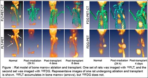
FLT imaging results were correlated with marrow histology and clinical survival in treated and control groups. Six of 6 irradiated control rats died with marrow aplasia during the second week following 950 cGy. Sub-lethally irradiated and transplanted rats animals showed clear evidence of definitive recovery as early as 6 days post irradiation or 4 days post transplantation respectively. FLT activity of all major marrow sites was easily identified and was superior to FDG images. Findings correlated with histologic evidence of early marrow repopulation and survival. Figure 1 illustrates FLT and FDG imaging performed in normal, post radiation and post transplanted rats.
We conclude that FLT imaging represents a practical noninvasive technique to evaluate marrow injury and early recovery following radiation and hematopoietic transplantation.
No relevant conflicts of interest to declare.
Author notes
Asterisk with author names denotes non-ASH members.


This feature is available to Subscribers Only
Sign In or Create an Account Close Modal