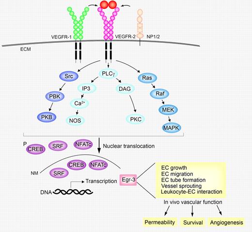
Among all known angiogenic molecules, vascular endothelial growth factor (VEGF) is probably the most extensively characterized growth factor that displays broad vascular functions including vasculogenesis, angiogenesis, vascular permeability, vascular survival, and vascular remodeling.1,2 These vascular functions of VEGF are mainly mediated by VEGF tyrosine kinase receptors (TKRs) expressed in endothelial cells (ECs), although non-TK neuropilin receptors may modulate TKR-activated signaling pathways (see figure).3 VEGF receptor-2 (VEGFR2; KDR for human and Flk-1 for mouse) is the key receptor that transduces active signals to execute VEGF-initiated endothelial activities. Despite the available information on this well-characterized signaling system, molecular players that translate VEGF-triggered signals into functional activities remain relatively poorly understood.
Involvement of Egr-3 in the VEGF-triggered signaling pathways in ECs. Upon binding to VEGFR2, VEGF activates several signaling pathways in ECs, leading to translocation of the activated SRF, NFATc, and CREB into the cell nucleus. SRF, NFATc, and CREB bind to the promoter elements of the egr-3 gene to transcriptionally increase expression levels of Egr-3, which executes VEGF-induced vascular functions including EC proliferation, migration, tube formation, vascular sprouting, and leukocyte-EC adhesion. These in vitro endothelial activities are essential processes for VEGF-induced in vivo angiogenesis, vascular survival, and vascular permeability.
Involvement of Egr-3 in the VEGF-triggered signaling pathways in ECs. Upon binding to VEGFR2, VEGF activates several signaling pathways in ECs, leading to translocation of the activated SRF, NFATc, and CREB into the cell nucleus. SRF, NFATc, and CREB bind to the promoter elements of the egr-3 gene to transcriptionally increase expression levels of Egr-3, which executes VEGF-induced vascular functions including EC proliferation, migration, tube formation, vascular sprouting, and leukocyte-EC adhesion. These in vitro endothelial activities are essential processes for VEGF-induced in vivo angiogenesis, vascular survival, and vascular permeability.
In an effort to crack the transcriptional code upon activation of VEGFRs, Suehiro et al employed DNA microarray to analyze the global gene-expression profiles of VEGF-activated human ECs.4 Interestingly, early growth response-1 (Egr-1) and Egr-3 transcriptional factors were markedly up-regulated in ECs after only 1 hour of stimulation with VEGF. Quantitative polymerase chain reaction analysis showed a more than 300-fold increase of Egr-3 mRNA relative to a 32-fold increase of Egr-1 in VEGF-stimulated endothelial cells. Moreover, VEGF-induced Egr-3 expression was sustained for a longer period compared to Erg-1.
Previous work has linked Egr-1 to development and progression of several vascular diseases including ischemic lung injury, atherosclerosis, and tumor growth.5 However, vascular functions of Egr-3 remain poorly understood. To gain further insights on signaling events leading to Egr-1 and Egr-3 up-regulation, various inhibitors targeting specific signaling components were used as blockades, which include VEGFR2, mitogen-activated protein (MAP) kinase (MAPK), c-Jun N-terminal kinases (JNK), Ca++ influx, p38MAPK, protein kinase C (PKC), PKC-δ, phosphoinositide 3-kinase (PI3K), protein kinase A (PKA), and calcineurin. It appeared that VEGFR2-activated signaling components including MAPK, JNK, and Ca++ influx are required for induction of both Egr-1 and Egr-3, whereas PKC, PI3K, calcineurin, PKC-δ, and PKA are essential only for up-regulation of Egr-3, but not for Egr-1. Thus, induction of Egr-1 and Egr-3 in ECs by VEGF involves overlapping but distinct signaling pathways.
In an Egr-3 promoter-driven luciferase reporter system, the authors showed that the VEGF response element was located between −515 and −37–base pair 5′ flanking sequences that contain consensus motifs for nuclear factor of activated T-cells (NFAT), cAMP-responsive element (CRE), and serum response element (SRE). Mutagenesis studies showed that each of NFAT, CRE, and SRE elements was vital to retain VEGF-induced promoter activity.
Knockdown experiments with siRNA against Egr-3 demonstrated virtually complete ablation of VEGF-induced Egr-3 protein expression in ECs. Inversely, overexpression of Egr-3 in ECs led to elevated levels of EC growth-, adhesion- and migration-associated proteins including vascular cell adhesion molecule-1 (VCAM-1), protein phosphatase slingshot homolog 1 (SSH-1), and chemokine (C-X-C motif) ligand 1 (CXCL-1). Notably, knockdown of Egr-3 by siRNA significantly attenuated VEGF-induced EC proliferation, migration, tube formation, and vascular sprouting in vitro, validating the functional impact of Egr-3 activation on angiogenesis. In addition, inhibition of Egr-3 by siRNA resulted in impairment of VCAM-1–dependent leukocyte adhesion to ECs.
Consistent with in vitro findings, delivery of an adenovirus carrying an inhibitory microRNA against Egr-3 (Ad-miEgr-3) in vivo sufficiently attenuated VEGF-induced angiogenesis in a Matrigel assay. Similarly, administration of Ad-miEgr-3 in a mouse melanoma xenograft model resulted in marked antiangiogenic effect, leading to significant inhibition of tumor growth. Moreover, antitumor activity was also correlated with reduction of leukocyte infiltration into tumor tissues, suggesting that antiangiogenic and anti-inflammatory effects may mutually contribute to antitumor activity of Ad-miEgr-3.
Taken together, the extensive work by Suehiro et al has shed new mechanistic insights on the VEGF-induced signaling pathways in ECs. In a series of well-controlled in vitro and in vivo experimental settings, the authors provide compelling evidence that the transcriptional factor Egr-3 as a crucial component executes the VEGF-triggered endothelial and angiogenic activities. In the view of the ongoing clinical practice of anti-VEGF drugs for cancer therapy,6,7 mechanistic understanding of the VEGF-triggered signaling events is fundamental for defining new therapeutic targets, improvement of current available drugs, conquering drug resistance, and selection of reliable biomarkers. On the basis of the present study and other emerging evidence, one could rationally speculate that targeting Egr-3 might be an effective and valid approach for cancer therapy. However, at present a few unanswered questions remain. What is the advantage of targeting Egr-3 compared with currently available anti-VEGF drugs? Is Egr-3 involved in mediating VEGF-induced vascular permeability? How specific is it for ECs if anti-Egr-3 agents are developed for therapeutic use? Is Egr-3 required for endothelial cell survival to maintain physiologic functions of the vasculature? If so, inhibition of Egr-3 might encounter severe adverse effects. Would targeting Egr-3 be an alternative solution to resolve anti-VEGF drug resistance issue? These clinically relevant issues warrant further investigation.
Conflict-of-interest disclosure: The author declares no competing financial interests. ■


This feature is available to Subscribers Only
Sign In or Create an Account Close Modal