Abstract
Abstract 1044
Acute myeloid leukemia (AML) represents approximately 15% of cases of childhood leukemia. In children, overall survival rates have improved to 65% but early relapse and poor response to induction therapy are significant reasons for failure. The classification of AML has evolved from the FAB system based on morphology, to the more prognostically useful 2008 WHO Classification based upon morphology, cytogenetics and molecular features (Vardiman JW et al, Blood. 114 p.937 (2009)). As the classification relied heavily on data from adults, the epidemiology and prognostic significance of these categories in children remains to be fully explored. In the current study we review 106 consecutive cases of AML diagnosed between 1993–2010 at Lucile Packard ChildrenÕs Hospital and classify them according to the 2008 WHO AML categories.
A retrospective chart review was performed. Data on karyotype, treatment, remission status or death was recorded. Bone marrow morphology was reviewed for all cases lacking a recurrent genetic abnormality and DNA was recovered from archived bone marrow aspirate smears for PCR analysis of NPM1, CEBPA and FLT-3 ITD.
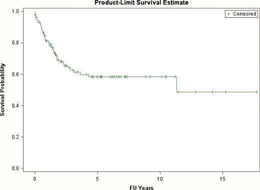
Our cohort consisted of 54 (51%) females and 52 (49%) males. Overall survival at 4 years was 58.3% (Figure 1). The breakdown of subgroups according to the WHO 2008 Classification is outlined in Table 1.
Classification of 106 cases of pediatric AML
| Classification . | No. of pts . | % of total (106) . |
|---|---|---|
| AML with recurrent genetic abnormalities | 52 | 49 |
| AML with t(8;21)(q22;q22); RUNX1-RUNX1T1 | 15 | 14 |
| AML with inv(16)(p13.1q22) or t(16;16)(p13.1;q22);CBFB-MYH11 | 3 | 3 |
| Acute promyelocytic leukemia with t(15;17)(q22;q12);PML-RARA | 10 | 9 |
| AML with t(9;11)(p22;q23);MLLT3-MLL | 8 | 8 |
| AML with t(6;9)(p23;q34); DEK-NUP214 | 2 | 2 |
| AML with inv(3)(q21q26.2) or t(3;3)(q21;q26.2); PRN1-EVI1 | 0 | 0 |
| AML (megakaryoblastic) with t(1;22)(p13;q13); RBM15-MKL1 | 4 | 4 |
| Provisional entity: AML with mutated NPM1/FLT-3 wild type | 7 | 7 |
| Provisional entity: AML with mutated NPM1/FLT-3 ITD | 2 | 2 |
| Provisional entity: AML with mutated CEBPA | 1 | 1 |
| AML with myelodysplasia-related changes | 12 | 11 |
| Therapy-related myeloid neoplasms | 11 | 10 |
| AML NOS | 17 | 16 |
| AML with minimal differentiation | 0 | 0 |
| AML without maturation | 2 | 2 |
| AML with maturation | 0 | 0 |
| Acute myelomonocytic leukemia | 2 | 2 |
| Acute monoblastic /moncytic leukemia | 2 | 2 |
| Acute monoblastic/monocytic leukemia with t(v;11q23) | 6 | 6 |
| Acute erythroid leukemia, with t(v;11q23) | 1 | 1 |
| Acute megakaryoblastic leukemia | 4 | 4 |
| Acute leukemia of ambiguous lineage | 1 | 1 |
| Myeloid sarcoma, not otherwise classified | 1 | 1 |
| Myeloid leukemia associated with Down Syndrome | 12 | 11 |
Overall Survival of Pediatric AML at Lucile Packard ChildrenÕs Hospital
Overall Survival of Pediatric AML at Lucile Packard ChildrenÕs Hospital
Our results vary from the adult literature, reflecting the increased prevalence of AML with recurrent genetic abnormalities, with some categories occurring almost exclusively in children, such as t (1;22) and t(9;11). Data from cases without one of the seven recurrent cytogenetic abnormalities shows a similar frequency to that seen other pediatric series (Manola. Euro J Haematol 83 p.391 (2009)). Twelve cases (11%) fall under the diagnosis of AML with myelodysplasia related changes, the majority of which share an MDS-related karyotype. This category was previously considered rare in children. Two cases of AML with mutated NPM1 also met diagnostic criteria for the AML-MRC category, including one with multilineage dysplasia, and one with a history of prior MDS. Cases of AML with variant 11q23 were nearly as common as those with the t(9;11). Cases of biphenotypic leukemia were rare (less than 1%), reflecting the more stringent criteria now utilized. Ten percent of cases are therapy-related AML, three times higher than the adult cohort at Stanford (Weinberg et al. Blood 113 p. 1906(2009)). This highlights a serious issue facing pediatric oncologists and their patients.
Our pediatric retrospective cohort highlights that important differences exist between adult and pediatric AML. When compared with adults, recurrent genetic abnormalities are more frequently represented as well as treatment related AML. The data also indicate that morphology with myelodysplastic changes is much more frequent than previously thought.
KLD and AHM contributed equally to this work.
No relevant conflicts of interest to declare.
Author notes
Asterisk with author names denotes non-ASH members.


This feature is available to Subscribers Only
Sign In or Create an Account Close Modal