Abstract
Conjugation of TLR agonists to protein or peptide antigens has been demonstrated in many studies to be an effective vaccine formula in inducing cellular immunity. However, the molecular and cellular mediators involved in TLR-induced immune responses have not been carefully examined. In this study, we identify Type I IFN and IL-12 as critical mediators of cross-priming induced by a TLR7 agonist-antigen conjugate. We demonstrate that TLR7-driven cross-priming requires both Type I IFN and IL-12. Signaling through the IFN-αβR was required for the timely recruitment and accumulation of activated dendritic cells in the draining lymph nodes. Although IL-12 was indispensable during cross-priming, it did not regulate DC function. Therefore, the codependency for these 2 cytokines during TLR7-induced cross-priming is the result of their divergent effects on different cell-types. Furthermore, although dermal and CD8α+ DCs were able to cross-prime CD8+ T cells, Langerhans cells were unexpectedly found to potently cross-present antigen and support CD8+ T-cell expansion, both in vitro and in vivo. Collectively, the data show that a TLR7 agonist-antigen conjugate elicits CD8+ T-cell responses by the coordinated recruitment and activation of both tissue-derived and lymphoid organ-resident DC subsets through a Type I IFN and IL-12 codependent mechanism.
Introduction
Activation of the innate immune system is a prerequisite to initiating adaptive immune responses. A major pathway eliciting these responses is the recognition of foreign bodies through Toll-like receptors (TLR), which results in the activation of antigen presenting cells (APC) and the production of a variety of pro-inflammatory mediators.1 Previously, we showed that conjugation of a synthetic agonist targeting TLR7 to protein antigens results in a highly immunogenic vaccine that potently generates protective CD8+ T-cell responses.2 TLR7 is an intracellular receptor that recognizes single-stranded RNA molecules and detects RNA viruses such as the influenza virus. Stimulation of TLR7 has been shown in both mice and humans to result in vigorous production of multiple pro-inflammatory cytokines, including Type I IFN and IL-12.3 The induction of Type I IFN and IL-12 is of particular interest given abundant evidence in the literature establishing these 2 cytokines as critical mediators of CD8+ T-cell activation.4,5
Type I IFN comprises a group of various IFN proteins, notably IFNα and IFNβ. IFNs are induced primarily during viral infections and have been shown to promote natural killer (NK), Type I helper T-cell (Th1), and CTL responses,4 which are critical to combat viral infections through the elimination of virus-infected cells. Similarly, IL-12 also promotes the development of Th1 and CTL-mediated immunity.6-8 However, the production of IL-12 is primarily associated with bacterial and parasitic infections.7,9 The role of IL-12 during viral infections remains unclear as some reports indicate that CD8+ T-cell responses elicited by most viruses are IL-12 independent.6,10 Furthermore, previous studies have shown that the presence of Type I IFN can actively suppress the production of IL-12 during viral infections,8 which suggest an antagonistic role of Type I IFN in the induction of IL-12. Indeed, CD8+ T-cell responses have been observed when IL-12 production is rescued through the blockade of Type I IFN.8 That said, some viruses, such as HSV-2 and MCMV, elicit the production of both Type IFN and IL-12.8,11,12 Therefore, the nature of the relationship between these 2 cytokines is more complex than has been reported thus far and the mechanisms by which they coordinate cellular immune responses remain unknown.
In this study we examined what roles Type I IFN and IL-12 play during a TLR7-induced CD8+ T-cell response generated in the presence of both cytokines. Using a TLR7 agonist-protein conjugate, we demonstrate that cross-priming of CD8+ T cells requires both Type I IFN and IL-12 for the purposes of DC and T-cell activation, respectively. Further, Type I IFN regulated the recruitment and accumulation of Langerhans cells (LC) and CD8α+ DCs in the draining lymph nodes (dLN) and elicited cross-presentation from both subsets. Collectively, our data suggest that TLR7 mediates cross-priming of CD8+ T cells through regulation of recruitment and cross-presentation by both tissue-derived and lymphoid organ-resident DC subsets in a Type I IFN-dependent manner.
Methods
Mice and immunizations
C57BL/6, C57BL/6 CD45.1+ (SJL), and IL-12 Rβ1−/− mice were obtained from The Jackson Laboratory or bred at National Jewish Health. TLR7−/− mice were provided by Dr Richard Flavell (Yale University School of Medicine, New Haven, CT), MyD88−/− mice were a gift from Dr Doug Golenbock (University of Massachusetts Medical School, Worcester, MA) and Type I IFN receptor−/− mice were provided by Dr Michel Aguet (ISREC, Lausanne, Switzerland) and backcrossed to B6 for at least 7 generations. All animal studies were approved beforehand by the National Jewish Health IACUC. Mice were immunized subcutaneously in the lower/upper flanks or in the footpad with the conjugated form of whole ovalbumin protein (OVA) that had been covalently linked to the TLR7 agonist, 3M012 (OVA-3M012). OVA was purchased from Sigma-Aldrich and any contaminating endotoxins were eliminated using Triton X-114 and SM-2 Bio-Beads (Bio-Rad Labs) as previously described.13 In experiments involving IL-12p70 blockade, 500 μg of anti–IL-12 (C17.8) antibody was administered intraperitonealy twice, each one at 24 and 2 hours before immunization. The TLR7 agonist, 3M012, was obtained through material transfer agreements with 3M Pharmaceuticals.
Primary and secondary CD8+ T-cell responses
Mice were bled either 7 days (primary) or 5 days (boost) following injections and the PBLs were then stained with PE-conjugated H-2Kb tetramers loaded with the OVA peptide, SIINFEKL, for 90 minutes at 37°C. OVA-specific CD8+ T cells were identified by gating on CD8+ CD4− B220− events, then gating on tetramer staining events that were positive for the activation marker CD44. Boost injections were administered 30-60 days after the primary injection.
Kinetics of antigen uptake, migration, and activation
Mice were immunized in the footpad with 5 μg of the Alexa-488–labeled OVA-TLR7a conjugate. At the indicated time points after immunization, DCs were isolated from draining LNs using Collagenase D and DNase as previously described.3 The uptake of antigen by DCs was determined by examining the frequency of CD11chigh events (pDCa-1− and B220−) that were positive for Alexa-488 OVA. The magnitude of uptake on a per cell basis was determined by measuring the mean fluorescence intensity of Alexa-488 in CD11chigh OVA+ events.
Adoptive transfer of OTI T cells
OTI transgenic T cells were isolated from spleen cell suspensions by staining the irrelevant cells with PE-coupled anti-CD4, CD11b, CD11c, CD19, DX5, NK1.1, IA/IE mAbs. These cells were then removed by adding anti-PE microbeads (Miltenyi Biotec) and passing the samples through a magnetic column. The negative (flow-through) fraction was collected and checked for purity by flow cytometry. To mimic the endogenous precursor frequency of OVA-specific CD8+ T cells, only 500 of the purified OTI T cells were transferred into recipient mice. The cells were resuspended in PBS and injected intravenously into B6, IL-12Rβ1−/− or IFN-αβR−/− mice. Three days after cell transfer, the recipient mice were immunized with the OVA-TLR7a conjugate, killed 7 days later, and harvested the blood and spleen to stain with tetramer as described above. The transferred OTI T cells and the recipient cells were distinguished based on the expression of the congenic markers CD45.1 and CD45.2, respectively.
Generation of IL-12Rβ1−/− and IFN-αβR−/− bone marrow chimeras
Single-cell suspensions of bone marrow were prepared from mice treated 4 days prior with Fluorouracil. Cells from B6 mice were combined with equal numbers of cells from either IL-12Rβ1−/− or IFN-αβR−/− mice. A total of 2.5 × 10⁵ bone marrow cells from each mouse type were combined and injected intravenously into lethally irradiated B6 mice (900 rads). Hematopoietic chimerism was determined by staining blood samples with anti-CD45.1 and CD45.2 mAbs and analyzing by flow cytometry.
Cross-priming and cross-presentation assays
DCs were isolated using a magnetic bead enrichment method with anti-CD11c (N418) PE mAb and anti-PE microbeads (Miltenyi Biotec) followed by an additional sort through the MoFlow cell sorter. Purity of the sorted fractions was assessed by flow cytometry and confirmed to be ∼ 80%-90%. In experiments where multiple DC subsets were sorted, the CD11c positive fraction after the magnetic bead enrichment process was stained with mAbs specific for the various surface markers outlined in Figure 5A and supplemental Figure 3, available on the Blood Web site (see the Supplemental Materials link at the top of the online article), then sorted on the MoFlow cell sorter. Purified DCs were incubated at the indicated cell numbers with one of the following: (1) in experiments measuring the ability of sorted DCs to cross-prime naive CD8+ T cells, DCs were coincubated with 1.0 × 10⁴ purified and CFSE-labeled OTI T cells, or (2) in experiments measuring the level of antigen cross-presentation directly ex vivo, DCs were coincubated with 5.0 × 10⁵ OTI effector T cells purified from a 5-day stimulation culture. In the first case, degree of CFSE dilution in OTI T cells was analyzed after 3 days in culture with the sorted DCs. In the second case, the magnitude of IFNγ production in the effector OTI T cells after 4-6 hours in culture with the sorted DCs was assessed and used as a measure of the level of antigen cross-presentation by DCs. This was quantified as percent maximum IFNγ response, which was calculated based on the percentage of the maximal OTI IFNγ response (achieved by SIINFEKL-pulsed DCs) that is induced by the sorted DCs.
Statistical analyses
Statistical analyses were performed in Prism Version 4.0 (GraphPad Software Inc) using Student 2-tailed t tests. Asterisk symbols (*) were used to mark the statistical significance of a given difference in sample means and summarize the P value in the following manner: *P < .05, **P < .005, and ***P < .0005. Unless otherwise indicated, shown values of bar graphs are the mean ± SEM.
Results
TLR7-driven expansion of CD8+ T cells is codependent on Type I IFN and IL-12
We previously demonstrated the potency by which a TLR7 agonist-antigen conjugate generates both CD4+ and CD8+ T-cell responses.14 More recently, we examined key differences between the conjugated and free TLR7 agonist in their ability to promote CD8+ T-cell responses.2 Given that stimulation of DCs using a TLR7 agonist leads to a rapid production of both Type I IFN and IL-12,3 we focused on the potential roles these 2 major inflammatory mediators may have on cross-priming. We immunized IFN-αβR–deficient mice or mice treated with IL-12 blocking antibodies using a conjugated complex of TLR7 agonist linked to the ovalbumin protein (OVA-TLR7a) as previously described.2,14 In IFN-αβR−/− mice, the OVA-TLR7a conjugate failed to generate any detectable CD8+ T-cell responses (Figure 1A). Interestingly, CD8+ T-cell responses were also abrogated in IL-12 Rβ1−/− (data not shown) or IL-12–blocked mice (Figure 1A). Thus, OVA-TLR7a conjugate-induced CD8+ T-cell responses are codependent on Type I IFN and IL-12. This codependency is in sharp contrast to many studies of viral infections which show an antagonistic relationship between Type I IFN and IL-12 in vivo.8 In the context of a TLR7-driven CD8+ T-cell response, however, both cytokines appear to play critical and nonredundant roles.
TLR7-driven expansion of CD8+ T cells is codependent on Type I IFN and IL-12. (A) B6 and IFN-αβR−/− mice were either untreated or injected with blocking anti–IL-12p40 mAb (C17.8) at both 24 and 2 hours before immunization with 10 μg of the OVA-TLR7a conjugate. (B) B6 mice with or without IL-12 blockade and IFN-αβR−/− mice were immunized with 10 μg of the OVA-TLR7a conjugate in the footpad. A boost injection of the conjugate was given using the same dose at 30 days post the primary injection. Blood was obtained from the tail vein on (A) day 7 post primary and (B) day 5 post boost injections and stained for OVA-specific CD8+ T cells using peptide loaded H-2Kb tetramers. The data shown are representative of 3 independent experiments. The shown dot plots are representative of 3 mice per mouse group in each experiment. Graphed values of the frequency of OVA-specific CD8+ T cells are expressed as the mean ± SEM. Statistical analyses (*) were performed as described in “Statistical analyses.”
TLR7-driven expansion of CD8+ T cells is codependent on Type I IFN and IL-12. (A) B6 and IFN-αβR−/− mice were either untreated or injected with blocking anti–IL-12p40 mAb (C17.8) at both 24 and 2 hours before immunization with 10 μg of the OVA-TLR7a conjugate. (B) B6 mice with or without IL-12 blockade and IFN-αβR−/− mice were immunized with 10 μg of the OVA-TLR7a conjugate in the footpad. A boost injection of the conjugate was given using the same dose at 30 days post the primary injection. Blood was obtained from the tail vein on (A) day 7 post primary and (B) day 5 post boost injections and stained for OVA-specific CD8+ T cells using peptide loaded H-2Kb tetramers. The data shown are representative of 3 independent experiments. The shown dot plots are representative of 3 mice per mouse group in each experiment. Graphed values of the frequency of OVA-specific CD8+ T cells are expressed as the mean ± SEM. Statistical analyses (*) were performed as described in “Statistical analyses.”
Direct stimulation of T cells through Type I IFN receptor is not required.
Type I IFN has recently been shown to have an important role in direct stimulation of CD8+ T cells in response to certain antigenic challenges,15 most notably, in response to vaccination with antigen and recombinant IFNα.4,16 Based on these results, we initially expected that the IFN-dependency of the TLR7-mediated T-cell response was a result of direct IFN stimulation of antigen-specific T cells. However, IFN-αβR–deficient mice were able to mount a readily detectable secondary response to the TLR7 agonist-antigen conjugate (Figure 1B). Further, IFN-αβR+ OTI TCR transgenic cells responded poorly when transferred into a conjugate immunized IFN-αβR−/− host (supplemental Figure 1), indicating that the quantity of the T-cell response is not dictated by IFN-αβR expression on the T cells. In contrast, blockade of IL-12 before both primary and secondary immunizations substantially limited responses to immunization with the conjugate (Figure 1B). Furthermore, in the same OTI T-cell adoptive transfer experiment as described above, the transferred IL-12Rβ1+ OTI T cells exhibited comparable levels of expansion in the IL-12 Rβ1−/− recipients as in the WT hosts (supplemental Figure 1). Taken together, these data suggest that IFN-αβR is not required on CD8+ T cells and IL-12 Rβ1 is not required on DCs. Thus, the codependence of the CD8+ T-cell responses on these 2 cytokines is likely because of their differing roles for stimulation of APC or CD8+ T cells.
Type I IFN is required for efficient accumulation, activation, and cross-presentation by DC in skin-draining lymph nodes
We next sought to clarify how Type I IFN and IL-12 operated in vivo by focusing on DC function and events preceding the activation of CD8+ T cells. To assess the impact of these cytokines on DC function, we fluorescently labeled the OVA-TLR7a conjugate and tracked its uptake, movement, and presentation by DCs in WT B6, IFNαβR−/−, or IL-12–blocked mice.
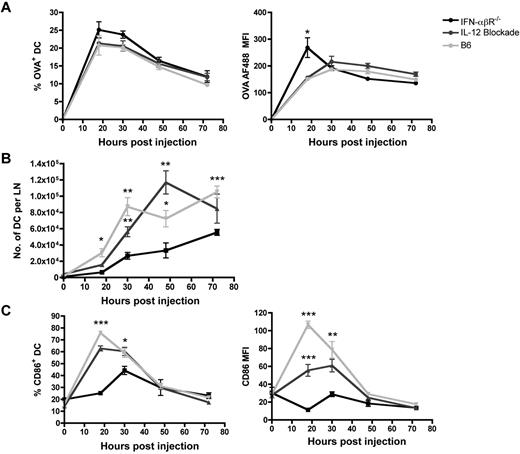
As early as 18 hours post immunization, a significant fraction of DCs had taken up the fluorescently labeled OVA-TLR7a conjugate (Figure 2A). The magnitude of antigen uptake peaked between 18 and 30 hours post immunization and a steady decline in the level of antigen-bearing DCs was observed afterward (Figure 2A). Relatively similar frequencies of antigen-bearing DCs, as well as similar amounts of antigen uptake per DC, were generally found in both IFNαβR−/− and IL-12–blocked mice at all times examined, suggesting that the rate and duration of antigen uptake are not significantly influenced by either Type I IFN or IL-12. It is worth noting that in some experiments, we observed some degree of reduced antigen uptake in the IFNαβR−/− mice (data not shown). This is consistent with recently published results,17 the likelihood of observing this difference probably correlating with the degree to which the TLR7 agonist-antigen conjugate was aggregated. However, the primary T-cell response in the IFNαβR−/− host is always substantially reduced, whether or not any reduction in antigen uptake by the DCs is observed. Thus, the level of antigen uptake does not predict the magnitude of the T-cell response. We conclude from these data that the underlying cause of the reduced T-cell response in the IFNαβR−/− is unlikely to be related to variations in antigen uptake.
Type I IFN is required for efficient accumulation and activation of DCs in the dLN. B6, IFN-αβR−/−, and IL-12–blocked mice were immunized in the footpad with 5 μg of OVA-TLR7a conjugate that had been labeled with the fluorescent dye, Alexa-Fluor 488. At the indicated time points after immunization, popliteal LNs draining the foot were harvested, minced, and digested with collagenase/DNase. The frequency of cells or the mean fluorescence intensity values shown were gated on (A) CD11c+ Alexa-Fluor 488+ cells, (B) CD11c+ cells, (C) or CD11c+ CD86+ cells. The data shown are representative of 2 independent experiments, 3 mice per group. Statistical analyses (*) were performed as described in “Statistical analyses.” The summary of P values, as indicated by (*), denote statistical significance of differences between the means of the WT or IL-12–blocked mouse and the means of IFN-αβR−/− mouse.
Type I IFN is required for efficient accumulation and activation of DCs in the dLN. B6, IFN-αβR−/−, and IL-12–blocked mice were immunized in the footpad with 5 μg of OVA-TLR7a conjugate that had been labeled with the fluorescent dye, Alexa-Fluor 488. At the indicated time points after immunization, popliteal LNs draining the foot were harvested, minced, and digested with collagenase/DNase. The frequency of cells or the mean fluorescence intensity values shown were gated on (A) CD11c+ Alexa-Fluor 488+ cells, (B) CD11c+ cells, (C) or CD11c+ CD86+ cells. The data shown are representative of 2 independent experiments, 3 mice per group. Statistical analyses (*) were performed as described in “Statistical analyses.” The summary of P values, as indicated by (*), denote statistical significance of differences between the means of the WT or IL-12–blocked mouse and the means of IFN-αβR−/− mouse.
In contrast, the absence of Type I IFN did significantly affect the accumulation of antigen-bearing DCs in the dLN (Figure 2B). IFN-αβR−/− mice lacked the rapid accumulation of DCs over the first 24 hours seen in both the WT and IL-12–blocked mice, resulting in an ∼ 4-fold lower accumulation of DC in the IFN-αβR−/−. Therefore, the ability of the OVA-TLR7a conjugate to induce rapid accumulation of DCs in the dLN appeared to be mediated by a Type I IFN-dependent, but an IL-12–independent mechanism.
Similarly, a differential requirement of Type I IFN and IL-12 was observed for DC activation in response to the TLR7 stimulus. Only 30% of the DC in IFN-αβR−/− mice up-regulated the activation marker CD86 18 hours after immunization, compared with nearly 80% of the DC in WT and IL-12–blocked mice (Figure 2C). Furthermore, IFN-αβR−/− DCs displayed a > 10-fold reduction in the surface level of CD86 expression (mean fluorescent intensity-MFI) relative to the levels in WT or IL-12–blocked DCs (Figure 2C). Similar results were obtained examining CD80 or MHC class II (I-Ab) as an alternative indicator of DC activation (data not shown). Taken together, our data indicate that Type I IFNs are crucial factors regulating the accumulation of DC in the dLN and their maturation into activated APC during a TLR7-driven response.
TLR7-induced cross-presentation is mediated by Type I IFN signaling in DCs
A key rate-limiting step in DC function is the presentation of exogenously-derived antigens on MHC class I molecules, a process termed cross-presentation. To determine the role of Type I IFN or IL-12 in this process, DCs from immunized mice were isolated and cocultured with in vitro-differentiated effector OTI cells. We then monitored the IFNγ response after 4 hours in culture as a measure of the level of antigen that was being presented by the freshly isolated DC. It is important to note that this assay is not influenced by the context of costimulation or accessory function of the DC and is simply a functional readout as to whether a DC has peptide/MHC complexes on its cell surface.18,19
Both WT DCs and DCs from IL-12–blocked mice were able to efficiently stimulate the effector OTI T cells in a DC-dose dependent manner (Figure 3A-B), indicating that IL-12 has no impact on cross-presentation. The low yield of DCs from the IFN-αβR−/− mice (Figure 2B) precluded our ability to perform a dose response with increasing numbers of DCs for that strain. We therefore analyzed the IFNγ response of the OTI T cells incubated with the same number of DCs from each host. When normalized back to the maximal response elicited by peptide-pulsed WT DCs, IFN-αβR−/− DCs exhibited an appreciable deficit in their capacity to stimulate OTIs (Figure 3C). Thus, on a per cell basis, IFN-αβR−/− DCs display a substantially reduced ability to present exogenously provided antigen in the context of a TLR7-agonist conjugate. These results suggest that Type I IFNs, but not IL-12, are crucial for cross-presentation by DCs induced by TLR7-mediated signals.
Type I IFN, but not IL-12, is required for cross-presentation. WT, IFN-αβR−/− (A), or IL-12– blocked mice (B,C) were immunized in the footpad with 50 μg OVA-TLR7a conjugate as outlined in panel A. Twenty-four hours after immunization, popliteal LN were harvested as before, the cell suspensions from each mouse group pooled, and the DC purified by flow sort based on CD11c expression. Sorted DCs were incubated at (C) 1 × 10⁶ cells per well or at (B) the indicated titration of cell numbers (DCs from WT and IL-12–blocked mice) with 0.5 × 10⁶ effector OTI T cells differentiated in 5 days of peptide-pulsed culture. The cells were coincubated for 4-6 hours in the presence of brefeldin A and then stained for intracellular IFNγ. Production of IFNγ by OTI T cells was assessed by gating on expression of the congenic marker CD45.1. The level of IFNγ response by the OTI T cells was expressed as a percent of the maximal production of IFNγ induced by DCs pulsed with the SIINFEKL peptide. The data shown are representative of 2 independent experiments.
Type I IFN, but not IL-12, is required for cross-presentation. WT, IFN-αβR−/− (A), or IL-12– blocked mice (B,C) were immunized in the footpad with 50 μg OVA-TLR7a conjugate as outlined in panel A. Twenty-four hours after immunization, popliteal LN were harvested as before, the cell suspensions from each mouse group pooled, and the DC purified by flow sort based on CD11c expression. Sorted DCs were incubated at (C) 1 × 10⁶ cells per well or at (B) the indicated titration of cell numbers (DCs from WT and IL-12–blocked mice) with 0.5 × 10⁶ effector OTI T cells differentiated in 5 days of peptide-pulsed culture. The cells were coincubated for 4-6 hours in the presence of brefeldin A and then stained for intracellular IFNγ. Production of IFNγ by OTI T cells was assessed by gating on expression of the congenic marker CD45.1. The level of IFNγ response by the OTI T cells was expressed as a percent of the maximal production of IFNγ induced by DCs pulsed with the SIINFEKL peptide. The data shown are representative of 2 independent experiments.
TLR7 activation engages multiple DC subsets to cross-prime CD8+ T cells ex vivo.
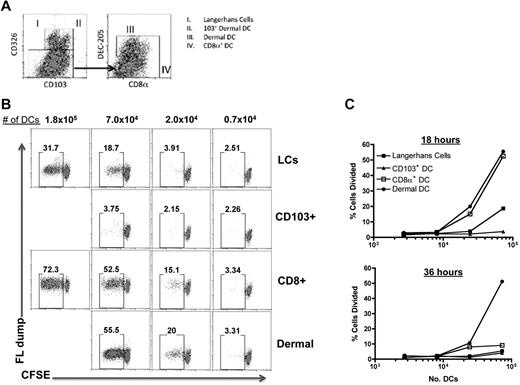
DCs in skin-dLN can be divided into at least 6 different populations: Langerhans cells, dermal DCs, CD103+ Langerin+ Dermal DCs, LN-resident CD8α+ DC, LN-resident CD11b+ DC, and plasmacytoid DC. While the capacity of LN-resident CD8α+ DC to mediate cross-priming is well-established, evidence that other DC subsets are capable of cross-priming are beginning to emerge.20 We therefore next examined the ability of individual DC subsets to prime naive CD8+ T cells ex vivo. DC subsets were purified based on the expression of CD11c and a unique combination of CD103, CD326, CD8α, CD11b, and DEC-205 (Figure 4A): LC were CD103−CD326+DEC-205highCD8α−CD11b+, CD103+ dermal DC were CD103+CD326intermediateDEC-205highCD8α−CD11b+,21,22 LN-resident CD8α+ DC were CD326−DEC-205intermediateCD8α+CD11b−, and LN-resident CD11b+ DC were CD326−DEC-205−CD8α−CD11bhigh.21,23,24 We confirmed our gating strategy and found that indeed the DEC-205high population of cells were Langerin+. A small number of the dermal DCs were positive for langerin expression (supplemental Figure 3), consistent with the known expression of langerin by the CD103+ dermal DCs.21 Furthermore, all migratory DC subsets (LC, CD103+ langerin+ dermal DC, dermal DCs) were MHC class IIhigh (supplemental Figure 3). Using this strategy, we sorted for the major migratory DC subsets as well as the CD8α+ DCs and monitored their ability to induce naive OTI CD8+ T-cell proliferation in vitro. On coculture, we found that the LN-resident CD8α+ DCs and dermal DCs were the most efficient DC subsets to induce CD8+ T-cell proliferation (Figure 4B-C). Curiously, we found that the CD103+ dermal DC subset did not exhibit this function.20 Perhaps even more surprising however, LCs stimulated a significant level of CD8+ T-cell proliferation (Figure 4B-C), being only ∼ 3-fold less potent than CD8α+ or dermal DCs. Given the substantial literature regarding the inability of LCs to initiate CD8+ T-cell responses,20,25-28 these data suggest a novel capacity for LCs to mediating cross-priming of CD8+ T cells in response to a TLR7 stimulus.
TLR7a-conjugate vaccination engages multiple DC subsets to cross-prime CD8+ T cells. WT mice were subcutaneously immunized with 50 μg of the OVA-TLR7a conjugate, and the dLNs were harvested 18 or 36 hours after immunization. DCs were isolated as described above, and the indicated DC subsets were sorted using the MoFlow cell sorter on the basis of surface marker expression as outlined in (A). Sorted cells were then cocultured either at the indicated titration of cell numbers (B,C) with 1.0 × 10⁴ purified and CFSE-labeled OTI T cells. After 3 days, OTI T cells were harvested from the culture and the dilution of CFSE was assessed by flow cytometry, gating on CD8+, CD3+, and B220- cells. The data shown are representative of 2 independent experiments.
TLR7a-conjugate vaccination engages multiple DC subsets to cross-prime CD8+ T cells. WT mice were subcutaneously immunized with 50 μg of the OVA-TLR7a conjugate, and the dLNs were harvested 18 or 36 hours after immunization. DCs were isolated as described above, and the indicated DC subsets were sorted using the MoFlow cell sorter on the basis of surface marker expression as outlined in (A). Sorted cells were then cocultured either at the indicated titration of cell numbers (B,C) with 1.0 × 10⁴ purified and CFSE-labeled OTI T cells. After 3 days, OTI T cells were harvested from the culture and the dilution of CFSE was assessed by flow cytometry, gating on CD8+, CD3+, and B220- cells. The data shown are representative of 2 independent experiments.
Cross-presentation induced by TLR7 is mediated by LCs and CD8α+ DCs
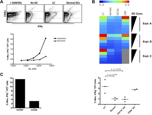
It remained a possibility that the capacity of LCs to elicit CD8+ T-cell proliferation may be an in vitro artifact specific to our unique vaccination method, so we measured each DC subset's presentation of antigen directly ex vivo. Migratory DCs from immunized mice were flow-sorted into either the LC or dermal DC subsets on the basis of the surface markers described previously (supplemental Figure 3). As in Figure 3 above, sorted DCs were coincubated with OTI effector T cells and their production of IFNγ used as a functional readout for antigen presentation (Figure 5A). LCs effectively stimulated the IFNγ production from the OTI cells across a broad range of DC numbers, being approximately 10-fold more potent than the dermal DCs (Figure 5A). This indicates that within 24 hours of immunization, LCs in the dLNs are more potent in cross-presentation of antigen in response to the TLR7 agonist-antigen conjugate than dermal DCs. Similar results were consistently observed in 6 independent experiments. In fact, LCs were, on a per cell basis, on par with the LN-resident CD8α+ DCs and far superior to LN-resident CD11b+ DCs, (Figure 5A-B). The results were the same when we used an alternative sorting approach for LCs based on expression of the cell surface marker CD326 (Ep-CAM).29 The OTI stimulation by the CD326+ cells was dramatically higher than that achieved by the CD326− fraction. This was not because of the activity of CD326+CD103+langerin+ dermal DCs, because subsequent sorting experiments which specifically isolated this subset revealed that CD103+ DCs possessed no capacity to cross-present antigen (Figure 4 and data not shown). Thus, when DCs are examined directly ex vivo, LCs and CD8α+ DCs cross-present antigen in response to TLR7 stimulation.
Cross-presentation induced by TLR7 is mediated by LCs and CD8α+ DCs. B6 mice were immunized in the footpad with 50 μg of the OVA-TLR7a conjugate. Twenty-four hours after immunization, popliteal LN were harvested as before, the cell suspensions from each mouse pooled, and the DCs were purified using a flow sorter (MoFlow) based on CD11c expression and the markers outlined in Figure 5A and supplemental Figure 3. (A) DCs were incubated at the indicated cell number and or (C) at 2.5 × 10⁵ cells per well with 0.5 × 10⁶ effector OTI T cells stimulated in 5 days of peptide-pulsed culture. The cells were coincubated for 4 hours in the presence of brefeldin A and then stained for intracellular IFNγ. Production of IFNγ by OTI T cells was assessed by gating on expression of the congenic marker CD45.1. The level of IFNγ response by the OTI T cells was expressed as a percent of the maximal production of IFNγ induced by DCs pulsed with the SIINFEKL peptide. (B) Sorted DCs were available in limited and varied frequencies across the 4 DC subsets as indicated. To normalize for differing range of cell frequencies and to enable comparison of OTI stimulation by various DC subsets, linear regression curves of the OTI response to titrating numbers of DCs for each subset were determined and the magnitudes of the corresponding OTI IFNγ response were normalized to a unified range of DC numbers. To compare the level of cross-presentation by DCs across 3 independent experiments, each DC subset incubated at a given cell concentration with OTI T cells was assigned a color in a heat map representing relative levels of OTI stimulation by each of the 4 DC subsets indicated. A graphical representation of the magnitudes of OTI IFNγ response is also shown for the highest concentration of DCs used in each experiment. The data shown are representative of (A) 6 and (C) 2 independent experiments. Statistical analyses (*) were performed as described in “Statistical analyses.”
Cross-presentation induced by TLR7 is mediated by LCs and CD8α+ DCs. B6 mice were immunized in the footpad with 50 μg of the OVA-TLR7a conjugate. Twenty-four hours after immunization, popliteal LN were harvested as before, the cell suspensions from each mouse pooled, and the DCs were purified using a flow sorter (MoFlow) based on CD11c expression and the markers outlined in Figure 5A and supplemental Figure 3. (A) DCs were incubated at the indicated cell number and or (C) at 2.5 × 10⁵ cells per well with 0.5 × 10⁶ effector OTI T cells stimulated in 5 days of peptide-pulsed culture. The cells were coincubated for 4 hours in the presence of brefeldin A and then stained for intracellular IFNγ. Production of IFNγ by OTI T cells was assessed by gating on expression of the congenic marker CD45.1. The level of IFNγ response by the OTI T cells was expressed as a percent of the maximal production of IFNγ induced by DCs pulsed with the SIINFEKL peptide. (B) Sorted DCs were available in limited and varied frequencies across the 4 DC subsets as indicated. To normalize for differing range of cell frequencies and to enable comparison of OTI stimulation by various DC subsets, linear regression curves of the OTI response to titrating numbers of DCs for each subset were determined and the magnitudes of the corresponding OTI IFNγ response were normalized to a unified range of DC numbers. To compare the level of cross-presentation by DCs across 3 independent experiments, each DC subset incubated at a given cell concentration with OTI T cells was assigned a color in a heat map representing relative levels of OTI stimulation by each of the 4 DC subsets indicated. A graphical representation of the magnitudes of OTI IFNγ response is also shown for the highest concentration of DCs used in each experiment. The data shown are representative of (A) 6 and (C) 2 independent experiments. Statistical analyses (*) were performed as described in “Statistical analyses.”
Type I IFN is required for efficient recruitment and accumulation of LCs and CD8α+ DCs in skin-draining LNs
Because both CD8α+ DCs and LCs are the most potent cross-presenting DC subsets, and since DC recruitment (Figure 2) and cross-presentation (Figure 3) are dependent on Type I IFN, it stood to reason that the recruitment of these 2 DC subsets may be disproportionally affected in the IFN-αβR−/− hosts. We therefore examined the frequency of individual DC subsets in the dLN of WT, IL-12–blocked, and IFN-αβR−/− mice at various time points post immunization with the OVA-TLR7a conjugate. Although the rate of accumulation in the dLN differed between the DC subsets, all peaked at 30 hours post immunization in WT and IL-12–blocked mice and the numbers remained steady through 72 hours (Figure 6A). However, in IFN-αβR−/− mice, the numbers of LC and CD8α+ DCs in the dLN were dramatically lower than in WT mice (Figure 6A). At 18 hours after immunization, LCs and CD8α+ DCs displayed a > 20-fold decrease in cell frequency, while LN-resident CD11b+ and dermal DC subsets did not exhibit any striking differences in the absence of either Type I IFN or IL-12. Despite its impact on LC and CD8α+ DC recruitment and/or accumulation, neither Type I IFN nor IL-12 dramatically influenced the efficiency of uptake of our fluorescently labeled TLR7-conjugate by any particular DC subset (Figure 6B). As mentioned above (Figure 2), this particular parameter varied (2- to 3-fold at most) depending on the level of aggregation of the conjugate, again consistent with recently published results.17 However, our data reveal that a differential influence on antigen uptake by these DC subsets was not necessary in order to observe a deficit in the subsequent CD8+ T-cell responses. Thus, there is a striking correlation of the sensitivity of LCs and CD8α+ DC subsets to Type I IFN and the capacity of these DC subsets to cross-prime/cross-present (Figures 4–5) and accumulate in the dLN (Figure 6).
Type I IFN is required for efficient recruitment and accumulation of LCs and CD8α+ DCs in skin-draining LNs. B6, IFN-αβR−/−, and IL-12–blocked mice were immunized in the footpad with 5 μg of OVA-TLR7a conjugate labeled with the fluorescent dye, Alexa-Fluor 488. At the indicated time points after immunization, popliteal LN draining the foot were harvested, minced, and digested with collagenase/DNase. Cells were then stained for the various markers shown and identified using the gating strategy outlined in Figure 5A and supplemental Figure 3. The frequency of each DC subset (A) or the proportion of cells bearing the antigen in each DC subset (B) are shown. The data shown are representative of 2 independent experiments using 3 mice per group. Statistical analyses (*) were performed as described in “Statistical analyses.” The summary of P values, as indicated by (*), denote statistical significance of differences between the means of the WT or IL-12Rβ1−/− mouse and the means of IFN-αβR−/− mouse.
Type I IFN is required for efficient recruitment and accumulation of LCs and CD8α+ DCs in skin-draining LNs. B6, IFN-αβR−/−, and IL-12–blocked mice were immunized in the footpad with 5 μg of OVA-TLR7a conjugate labeled with the fluorescent dye, Alexa-Fluor 488. At the indicated time points after immunization, popliteal LN draining the foot were harvested, minced, and digested with collagenase/DNase. Cells were then stained for the various markers shown and identified using the gating strategy outlined in Figure 5A and supplemental Figure 3. The frequency of each DC subset (A) or the proportion of cells bearing the antigen in each DC subset (B) are shown. The data shown are representative of 2 independent experiments using 3 mice per group. Statistical analyses (*) were performed as described in “Statistical analyses.” The summary of P values, as indicated by (*), denote statistical significance of differences between the means of the WT or IL-12Rβ1−/− mouse and the means of IFN-αβR−/− mouse.
Depletion of LCs in vivo reduces TLR7-induced CD8+ T-cell expansion
We next sought to confirm these observations in vivo through the use of mice deficient in LCs or CD8α+ DCs. To explore the role of the CD8α+ DCs, we used the Batf3−/− hosts, which are deficient in both CD8α+ DCs as well as the dermal CD103+ DCs.30 To explore the role of the LCs in vivo, we used Lang-DTR mice, which, on administration of diptheria toxin (DT), results in the deletion of Langerin-expressing LCs and CD103+ DCs.21,22,31 The OVA-TLR7a conjugate vaccine was administered to the Batf3−/− mice and the Lang-DTR mice with or without prior DT injection, and the primary CD8+ T-cell response was measured as described above.
As previously observed, OVA-specific CD8+ T cells were evident in WT controls in both the peripheral blood (Figure 7A) and dLN (Figure 7B). Somewhat surprisingly, CD8+ T-cell responses were detectable in both the LC-depleted (Lang-DTR + DT) and the CD8α+ DC-deficient (Batf3−/−) hosts. In particular, the percentage of OVA-specific CD8+ T cells in the blood (Figure 7A) or the dLN (Figure 7B) of the Batf3−/− hosts was not statistically different from that seen in the WT host. In contrast, the response in the DT treated Lang-DTR mice was on average 2- to 4-fold lower than that found in the WT or non-DT treated Lang-DTR hosts (Figure 7A-B). Though variable, examination of the results across multiple experiments confirmed a statistically significant reduction in the CD8+ T-cell expansion after LC depletion (Figure 7C). Because both the Batf3−/− and the DT-treated Lang-DTR mice also have a deficiency in the CD103+ dermal DCs,21,22,31 these data indicate a role for LCs, in vivo, in the generation of CD8+ T cells from the TLR7 agonist-antigen conjugate. The deletion of LCs was confirmed by FACS analysis staining for the presence or absence of CD11c+CD326+ cells (not shown). In addition, the Batf3−/− mice were confirmed as deficient in CD8α+ DCs both by FACS staining (not shown) and by their inability to respond to a different form of whole protein vaccination (polyIC + antiCD4032 ; supplemental Figure 4). Collectively the data are consistent with the interpretation that the loss of the LCs confers a larger deficit in the responding CD8+ T cells than the loss of the CD8α+ DCs. These in vivo data are thus consistent with our in vitro findings that a TLR7 agonist based vaccine is unique in its capacity to effectively recruit LCs to cross-present antigen to CD8+ T cells.
Depletion of LC reduces TLR7-induced CD8+ T-cell expansion in vivo. Lang-DTR mice or Batf3−/− mice were immunized in the footpad with 20 μg of the OVA-TLR7a conjugate. Lang-DTR mice were left untreated (no DT) or treated with 1μg DT injected intraperitoneally 24 hours before immunization (+DT). On day 6 after immunization, blood obtained from the tail vein and cell suspensions from the popliteal LN were stained for OVA-specific CD8+ T cells using peptide loaded H-2Kb tetramers as before. (A) Representative dot plots (showing live, CD8+, B220- events) of tetramer staining from the peripheral blood from the indicated hosts. (B) Total number of tetramer+CD8+ T cells in the draining popliteal LN from the indicated hosts. Panels A and B are representative of 3 independent experiments. Statistical significance (Student t test), to the degree indicated by the asterisks (see “Statistical analyses”), was observed between the DT treated Lang-DTR mice and both nonDT treated controls and BatF3−/− hosts. (C) Normalized data from 2 independent experiments, using 3-7 mice per group per experiment. The number of tetramer+ cells in individual mice was divided by the average number of tetramer+CD8+ cells derived from the WT (B6 or nonDT treated Lang-DTR) mice in the given experiment. This percent of WT control was then plotted for both strains of genetically modified mice as well as for the WT mice to indicate the variability in the responses across strains and across experiments. Statistical significance (Student t test) was observed between the DT treated Lang-DTR and both non-DT–treated controls and Batf3−/− hosts.
Depletion of LC reduces TLR7-induced CD8+ T-cell expansion in vivo. Lang-DTR mice or Batf3−/− mice were immunized in the footpad with 20 μg of the OVA-TLR7a conjugate. Lang-DTR mice were left untreated (no DT) or treated with 1μg DT injected intraperitoneally 24 hours before immunization (+DT). On day 6 after immunization, blood obtained from the tail vein and cell suspensions from the popliteal LN were stained for OVA-specific CD8+ T cells using peptide loaded H-2Kb tetramers as before. (A) Representative dot plots (showing live, CD8+, B220- events) of tetramer staining from the peripheral blood from the indicated hosts. (B) Total number of tetramer+CD8+ T cells in the draining popliteal LN from the indicated hosts. Panels A and B are representative of 3 independent experiments. Statistical significance (Student t test), to the degree indicated by the asterisks (see “Statistical analyses”), was observed between the DT treated Lang-DTR mice and both nonDT treated controls and BatF3−/− hosts. (C) Normalized data from 2 independent experiments, using 3-7 mice per group per experiment. The number of tetramer+ cells in individual mice was divided by the average number of tetramer+CD8+ cells derived from the WT (B6 or nonDT treated Lang-DTR) mice in the given experiment. This percent of WT control was then plotted for both strains of genetically modified mice as well as for the WT mice to indicate the variability in the responses across strains and across experiments. Statistical significance (Student t test) was observed between the DT treated Lang-DTR and both non-DT–treated controls and Batf3−/− hosts.
Discussion
In this study, we characterized the mechanism of a protein-conjugated TLR7 agonist in inducing cellular immunity. Our data identify 2 major innate cytokines, Type I IFN and IL-12, as critical factors mediating this process. Although these 2 cytokines have long been associated with the promotion of Th1 and CTL responses, our study reveals a novel codependent mechanism by which TLR7 signaling mediates cross-priming. Imidazoquinoline-based TLR7 agonists such as we use here induce potent IFN production exclusively from the pDC subset.33 Similarly, we and others3,34 have shown that imidazoquinolines induce IL-12 production primarily from the CD11b+ DC subset. Our present data demonstrate that both of these cytokines are necessary to induce cross-priming from still other DC subsets: CD8α+ DC, dermal DC, and LCs. Thus, the data suggest that the coordinated response of at least 4 separate DC subsets is necessary for the induction of productive CD8+ T-cell responses from a TLR7 agonist. Several studies have reported similarity between the activity of TLR7 agonists in mouse and TLR7/8 agonists in human with regards to eliciting Type I IFN and IL-12 from pDCs and mDC subsets, respectively.35 Furthermore, the human CD141+ DC subset was recently identified as the equivalent to the mouse CD8α+ DC subset, both with respect to TLR expression and functional capacity to cross present antigen to CD8+ T cells.36 These data, in conjunction with our findings here, increase our confidence that the clinical use of this TLR7/8 agonist-conjugate formulation may be as successful in the clinic as it is in mice.
We found no appreciable role for IL-12 in any aspect of DC activation or antigen presentation. Our results, albeit indirect, suggest that the likely role for IL-12 is for a direct stimulation of CD8+ T cells, a prediction with precedent in the literature.5,37 In contrast, we found that Type I IFN was critical for the rapid recruitment and accumulation of DC in dLN after immunization with the OVA-TLR7a conjugate. Importantly, LCs were the only migratory DC subset in which its migratory activity was regulated by Type I IFN (Figure 6A). In further support of our findings, imiquimod, a more widely used form of TLR7 agonist, was previously shown to enhance the migration of LC when applied topically on the surface of the skin.38 However, this observation detected only marginal differences in cell number and was based on the expression of a single marker, CD1a, to identify LC. With the recent discovery of Langerin+ dermal DC joining epidermal LC and dermal DC on the list of migratory DC subsets,21,22,31 a combinatorial use of multiple surface markers is required to carefully distinguish one particular subset from another.
In addition to the sensitivity of LC to Type I IFN, we found LCs that migrated to the dLN also cross-presented the conjugated protein antigen to CD8+ T cells (Figures 4,5,7). LCs are known for their ability to capture extracellular materials from the skin and transfer antigens to the dLN. However, whether LCs play a direct role in T-cell priming is a debated subject. Several studies have shown that LCs present skin-associated antigens which induces tolerogenic effects under steady state and noninflammatory conditions.39,40 Other studies have shown their dispensability during CTL priming in response to HSV infections in the skin or direct presentation of skin-associated antigens.25-27 Thus, the question of whether LC directly cross-present exogenously derived antigens remains unclear and likely related to the model of infection or precise method of stimulation. The disparity on the role of LC in our study and previous findings may be because of a combination of differences in the experimental methods and models used. For example, some studies used LC from skin explants that use cytokines and chemokines to induce migration and maturation in vitro, while others used the assumed radioresistant property of LC to examine their role using bone-marrow chimeric mice. In addition, there were differences in the type of immunogen used to trigger DC activation, which could vary in the type of cells targeted for activation, migration, and antigen presentation. Our cross-presentation assay uses DCs that have undergone all their maturation, migration, and localization in vivo, and only examines their function ex vivo in a short assay that detects whether a particular DC subset is cross-presenting antigen at the time the cells were harvested.
This contrasts sharply with the standard cross-priming assay where the sorted DC subsets are incubated with naive T cells for multiple days in vitro, which may result in the capacity of an antigen bearing DC to cross-present antigen in a fashion divergent from its in vivo function. Factors such as DC survival, the capacity to express specific costimulatory markers or cytokines, and/or the capacity to induce cross-presentation pathways may well be unduly influenced by whatever in vitro conditions are used. For example, we and others have observed that DCs in vitro induce the expression of the TNF ligand CD70 much more readily than DCs in vivo.32,41,42 Given the potency with which CD70 augments T-cell responses,32,41-43 particularly with respect to T-cell survival,44 the success or failure of any specific DC subset to survive and express this one TNF ligand in vitro may impact the conclusion that can be made from the assay. Indeed, the fact that the dermal DC subset was one of the more potent subsets at cross-priming in the standard assay, but was ineffective in our cross-presentation assay may well be related to one or more of the concerns listed above. While the specific causes underlying this difference with regard to dermal DCs remain unclear, the fact that we observed activity from the LCs in both the standard cross-priming assay and in our cross-presentation assay further lends confidence to our conclusion that the TLR7a-antigen conjugate may be unique in its ability to effectively mobilize LCs to participate in mediating the subsequent CD8+ T-cell response. Our observations in Batf3−/− and DT-treated Lang-DTR hosts (Figure 7) are consistent with this prediction as well. Indeed, the statistical significance with which the CD8+ T-cell response is reduced, albeit modestly, in the DT-treated Lang-DTR mice compared with the Batf3−/− mice indicates that, if anything, the LCs may play a larger role in cross priming CD8+ T-cell responses in vivo in response to the TLR7 agonist-antigen conjugate than the CD8α+ DCs.
It is interesting to note that the efficacy of the TLR7 agonist in the clinic (Aldara) has largely been confined to topical administration of the compound, such as in basal cell carcinoma, carcinoma in situ, and actinic keratosis.45,46 It is temping to speculate that the success of Aldara against these conditions is related to the unique ability of TLR7 to enlist LCs into the development of cellular immunity. Information on the distribution of TLR7 expression among the various DC subsets is not yet complete. In particular, whether any of the migratory DC subsets derived from the skin express TLR7, particularly after activation via Type I IFN, is unknown. Previous studies have detected TLR7 expression almost exclusively in pDC and CD8α− DCs in the mouse.34 However, the CD8α− DC subsets encompass LN-resident CD11b+ DC and probably at least some of the migratory DC subsets in skin-dLN. Furthermore, all previous studies on TLR expression by various DC subsets were performed on steady state DCs isolated from unprimed mice, and there is no available data on the TLR expression profile of DC subsets after vaccination. This is particularly of interest for a TLR7 agonist-based vaccine, as there is precedence for the induction of TLR7 expression in several cell types, such as DCs and even keratinocytes, after Type I IFN stimulation.47 Indeed, we have observed a dramatic up-regulation of TLR7 expression in the dLN of mice injected subcutaneously with recombinant IFNα (J.Z.O. and R.M.K., unpublished results, June 2010). Given published results regarding a requirement for the TLR stimulus and the antigen to be localized to the same endosomal compartment to achieve effective presentation of the antigen,48-50 it is tempting to speculate that IFNα induces TLR7 expression in the LCs which is then used by the conjugate vaccine to induce cross-presentation. This possibility is currently under investigation.
The use of TLR agonists to induce adaptive responses is now a widely used application in many studies. However, most of these studies depend on mechanisms that induce CTL responses by way of additive or synergistic effects arising from concurrently targeting multiple TLRs or the addition of agonistic anti-CD40 antibodies. In this study, we demonstrated the efficacy of conjugating an agonist for one TLR to a model protein antigen as an alternative vaccine formulation. Collectively, our study highlights the versatility of TLR7 agonist conjugates for using multiple pathways in bridging the innate and adaptive immune systems.
The online version of this article contains a data supplement.
The publication costs of this article were defrayed in part by page charge payment. Therefore, and solely to indicate this fact, this article is hereby marked “advertisement” in accordance with 18 USC section 1734.
Acknowledgments
The authors thank Ken Murphy from Washington University (St Louis, MO) for supplying the BatF3−/− mice, and Dan Kapplan (University of Minnesota, Minneapolis, MN) and Bernard Malissen (Université de la Méditerranée, Marseille, France) for supplying the Lang-DTR mice. They thank 3M Pharmaceuticals for supplying the TLR7-conjugate molecules. Finally, they thank Bob Seder at the National Institutes of Health Vaccine Research Center (Bethesda, MD) for years of collaboration and conversation on this topic.
J.Z.O. and M.A.B. were supported by pre-doctoral and postdoctoral training grants, respectively, from the Cancer Research Institute. This work was additionally supported by grants from the National Institutes of Health (AI06877 and AI066121) and the DoD (W81XWH-07-1–0550).
National Institutes of Health
Authorship
Contribution: J.Z.O. designed research, performed experiments, conducted analysis and wrote and edited the manuscript; J.S.K. and M.A.B. performed experiments and edited the manuscript; and R.M.K. directed the research, performed experiments, conducted analysis, and wrote and edited the manuscript.
Conflict-of-interest disclosure: The authors declare no competing financial interests.
The current affiliation for J.Z.O. is Emory Vaccine Center, Emory University, Atlanta, GA.
Correspondence: Ross M. Kedl, Associate Professor, Dept of Immunology, University of Colorado Denver, New Jewish Health, Goodman Bldg K825, Denver CO, 80206; e-mail: ross.kedl@ucdenver.edu.


This feature is available to Subscribers Only
Sign In or Create an Account Close Modal