Abstract
Abstract 1760
Hairy cell leukemia (HCL) has a unique pattern of tissue infiltration by the HCL cells, causing splenomegaly and marrow infiltration, but rarely affecting the lymph nodes, or causing a significant leukemic involvement. Chemokine receptors and adhesion molecules play key roles in normal B cell trafficking and homing to different tissue microenvironments, and expression of these molecules on hairy cells is thought to function in a similar fashion, regulating the trafficking of hairy cells between the blood, spleen, and bone marrow. In this study, we profiled the expression and function of chemokine receptors (CXCR4, CXCR5, CXCR3, CCR7) and adhesion molecules (CD49d/VLA-4, CD54/ICAM-1, CD44, and CD62L) in primary HCL cells and HCL cell lines (Bonna-12, HC-1 and ESKOL). Primary HCL cells from 9 different patients displayed high levels of CD49d/VLA-4, CD54/ICAM-1, CXCR4, CD44, CD103 and CD11c; only in few cases we found a small sub-population of cells expressing CXCR5. Cell adhesion molecules like CD62L and the chemokine receptors CCR7 that are critical for entry and positioning in lymph nodes were not expressed in any of the analyzed samples. Similar immunophenotypic profiles were found in the HCL cell lines, with the exception of CD103 and CD11c, which were not expressed on the HCL cell lines.
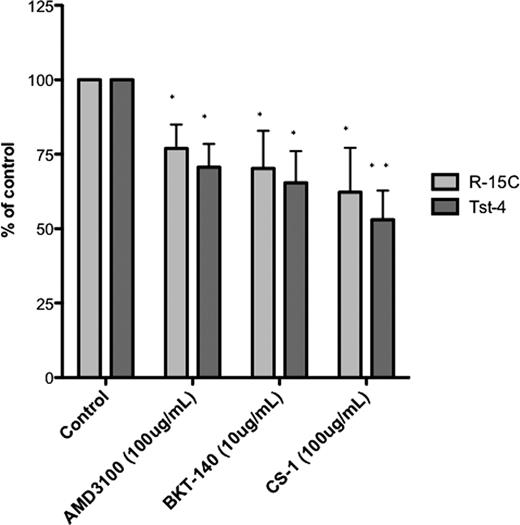
Bone marrow stroma cells (BMSC) constitutively secrete CXCL12 and provide ligands for VLA-4 integrins, and thereby may attract and retain HCL cells in the marrow. Also, these two molecules can be clinically targeted using small molecule antagonists or monoclonal antibodies (mAbs). Therefore, we analyzed HCL cell migration and adhesion towards BMSC (R-15C and Tst-4) after 4 hours of co-culture. As documented via phase contrast microscopy and quantified by flow cytometry, HCL cell lines and primary HCL cells showed abundant adhesion and migration beneath BMSC. Next, we examined whether blocking of CXCR4 using the CXCR4 antagonists AMD3100 and BKT-140 or blocking of VLA-4 integrins using CS-1 peptides affects this adhesion of HCL cells to BMSC. Pre-incubation with AMD3100 significantly reduced BMSC adhesion of hairy cells to levels that were 70.6 ±7.8% (p<0.05) of controls using Tst-4 BMSC. Similar results were found when hairy cells were pre-incubated with the CXCR4 antagonist BKT-140 (65.4 ± 10.6%, p<0.05, n=7). Interestingly, when hairy cells were pretreated with a cyclic peptide inhibitor with a minimal VLA-4 binding motif “LDV” (CS-1 peptide), a significant reduction of HCL cell adhesion was also found, reducing HCL cell adhesion to 53.0 ± 9.7% of controls (p< 0.01) with Tst-4 BMSC; similar results were found using R-15C BMSC, as shown in the Figure.
In a preliminary study, with 4 cases analyzed, we also found that co-culture of primary HCL cells with BMSC improved HCL cell survival, as assessed by staining with DiOC6 and PI. BMSC co-culture increased the proportion of viable HCL cells to 134 ± 13% (n=4) of the control (HCL in culture without BMSC) on day 2 and to 252 ± 72% (n=2) on day 4.
Collectively, our studies provide insight into the molecular interactions between HCL cells and BMSC and provide an explanation for the distinct pattern of tissue infiltration in HCL. Also, they provide a rationale for further preclinical development of CXCR4 and VLA-4 antagonists in HCL.
No relevant conflicts of interest to declare.
Author notes
Asterisk with author names denotes non-ASH members.


This feature is available to Subscribers Only
Sign In or Create an Account Close Modal