Abstract
This prospective study investigated the efficacy, safety, and response duration of low-dose rituximab (100 mg fixed dose for 4 weekly infusions) together with a short course of steroids as first- or second-line therapy in 23 patients with primary autoimmune hemolytic anemia (AIHA). The overall response was 82.6% at month +2, and subsequently stabilized to ∼ 90% at months +6 and +12; the response was better in warm autoimmune hemolytic anemia (WAIHA; overall response, 100% at all time points) than in cold hemagglutinin disease (CHD; average, 60%); the relapse-free survival was 100% for WAIHA at +6 and +12 months versus 89% and 59% in CHD, respectively, and the estimated relapse-free survival at 2 years was 81% and 40% for the warm and cold forms, respectively. The risk of relapse was higher in CHD and in patients with a longer interval between diagnosis and enrollment. Steroid administration was reduced both as cumulative dose (∼ 50%) and duration compared with the patient's past history. Treatment was well tolerated and no adverse events or infections were recorded; retreatment was also effective. The clinical response was correlated with amelioration biologic markers such as cytokine production (IFN-γ, IL-12, TNF-α, and IL-17), suggesting that low-dose rituximab exerts an immunomodulating activity. This study is registered at www.clinicaltrials.gov as NCT01345708.
Introduction
Autoimmune hemolytic anemia (AIHA) is an acquired autoimmune disease characterized by the production of Abs directed against autologous RBCs. AIHA is classified into warm- and cold-reactive Ab types, and can be either primary or secondary to lymphoproliferative disease, infections, immunodeficiency, and tumors.1-3 The degree of hemolysis, from fully compensated to fulminating, depends on the characteristics of the auto-Ab (class, quantity, specificity, thermal amplitude, ability to fix complement, and ability to react with tissue macrophages), on the activity of the reticulo-endothelial system, and on the efficacy of the erythroblastic response.2 Conventional therapy of warm AIHA (WAIHA) includes administration of corticosteroids as a first-line therapy, which is effective in roughly two-thirds of patients; those who are refractory or relapse after the initial response (approximately one-third) need additional second-line therapies that include splenectomy and immunosuppressive agents, which are reported to provide a 60%-75% and 40%-60% response rate, respectively. Conventional treatment with corticosteroids in cold hemagglutinin disease (CHD) induced responses in only 14% of patients, and additional treatments, including alkylating agents, IFN-α, and low-dose cladribine, have little efficacy.2,4-6
Rituximab is a chimeric mAb directed against CD20 that was first developed for the treatment of lymphoproliferative malignancies. The drug was also shown to be effective in rheumatoid arthritis, systemic lupus erythematosus, multiple sclerosis, and several hematologic autoimmune diseases such as primary immune thrombocytopenia (previously referred to as idiopathic thrombocytopenic purpura, ITP), acquired hemophilia, thrombotic thrombocytopenic purpura, and AIHA.7-11 Rituximab is effective in approximately 60% of WAIHA patients, suggesting that it may represent an alternative to splenectomy and/or immunosuppressive/cytotoxic therapy. In addition, it induces durable responses in CHD, offering an effective treatment to a group of patients with limited therapeutic options, and combination therapy with rituximab and oral fludarabine was proven to be very effective in CHD, resulting in a 75% response rate, complete remission in approximately 20% of patients, and more than 66 months estimated response duration.12
In these hematologic autoimmune diseases, rituximab was generally administered at the dose scheduled for the treatment of lymphomas (375 mg/m2/weekly for 4 weeks). In an attempt to minimize side effects and to reduce costs, and considering that the lymphocyte burden in autoimmune hematologic conditions is lower than the tumor mass in lymphoproliferative diseases, low-dose (LD) rituximab (100 mg fixed dose) has been used successfully in autoimmune cytopenias.13 A prospective study demonstrated that rituximab is effective in roughly 60% of ITP patients, but has only a moderate long-term effect14 ; furthermore, a randomized trial reported more pronounced sustained responses in ITP patients treated with LD-rituximab and steroids compared with steroids alone, but with similar response rates.15 Finally, a prospective study in patients with steroid-refractory AIHA and ITP showed that LD-rituximab plus alemtuzumab subcutaneously (10 mg on days 1-3) induced an overall response (OR) rate of 100%, with complete responses (CRs) in 58% of patients.16 The aim of this prospective study was to evaluate the efficacy, safety, and response duration of LD-rituximab associated with a short course of prednisone as first-line therapy in newly diagnosed WAIHA and CHD and as second-line therapy in WAIHA relapsed after standard oral prednisone. A further aim was to correlate the clinical response with cytokine production in cultures.
Methods
Patients
This phase 2, single-arm prospective multicenter study started in June 2008 and concluded in May 2011, and involved 3 Italian academic centers or hospitals. The study protocol was approved by the Ethical Committee of Human Experimentation of all participating institutions, is registered at www.clinicaltrials.gov, and patients gave informed consent in accordance with the Declaration of Helsinki (European Union Drug Regulating Authorities Clinical Trials number 2008-006713-25, NCT01345708). Patients were > 18 years of age and affected by WAIHA or CHD either newly diagnosed or relapsed after first-line treatment with oral prednisone. AIHA was defined by symptomatic anemia and a positive direct antiglobulin test (DAT) in the absence of underlying lymphoproliferative, infectious, or neoplastic disease (according to the single-center diagnostic criteria). Exclusion criteria were active bacterial, viral (HIV, HCV, or HBV), or fungal infection requiring systemic therapy, immunologic deficit (congenital or acquired), history of malignancies within 3 years before study entry, concomitant immunosuppressive or cytotoxic treatment, pregnancy, or any condition that would preclude the ability to give informed consent.
Treatment
Rituximab was administered at the fixed dose of 100 mg as an IV infusion on days +7, +14, +21, and +28, along with oral prednisone 1 mg/kg/d from day +1-+30, followed by tapering according to the following schedule: 10 mg/wk until 0.5 mg/kg/d, then 5 mg/wk until stop. Patients received oral acetaminophen 500 mg and IV chlorphenamine 10 mg as premedication therapy.
Assessments and outcome measures
Complete clinical examination, complete blood counts, hemolytic markers (reticulocytes, total and unconjugated bilirubin, lactate dehydrogenase, and haptoglobin), were performed at enrollment, at days +7, +14, +21, +28, and +35, monthly until month +6, and then every 3 months until the end of follow-up. DAT, hepatic, and renal function tests were performed at enrollment, at day +35, at month +3, and then every 3 months until the end of follow-up.
The primary objective of the study was to evaluate the efficacy in terms of OR, CR (defined as hemoglobin [Hb] > 12 g/dL and normalization of all hemolytic markers), and partial response (PR, defined as Hb = 10-12 g/dL or at least 2 g/dL increase in Hb, and no transfusion requirement). Further objectives were to evaluate the time to response, the duration of the response (sustained response, SR, defined as Hb > 10 g/dL in the absence of any treatment), and the relapse-free survival (RFS). Moreover, we assessed the safety profile as adverse event incidence up to 12 months from the beginning of therapy according to NCI-CTC Version 3.0. Finally, to verify a possible steroid-sparing effect of LD-rituximab, we compared the amount of past steroid therapy in previously relapsed patients with that administered in association with LD-rituximab at equal time intervals.
Immunologic investigations
To evaluate cytokine production (TNF-α, IFN-γ, IL-12, IL-4, and IL-17), heparinized blood samples from patients (n = 12) and controls (n = 16) were diluted 1:6 with RPMI 1640 medium (Gibco-BRL) and stimulated with 2 μg/mL of phytohemagglutinin (Sigma-Aldrich) in 24-well plates for 48 hours. Cytokine production was measured using commercially available ELISA kits (R&D Systems).
DAT for the detection of IgG and complement bound to RBCs was performed with the tube technique using the standard method with polyspecific anti–human globulin and monospecific anti-IgG and anti-C3 antisera (Ortho Clinicals Diagnostics).17
Statistical analysis
The Student t test and χ2 test were used to analyze continuous and categorical variables, respectively. RFS (%) was evaluated using the Kaplan-Meier method. Cox regression models were fitted to calculate the relapse hazard ratios (HRs) and 95% confidence intervals (95% CIs) according to select demographic and clinical variables. Multiple random intercept models were used to evaluate trend of continuous variables (eg, prednisone use, hematologic variables) in the periods before and after LD-rituximab treatment taking into account the positive correlations within subjects at follow-up visits.18 The average daily prednisone doses before and after LD-rituximab were compared considering each day of therapy prescription and also the days between follow-up visits. The sex and an indicator variable for the period (after/before start of therapy) were included in the random-effect models. The modifying effect of AIHA on prednisone administration was evaluated by including a product-term in the regression model and by performing 2 separate analyses for WAIHA and CHD. Multiple random-effect models were used to assess cytokine levels over time, including as covariates the AIHA and the time since diagnosis to start of rituximab therapy. All analyses were performed with Stata Version 11 software.19
Results
Clinical characteristics of patients at enrollment
Table 1 shows the clinical and serologic characteristics of patients at enrollment. WAIHA was more frequent than CHD. The latter patients were almost all females, relapsed after steroid treatment, and had a longer median interval from diagnosis to rituximab treatment. None of the patients was splenectomized. The median Hb value at enrollment was lower in WAIHA than in CHD patients (median, 8.65 g/dL; range, 4.4-13.4 and 9.4 g/dL; range, 7.1-12.2, respectively), and 6 of 14 (43%) WAIHA and 2 of 9 (22%) CHD patients showed Hb values lower than 8 g/dL. Hemolytic markers were abnormally elevated in almost all patients with no differences between WAIHA and CHD (see also baseline values, Table 2).
Clinical and serologic characteristics of patients at enrollment
| . | All patients (n = 23) . | WAIHA patients (n = 14) . | CHD patients (n = 9) . |
|---|---|---|---|
| Male, n (%) | 7 (30.4%) | 6 (43%) | 1 (11%) |
| Female, n (%) | 16 (69.6%) | 8 (57%) | 8 (89%) |
| Median age, y (range) | 56 (27-75) | 46.5 (27-75) | 61 (30-71) |
| Newly diagnosed patients, n (%) | 7 (30.4%) | 6 (43%) | 1 (11%) |
| Relapsed after steroid treatment, n (%) | 16 (69.6%) | 8 (57%) | 8 (89%) |
| Median interval from diagnosis to rituximab, mo (range) | 23 (0-276) | 8 (0-122) | 46 (7-276) |
Hematologic parameters of patients at enrollment and after rituximab treatment
| . | Enrollment (n = 14) . | Month 2 (n = 14) . | Month 6 (n = 14) . | Month 12 (n = 14) . |
|---|---|---|---|---|
| WAIHA, median (range) | ||||
| Hb, g/dL | 8.7 (4.4-13.2) | 13.1 (11.2-15.3) | 13.5 (10.4-17.1) | 13.3 (10.6-15.4) |
| Reticulocyte, × 109/L | 205 (40-320) | 90 (30-274) | 76 (40-250) | 83 (45-160) |
| LDH, U/L | 623 (314-1961) | 479 (341-1031) | 417 (276-605) | 403 (281-816) |
| Unconjugated bilirubin, mg/dL | 1.66 (0.4-6.4) | 0.5 (0.2-2.1) | 0.6 (0.2-1.3) | 0.63 (0.2-1.4) |
| CHD, median (range) | (n = 9) | (n = 9) | (n = 8) | (n = 4) |
| Hb, g/dL | 9.4 (7.1-12.2) | 9.8 (9.1-14.9) | 11.7 (9.2-14.5) | 11.6 (11.1-12.6) |
| Reticulocyte, × 109/L | 140 (52-269) | 101 (40-210) | 95 (72-130) | 90 (36-148) |
| LDH, U/L | 629 (420-2173) | 547 (257-1367) | 457 (273-718) | 414 (318-464) |
| Unconjugated bilirubin, mg/dL | 1.66 (0.6-3.6) | 1.34 (0.4-2.7) | 0.8 (0.7-1.3) | 0.7 (0.5-0.8) |
Normal ranges: Hb, 13.6-16.7 g/dL; reticulocytes, 16-84 × 109/L; lactate dehydrogenase (LDH), 240-480 U/L; unconjugated bilirubin, < 0.75 mg/dL. CHD values at month +6 are shown for 8 cases (excluding 1 relapsed at month +4) and at month +12 for 4 cases (excluding 2 further relapses, 1 of whom has not yet reached the follow-up, and 1 who died at month +7).
Response to therapy
All patients included in the study completed the therapeutic program receiving the 4 infusions of LD-rituximab as scheduled, and were subsequently followed up for a median of 15 months (range, 6-35); the follow-up was at least 12 months in 18 of 23 patients (78%), 18 months in 11 of 23 patients (48%), and 24 months in 7 of 23 patients (30%). ORs at months +2, +6, and +12 were 82.6%, 91.3%, and 84.2%, respectively. Considering separately warm and cold forms (Table 3), all cases of WAIHA responded at month +2 and maintained the response until months +6 and +12; response rates were lower in CHD, with an OR at month +2 in 55.6% of cases and a SR at month +6 and +12 in 77.7% and 66.7%, respectively; relapse rates were 11.1% and 33.3% at months +6 and +12, respectively. The median time to OR was 16 days (range, 6-62) for WAIHA and 19 days (range, 6-166) for CHD.
Response rate and outcome after rituximab therapy
| . | Month +2 . | Month +6 . | Month +12 . |
|---|---|---|---|
| WAIHA 14/23 (60.9%) | OR 14/14 (100%) | SR 14/14 (100%) | SR 13/13 (100%) |
| CR 11 (78.6%) | CR 10 (71.4%) | CR 9 (69.2%) | |
| PR 3 (21.4%) | PR 4 (28.6%) | PR 4 (30.8%) | |
| NR 0 (0%) | NR 0 (0%) | NR 0 (0%) | |
| Relapse rate 0 (0%) | Relapse rate 0 (0%) | Relapse rate 0 (0%) | |
| CHD 9/23 (39.1%) | OR 5/9 (55.6%) | SR 7/9 (77.7%) | SR 3/6 (50%) |
| CR 4 (44.4%) | CR 4 (44.4%) | CR 1 (16.6%) | |
| PR 1 (11.2%) | PR 3 (33.3%) | PR 2 (33.3%) | |
| NR 4 (44.4%) | NR 1 (11.15%) | NR 0 (0%) | |
| Relapse rate 0 (0%) | Relapse rate 1 (11.15%) | Relapse rate 2 (33.3%) |
NR indicates no response.
In univariate analysis, response was significantly associated with WAIHA (P = .023, P = .047, and P = .013 at months +2, +6, and +12, respectively), and the probability of CR at month +2 was associated with younger age (P = .023, χ2), higher weight (P = .013, χ2), and shorter interval between diagnosis and rituximab therapy (P = .077, χ2). In multivariate analysis, the thermal characteristic of hemolytic anemia emerged among age, weight, and interval between diagnosis and rituximab therapy as the only variable associated with a sustained response at month +6 (OR = 9.6; 95% CI, 0.9-105.1; P = .064), and +12 (OR = 21.9; 95% CI, 0.9-484; P = .05).
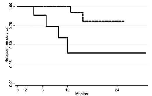
The cumulative RFS for all cases of AIHA at +6 and +12 months was 96% and 86%, respectively, and the estimated RFS at 2 years was 68%. Considering separately WAIHA and CHD, the cumulative RFS times at +6 and +12 months were significantly reduced in the latter (89% and 59% for CHD vs 100% for WAIHA at both time points, respectively, P = .015; Figure 1). The estimated RFS at +24 months was 40% and 81% for cold and warm cases, respectively. In addition, univariate Cox regression models showed that risk of relapse was higher in CHD (HR = 6.1; 95% CI, 1.1-33.8; P = .04) and in patients with longer interval between diagnosis and rituximab therapy (HR = 1.01; 95% CI, 1.00-1.02; P = .03); age, gender, and weight showed no relationship with relapse risk. Among 6 relapsed patients (4 CHD and 2 WAIHA, 3 in the first year after LD-rituximab and 3 subsequently), 1 CHD patient who relapsed at month +7 was retreated with a second LD-rituximab (achieving PR and SR at +9 and +13, respectively) and with a third cycle, achieving an ongoing SR at month +20; the patient was therefore able to stop steroids for several months between cycles. Another patient with WAIHA who had a severe relapse (Hb, 6.5 g/dL) at month +16 was retreated with LD-rituximab, achieving a PR until month +24.
RFS of 14 WAIHA (dotted line) and 9 CHD (continuous line) patients estimated with the Kaplan-Meier method.
RFS of 14 WAIHA (dotted line) and 9 CHD (continuous line) patients estimated with the Kaplan-Meier method.
Hematologic evaluation
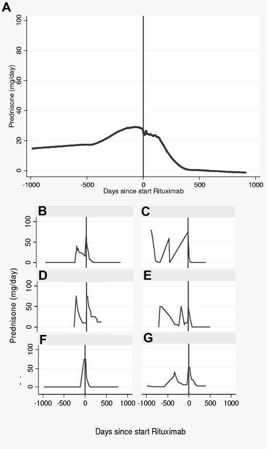
The main laboratory data of WAIHA and CHD are shown in Table 2. In WAIHA, hematologic parameters at month +2 were normalized in most patients: Hb levels significantly increased (P < .0001 vs enrollment), and hemolytic markers decreased (P = .005, P = .03, and P = .002, for absolute reticulocyte, lactate dehydrogenase, and unconjugated bilirubin, respectively); at months +6 and +12 hematologic parameters were stable. At variance, in CHD Hb levels increased to a lesser extent at month +2 (P = .02 vs enrollment) and hemolytic markers decreased but were still above normal ranges; at month +6, Hb levels further increased and hemolytic parameters were almost normalized; hematologic data at month +12 were stable in the 4 evaluable CHD cases. The increase of Hb and the reduction of hemolytic markers were confirmed by analyzing all hematologic data before and after LD-rituximab with multiple random effect models (Hb, +0.6 mg/dL, P < .001; unconjugated bilirubin, −0.5 mg/dL, P < .001; and reticulocytes, −36.3 × 10⁹/L, P < .001). Interestingly, this statistical analysis allowed the comparison of prednisone administrations (491 cumulative doses) before and after LD-rituximab in the 15 patients with an adequate follow-up pre-enrollment (795 ± 170 days, mean ± SEM): the average cumulative prednisone doses were reduced (by approximately 50%) after LD-rituximab compared with the pre-study treatment (3995 vs 7810 mg/d, P = .08, Figure 2); the average daily dosage was reduced as well (11.7 vs 17.2 mg/d, P = .10); however, the reduction was less evident in the 6 CHD patients (−0.6 mg/d, P = .05) than in the 9 WAIHA patient (−4.1 mg/d, P < .001). For the duration of steroid therapy, 9 of 14 (64%) WAIHA patients and 5 of 9 (56%) CHD patients had completely stopped steroids at +89 days (median values, range 58-150); moreover, the overall median duration was significantly reduced after LD-rituximab compared with pre-study treatment (150 days, range 58-548, 56% of time, vs 548 days, range 30-1459, 87% of time, P = .027).
Steroid treatment before and after LD-rituximab in AIHA patients. (A) Average locally weighted smoothed (lowess) of 15 AIHA patients relapsed after standard steroid treatment. (B-G) Typical examples of prednisone administration in 6 AIHA patients. (B-C) WAIHA responders to LD-rituximab. (D-E) WAIHA responders relapsed at month +13 and +16 after LD-rituximab. (F) CHD responder to LD-rituximab. (G) CHD nonresponder relapsed at month +10 after LD-rituximab.
Steroid treatment before and after LD-rituximab in AIHA patients. (A) Average locally weighted smoothed (lowess) of 15 AIHA patients relapsed after standard steroid treatment. (B-G) Typical examples of prednisone administration in 6 AIHA patients. (B-C) WAIHA responders to LD-rituximab. (D-E) WAIHA responders relapsed at month +13 and +16 after LD-rituximab. (F) CHD responder to LD-rituximab. (G) CHD nonresponder relapsed at month +10 after LD-rituximab.
Safety
Overall, rituximab therapy was well tolerated and no patients experienced the most frequently described infusion-related reactions. No infectious, hematologic, or extrahematologic complications were documented during follow-up. As a consequence of steroid therapy, 5 patients experienced increase of body weight (on average, 5 kg), and 1 patient worsening of preexisting hypertension that required temporary adjustment of therapy. An acute ischemic heart attack followed by fatal ischemic stroke occurred in 1 patient (who was in a CR/SR) 7 months after the end of rituximab therapy. No significant changes were observed for leukocyte or lymphocyte counts, or in liver and renal function tests (data not shown).
Immunologic assessment
All patients displayed a positive DAT at enrollment, 14 with anti-IgG (WAIHA) and 9 with anti-C antisera (CHD); at month +6, DAT was still positive in all patients, whereas at month +12, 2 of 18 patients became DAT negative (12.5%). Titers and scores were available in 8 patients who were still DAT+ and 2 showed a clear decrease of both parameters, 2 a little decrease, and 4 no significant changes.
Concerning Th-1 cytokine production, at enrollment, TNF-α was lower in patients compared with controls, although not significantly (946 ± 208 vs 1574 ± 139 pg/mL, mean ± SEM), and increased at months +3 and +6, reaching normal values (1289 ± 152 and 1591 ± 189 pg/mL, respectively). Likewise, IL-12 production was lower than normal values (697 ± 253 vs 1022 ± 38 pg/mL), and increased at months +3 and +6 (1130 ± 140 and 1295 ± 214 pg/mL, respectively); IFN-γ was also significantly reduced (1245 ± 412 in patients vs 2897 ± 55.3 pg/mL in controls, P = .04), and increased after therapy without reaching normal values. These findings were confirmed by multiple random-effect models (data not shown). Regarding Th-2 cytokines, at enrollment, IL-4 was higher in patients compared with controls (24.6 ± 8.6 vs 12.6 ± 1.2 pg/mL), diminished at month +3, reaching normal values (11.0 ± 3.4 pg/mL), but returned to baseline levels at month +6 (22.6 ± 11.6 pg/mL). Likewise, at enrollment, IL-17 was significantly increased compared with controls (37.8 ± 7.4 vs 5.6 ± 0.1 pg/mL, P = .03), and showed a slight reduction at month +6 (30.2 ± 6.2 pg/mL, P = .04 vs controls). Although the number of patients evaluated is too small to draw definite conclusions, IL-17 at month +6 was lower in responding patients (n = 7) than in nonresponding patients (n = 4; 19.6 ± 5.0 vs 43.9 ± 13.0 pg/mL, P = .068), whereas there was no difference in the other cytokines investigated when the 2 populations were considered separately.
B-cell depletion was evaluated in 3 patients (2 responders and 1 nonresponder): the median baseline value of CD20+ cells was 0.18 × 10⁹/L, (range, 0.09-0.49) compared with 0.03 × 10⁹/L (range, 0.01-0.14) at month +6, without any difference between responding and nonresponding cases. Serum IgG, IgA, and IgM levels were comparable at baseline and at month +6 (data not shown).
Discussion
First-line therapy of autoimmune hemolytic anemias is based on corticosteroids, which are reported to provide a response in 70%-85% of WAIHA patients; however, only one-third of patients remain in long-term remission once the drug is discontinued, and a further 50% require maintenance doses.3 This long-term steroid therapy, even at low doses, is known to have potentially harmful side effects. Second-line therapy with cytotoxic and immunosuppressive drugs such as azathioprine, cyclophosphamide, and cyclosporine is reported to provide a 40%-60% response rate, but their use may be associated with serious side effects, and the effectiveness of other options, such as IV immunoglobin, plasmapheresis, and danazol, is controversial.3-5 In CHD, corticosteroids are effective in only 1 of 6 of patients, and other treatments (eg, alkylating agents, IFN-α, and low-dose cladribine) have little efficacy.2,4-6 Therefore, we pursued the idea of developing a “potentially curative” therapy for AIHA combining 2 well-demonstrated, effective treatments, steroids and rituximab, at doses lower than those usually used. Our aim was to minimize side effects and costs and synergize the curative actions of these 2 drugs. Furthermore, the reduction of rituximab doses may decrease the risk of complications, such as the reactivation of viral infections and progressive multifocal encephalopathy, although a specific study is needed to address this issue. Progressive multifocal encephalopathy, the most harmful of these complications, has been reported after rituximab treatment (median time from last rituximab dose, 5.5 months), generally in heavily immunosuppressed lymphoma patients.20-24
The results of this prospective study show that low doses of rituximab (nearly one-seventh of the standard dose), together with a short course of prednisone, is effective in AIHA patients, with a sustained response in 90% of patients at 1 year and an estimated RFS in roughly two-thirds of patients at 2 years. These responses are greater than those reported in the literature for conventional steroids alone, and are undoubtedly superior compared with those of second-line therapy with cytotoxic and immunosuppressive drugs. The responses observed in the present study were associated with a significant increase of Hb levels and a reduction of hemolytic markers, even if only 2 patients became DAT negative. Treatment was well tolerated and no rituximab-related adverse events were recorded, including leucopenia/neutropenia and infections. Our results compare favorably with those reported with conventional doses of rituximab (effective in roughly 60% of patients, but with a great variability: range 40%-100%),7,11 and are similar to the more recent studies showing response rates of 80%-90% and a disease-free survival at 2 years in one-half of patients.25-28 Despite the small numbers, our results clearly show that response rates and RFS, as well as amelioration of hematologic parameters, were more evident in warm than cold forms of the disease, with virtually all patients with the former responding. Steroid therapy may contribute to improving and accelerating the response of WAIHAs, which are known to be more sensitive to steroids than CHD. Cold forms respond to a lesser extent to low doses of rituximab; likewise, the time to response was particularly prolonged in some patients with CHD, as observed previously with standard doses,7,11,28-31 supporting the idea that CHD is a different clinical entity from classic WAIHA.3,6 Consistently, the presence of a clonal B-cell lymphoproliferation was found by reexamining BM biopsies in more than two-thirds of patients otherwise classified as primary CHD, suggesting the existence of a continuous spectrum between CHD and lymphoproliferative diseases such as lymphoplasmacytic lymphoma, marginal zone lymphoma, and Waldenstrom macroglobulinemia.6 In agreement with the “clonality” of CHD, more aggressive therapy with rituximab and oral fludarabine was proven to be more effective than rituximab alone at standard doses (in three-fourths compared with one-half of cases).12 Finally, we found that the response to LD-rituximab was associated with a shorter disease duration, suggesting that it may be more appropriate as an earlier treatment; however, this finding may also reflect the longer disease duration of CHD, in which patients do worse compared with WAIHA.
An interesting finding of our study is that steroid administration during LD-rituximab was reduced, particularly in WAIHA, both in terms of cumulative dose (roughly 50%) and duration, compared with the patient's past history, suggesting a steroid-sparing effect of this treatment. Moreover, the sustained response that leaves the patient free of therapy for longer periods compared with the pretreatment history, and the possibility of obtaining a response with a retreatment, has already been reported with conventional doses of rituximab.7,11
It is well established that the response to rituximab is related to B-cell depletion, with the consequent inhibition of several B-cell pathologic activities, such as the production of auto-Abs, cytokine secretion, and APC function.9 B-cell depletion has been documented at low doses in ITP,14,15 and, although in limited cases, also in our series. However, it has been proposed recently that the drug exerts an immunomodulating activity; for example, in ITP, it normalizes the abnormal autoreactive T-cell response and rituximab-opsonized B cells block the macrophage Fc-receptor function, consequently reducing the sequestration of platelets in the spleen.32,33 To investigate this issue, we tested in vitro production of several cytokines. We focused on Th1 cytokines (IFN-γ, IL-12, and TNF-α), which are reported to be reduced in autoimmune diseases including AIHA34-39 ; in addition, we studied IL-17, which is produced by a novel Th-cell subset distinct from Th1 and Th2 cells, which are critical in inflammation and autoimmunity.38 IL-17 expression was reported to be increased at the site of inflammation in patients with rheumatoid arthritis, psoriasis, multiple sclerosis, and uveitis.40-43 Furthermore, in animal models, mice genetically deficient in IL-17A were less susceptible to collagen-induced arthritis and experimental autoimmune encephalitis, and neutralization of IL-17 by treatment with anti–IL-17 Abs in vivo protected mice from experimental autoimmune uveitis.40,44 Conversely, Th1 cytokines such as IFN-γ and IL-12 inhibit Th17 differentiation and therefore exert a protective role against IL-17–mediated autoimmune inflammation.45 We found that AIHA patients displayed reduced IFN-γ, IL-12, and TNF-α levels compared with healthy controls; however, levels of IL-17 were greatly increased, as has been found previously in other autoimmune diseases.40-43 Treatment with LD-rituximab has minor effects on IL-17, with only a slight reduction at month +6, which was more evident in responding patients; however, IFN-γ, IL-12, and TNF-α, which exert a protective role against IL-17–mediated autoimmune damage,45 increased over time, sometimes reaching normal values. These data suggest that LD-rituximab treatment, in addition to its B-cell–depleting activity, may provide a favorable immunomodulating effect that persists after steroid discontinuation.
In conclusion, LD-rituximab associated with a short course of steroid therapy is a safe and effective treatment, particularly in WAIHA and in patients with newly diagnosed disease, with response rates and sustained responses comparable to standard doses of rituximab. Treatment leaves patients free of therapy for longer periods and results in a steroid-sparing effect, although specific controlled studies should be designed to address all of these issues. Retreatment with LD-rituximab is also effective. LD-rituximab seems to be more appropriate in WAIHA than in CHD, raising the question of whether a higher dose is required for latter disease, which often shows BM clonal lymphoproliferation. Finally, LD-rituximab has immunomodulating effects, confirming that B-cell depletion is not the unique mechanism of action of the drug.
There is an Inside Blood commentary on this article in this issue.
The publication costs of this article were defrayed in part by page charge payment. Therefore, and solely to indicate this fact, this article is hereby marked “advertisement” in accordance with 18 USC section 1734.
Acknowledgments
This work was supported by the Fondazione IRCCS Ca' Granda Ospedale Maggiore Policlinico (grant RC 2011, 160-02).
Authorship
Contribution: W.B. designed the research, collected, analyzed, and interpreted the data, and wrote the manuscript; F.Z. collected the data and critically revised the manuscript; A. Zaninoni and F.G.I. performed the research and biologic investigations, partially wrote the manuscript, and collected data for statistical analysis; M.L.B. collected data; E.D.B. and B.F. collected data and partially wrote the manuscript; D.C. performed the statistical analysis, contributed to the interpretation of the data, and partially wrote the manuscript; A.C. and R.F. revised the manuscript; and A. Zanella contributed to the study design, interpreted the data, and wrote the manuscript.
Conflict-of-interest disclosure: F.Z. is on the advisory board for Roche. The remaining authors declare no competing financial interests.
Correspondence: Wilma Barcellini, MD, Unita Operativa Ematologia 2, Padiglione Granelli, Fondazione IRCCS Ca' Granda Ospedale Maggiore Policlinico, Via F. Sforza 35-20122 Milano, Italy; e-mail: wbarcel@policlinico.mi.it.


This feature is available to Subscribers Only
Sign In or Create an Account Close Modal