Hereditary hemochromatosis (HH) is a highly prevalent genetic disorder characterized by excessive parenchymal iron accumulation leading to liver cirrhosis, diabetes, and in some cases hepatocellular carcinoma. HH is caused by mutations in the genes encoding upstream regulators of hepcidin or more rarely in the hepcidin gene itself. A deficit in hepcidin results in intestinal iron hyperabsorption; however, the local effectors mediating the up-regulation of iron absorption genes are unknown. We hypothesized that HIF-2 could mediate high iron absorption rates in HH. We generated Hepc−/− mice (a murine model of hemochromatosis) lacking HIF-2 in the intestine and showed that duodenal HIF-2 was essential for the up-regulation of genes involved in intestinal iron import and the consequent iron accumulation in the liver and pancreas. This study highlights a role of HIF-2 in the dysregulation of iron absorption and chronic iron accumulation, as observed in patients with hemochromatosis.
Introduction
Hereditary hemochromatosis (HH) is a heterogeneous genetic disease characterized by excessive iron accumulation in the liver and parenchyma. Clinical manifestations include liver cirrhosis, diabetes, cardiomyopathy, arthropathy, hypermelanotic skin pigmentation, and hepatocellular carcinoma. HH is typically caused by mutations in genes encoding either upstream signaling molecules involved in the induction of hepcidin expression (HFE, transferrin receptor 2, and hemojuvelin) or more rarely in the hepcidin gene itself.1,2 Iron absorption in the duodenum is the only way to control iron entry in the body and is finely regulated in response to systemic iron requirements.3 At the apical brush border of duodenal enterocytes, duodenal cytochrome b (DCYTB) facilitates non-heme iron uptake by divalent metal transporter 1 (DMT1), whereas ferroportin (FPN) exports iron across the basolateral membrane.4
Hepcidin is the central regulatory molecule of systemic iron homeostasis5 and regulates cellular iron efflux by binding to FPN and inducing its internalization and subsequent degradation in the lysosome.6 Although hepcidin is known to act at a systemic level to regulate the rate of iron absorption by controlling the amount of iron exported across the basolateral membrane by FPN, the local effectors mediating the up-regulation of apical iron absorption genes in hemochromatosis are unknown. We and others have previously demonstrated that the hypoxia-inducible factor-2α (HIF-2α) transcription factor, and not HIF-1α, regulates DMT1, DCYTB, and FPN expression in the duodenum at basal level, iron deficiency, and in conditions of increased erythropoiesis.7,–9 HIF-1 and HIF-2 are heterodimeric transcriptional factors and central mediators of cellular and systemic adaptation to hypoxia. In the presence of oxygen, the HIF-α subunit is hydroxylated by oxygen- and iron-dependent prolyl hydroxylases and targeted to the proteasome after the binding to the von Hippel-Lindau protein. On hypoxia (or iron deficiency), HIF-α is stabilized and binds to the HIF-β constitutive subunit to induce the transcription of target genes.10
We hypothesized that HIF-2 could be a mediator of high iron absorption rates in HH and addressed this question by breeding the hepcidin knockout mice (Hepc−/−), a model of severe iron overload, with mice lacking HIF-2 in the intestinal epithelium.
Methods
Animals
Animal studies described here were reviewed and approved (Agreement P2.CP.151.10.) by the Président du Comité d'Ethique pour l'Expérimentation Animale Paris Descartes. We intercrossed mice homozygous for germline knockout of hepcidin11 and mice with loss of HIF-2α specifically in the intestinal epithelium Hif-2αlox/loxVillin-Cre+7, both in a C57BL/6J genetic background, to produce the Hepc−/−/Hif-2αlox/lox/Villin-Cre+ mouse strain (referred as Hepc−/−Hif-2αΔint). Finally, we interbred Hepc−/−HIF-2αΔint and Hepc−/−/Hif-2αlox/lox/VillinCre− mice (here referred as Hepc−/−). Male mice were analyzed at the age of 5 months and compared with control genotypes, including Hepc+/+Hif-2αlox/lox/VillinCre− and Hepc+/−Hif-2αlox/lox/VillinCre− (referred as controls [CTR]).
Reverse transcription and real-time quantitative PCR
RNA extraction, reverse transcription, quantitative PCR, and sequences of the primers used have been previously described.7 All samples were normalized to the threshold cycle value for cyclophilin.
Western blot
Frozen whole duodenal tissue was homogenized using a pestle and smash, and extraction of membrane proteins was performed as previously described.7 The following antibodies were used: DMT1 antibody recognizing both DMT1-IRE and non-IRE isoforms12 (kind gift of François Cannone-Hergaux), DCYTB antibody (Alpha Diagnostic DCYTB11-A), and FPN antibody (Alpha Diagnostic, MTP11-A).
Iron measurements and immunostaining
Plasma and tissue iron were quantified colorimetrically by a previously described method.7 For histology, tissues were fixed in 4% formaldehyde and embedded in paraffin and stained with Perls Prussian blue and nuclear fast red counterstain.
Statistical analysis
Statistical analysis was performed using GraphPad Prism Version 4.0, and the significance of experimental differences was evaluated by 1-way ANOVA followed by a Bonferroni posttest. Values in the figures are expressed as mean ± SEM.
Results and discussion
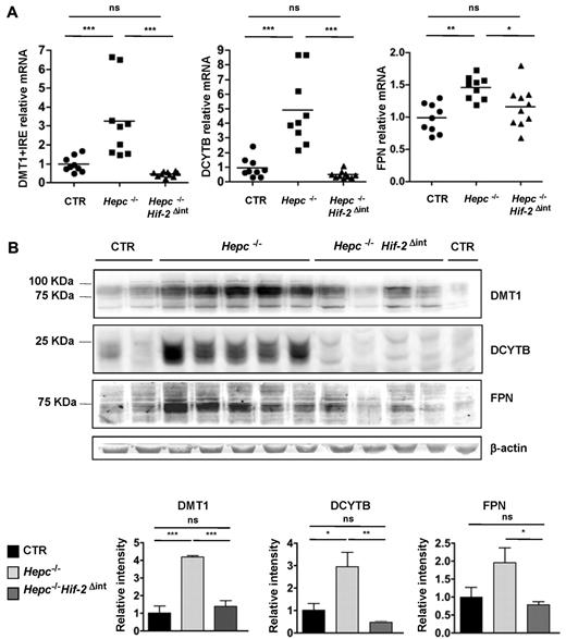
To test whether HIF-2 can mediate the up-regulation of iron absorption genes in HH, we generated Hepc−/− mice deleted for HIF-2α in the duodenum (Hepc−/−Hif-2αΔint mice). These mice do not exhibit any overt phenotypic abnormalities. We previously reported that DMT1, DCYTB, and FPN protein levels were increased in the duodenum of hepcidin-deficient mice (the Usf2−/− mouse model13 ). We confirmed this result (Figure 1B) and further demonstrated that Hepc−/− mice presented high levels of DMT1, DCYTB, and FPN mRNA (although to a lesser extent) compared with control mice (Figure 1A), suggesting that a transcriptional control of these genes takes place in the duodenum of these mice. The levels of DMT1, DCYTB, and FPN transcript and protein were fully attenuated in Hepc−/−Hif-2αΔint (Figure 1A-B) compared with Hepc−/− mice with levels not statistically different from wild-type mice. The duodenal deletion of HIF-2α decreased significantly FPN protein levels, despite the lack of hepcidin, which should prevent FPN degradation by systemic regulation.
Iron absorption genes are decreased in Hepc−/−Hif-2αΔint compared with Hepc−/− mice. (A) Relative mRNA expression of DMT1 + IRE, DCYTB, and FPN normalized to Cyclophilin in the duodenum of Hepc−/−Hif-2αΔint (●; n = 9) versus Hepc−/− (■; n = 9) and CTR (▴; n = 9) mice. (B) Western blot of FPN, DMT1, and DCYTB on membrane extracts of whole duodenum from Hepc−/−Hif-2Δint and Hepc−/− mice versus CTR littermates. Expression was normalized to β-actin. Results were quantified using ImageJ Version 1.43r software (http://rsb.info.nih.gov/ij/). All genotypes used contain the Hif-2αlox/lox allele. *P < .05. **P < .01. ***P < .001. ns indicates not significant.
Iron absorption genes are decreased in Hepc−/−Hif-2αΔint compared with Hepc−/− mice. (A) Relative mRNA expression of DMT1 + IRE, DCYTB, and FPN normalized to Cyclophilin in the duodenum of Hepc−/−Hif-2αΔint (●; n = 9) versus Hepc−/− (■; n = 9) and CTR (▴; n = 9) mice. (B) Western blot of FPN, DMT1, and DCYTB on membrane extracts of whole duodenum from Hepc−/−Hif-2Δint and Hepc−/− mice versus CTR littermates. Expression was normalized to β-actin. Results were quantified using ImageJ Version 1.43r software (http://rsb.info.nih.gov/ij/). All genotypes used contain the Hif-2αlox/lox allele. *P < .05. **P < .01. ***P < .001. ns indicates not significant.
We next asked whether the decrease of genes involved in iron absorption at the apical (DMT1 and DCYTB) and the basolateral membrane (FPN) was sufficient to prevent the hyperabsorption characteristic of the Hepc−/− mice. Interestingly, the double knockout presented a significantly decreased accumulation of nonheme iron in the liver and pancreas compared with Hepc−/− littermates. This was assessed both quantitatively (Figure 2A) and qualitatively by Perls' blue staining (Figure 2B). Plasma ferritin levels, reflecting parenchymal iron storage, were significantly diminished in Hepc−/− mice lacking duodenal HIF-2, compared with Hepc−/− mice (Figure 2C). However, plasma iron concentrations or transferrin saturation (Figure 2C) did not differ between the Hepc−/−Hif-2αΔint and Hepc−/− littermates, suggesting a contribution of the iron recycled from the spleen, an organ that is not affected by the deletion of Hif-2α.14 Indeed, most circulating iron is provided by macrophage iron recycling, and this process seems not affected in the Hepc−/−Hif-2αΔint mice compared with the Hepc−/− mice, as shown by the lack of detectable iron in the macrophages of the spleen in both models (Figure 2B). Interestingly, hematologic parameters (hemoglobin, hematocrit, mean corpuscular volume) were decreased in the Hepc−/−HIF-2αΔint mice compared with the Hepc−/− mice and not statistically different from wild-type mice (supplemental Figure 1, available on the Blood Web site; see the Supplemental Materials link at the top of the online article).
Iron parameters are decreased in the Hepc−/−Hif-2αΔint mice compared with Hepc−/− mice. (A) Quantification of liver (n = 12 per group) and pancreas (n = 6 per group) iron levels in Hepc−/−Hif-2αΔint (●) and Hepc−/− (■) versus CTR (▴) mice. (B) Perls' blue staining of the liver, pancreas, and spleen of CTR, Hepc−/−, and Hepc−/−Hif-2αΔint mice. One representative picture of each genotype is shown. Bars represent 200 μm. (10×/0.45, Nikon E800 microscope, CDD QICAM cooled camera [QImaging, QCapture Version 2.98.2 software [Qualitative Imaging Corporation]). (C) Plasma ferritin, plasma iron, and transferrin saturation in Hepc−/−Hif-2αΔint (▴; n = 9) versus Hepc−/− (■; n = 9) and CTR (● n = 9) mice. ***P < .001. ns indicates not significant.
Iron parameters are decreased in the Hepc−/−Hif-2αΔint mice compared with Hepc−/− mice. (A) Quantification of liver (n = 12 per group) and pancreas (n = 6 per group) iron levels in Hepc−/−Hif-2αΔint (●) and Hepc−/− (■) versus CTR (▴) mice. (B) Perls' blue staining of the liver, pancreas, and spleen of CTR, Hepc−/−, and Hepc−/−Hif-2αΔint mice. One representative picture of each genotype is shown. Bars represent 200 μm. (10×/0.45, Nikon E800 microscope, CDD QICAM cooled camera [QImaging, QCapture Version 2.98.2 software [Qualitative Imaging Corporation]). (C) Plasma ferritin, plasma iron, and transferrin saturation in Hepc−/−Hif-2αΔint (▴; n = 9) versus Hepc−/− (■; n = 9) and CTR (● n = 9) mice. ***P < .001. ns indicates not significant.
In conclusion, our data suggest that HIF-2 contributes to the intestinal iron hyperabsorption in a mouse model of HH but may not overcome all of the negative consequences of the abnormal iron metabolism. Associations between single nucleotide polymorphisms at Hif-2α locus and blood-related phenotypes have been recently demonstrated.15,16 It would be of interest to determine whether HIF-2α polymorphisms could be found associated with iron burden in hemochromatosis. Current treatments for iron overload disorders are limited to phlebotomy or, in case of severe anemia, cardiac failure, or poor tolerance, to chelation therapies.2 Here, we propose that therapeutic intervention on intestinal HIF-2α activity might be beneficial to reduce the rates of iron absorption and parenchymal iron overload.
The online version of this article contains a data supplement.
The publication costs of this article were defrayed in part by page charge payment. Therefore, and solely to indicate this fact, this article is hereby marked “advertisement” in accordance with 18 USC section 1734.
Acknowledgments
The authors thank Corinne Lesaffre and Maryline Favier at the Cochin Institute's Morphology and Histology facility, F. Canonne-Hergaux for his kind gift of antibodies against DMT1, and Jacques Mathieu for helpful advice.
This work was supported by Agence Nationale pour la Recherche (ANR-08-JCJC-0123 and ANR-08-GENO) and the European Research Council under the European Community's Seventh Framework Program (FP7/2011-2015 grant agreement 261296). M.M. was supported by Association pour la Recherche sur le Cancer (fellowship).
Authorship
Contribution: M.M., P.M., S.D., and J.-C.D. performed experiments; M.M., S.V., and C.P. wrote the manuscript; and all authors conceived, analyzed, and interpreted the experiments.
Conflict-of-interest disclosure: The authors declare no competing financial interests.
Correspondence: Carole Peyssonnaux, Institut Cochin, Département Endocrinologie Métabolisme et Cancer, Inserm U1016, Centre National de la Recherche Scientifique Unité Mixte de Recherche 8104, 24 rue du Faubourg Saint Jacques, 75014 Paris, France; e-mail: carole.peyssonnaux@inserm.fr.


![Figure 2. Iron parameters are decreased in the Hepc−/−Hif-2αΔint mice compared with Hepc−/− mice. (A) Quantification of liver (n = 12 per group) and pancreas (n = 6 per group) iron levels in Hepc−/−Hif-2αΔint (●) and Hepc−/− (■) versus CTR (▴) mice. (B) Perls' blue staining of the liver, pancreas, and spleen of CTR, Hepc−/−, and Hepc−/−Hif-2αΔint mice. One representative picture of each genotype is shown. Bars represent 200 μm. (10×/0.45, Nikon E800 microscope, CDD QICAM cooled camera [QImaging, QCapture Version 2.98.2 software [Qualitative Imaging Corporation]). (C) Plasma ferritin, plasma iron, and transferrin saturation in Hepc−/−Hif-2αΔint (▴; n = 9) versus Hepc−/− (■; n = 9) and CTR (● n = 9) mice. ***P < .001. ns indicates not significant.](https://ash.silverchair-cdn.com/ash/content_public/journal/blood/119/2/10.1182_blood-2011-09-380337/5/m_zh89991184890002.jpeg?Expires=1769138777&Signature=gbp5d3F2wiJUuc0~2qxTlCiMbAzO451byHLwrpNsmeW~ZzATSmmr8Snpn-itAHpeuHepgnCug~bBWuUm1OQppMn36ZqwE1Kxjl9JQ4CB8-dC6Z8dAIpOAQ3cl0wRzPJus6B9IKLrM7bDsU6sOLrgwz~ogb9s2aIgO7IKtQST3xsoQg1zBwvWzbZr1e639C-yI8RKXXyhAAp0B6r~iijZhGwC1bay7ovAp4NZ5cNeUXtWYJLaX5rSAB2cT~cPxBp2yuX0VGpzmXtZef3O4lox7Jy4BYxPZeOjKpSxK17c4RZrdWNN25fUM5auW8wJ0hQWNoIQlsP0dKqjZ1-T57leOg__&Key-Pair-Id=APKAIE5G5CRDK6RD3PGA)
This feature is available to Subscribers Only
Sign In or Create an Account Close Modal