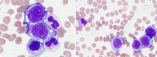
A 25-year-old woman presented with lymphadenopathy. Peripheral blood showed leukocytosis, anemia, and thrombocytopenia (white blood cell count, 102.9 × 10⁹/L; hemoglobin level, 76 g/L; and platelet count, 32 × 109/L). Peripheral smear demonstrated a predominant population of large cells with reticular chromatin, folded to irregular nuclear contours, and abundant basophilic cytoplasm. Many cells contained cytoplasmic vacuoles. By flow cytometry, these cells were within the monocyte gate (CD45/side scatter) and expressed the myeloid-associated marker CD13, in addition to CD56, HLA-DR, CD5, CD7 (partial), and cytoplasmic CD3 (dim). They were negative for CD33, CD34, CD117, CD19, CD3, CD4, and CD8. Cytochemically, the cells were negative for butyrate and chloroacetate esterases. A lymph node biopsy showed ALK-positive anaplastic large cell lymphoma (ALCL). Peripheral blood fluorescence in situ hybridization demonstrated ALK gene rearrangement in 97.5% of nuclei, confirming a leukemic phase of the ALK-positive ALCL.
ALCL is a peripheral T-cell lymphoma characterized by large pleomorphic lymphoid cells with abundant cytoplasm. Leukemic phase of ALCL is rare but associated with poor prognosis. It is most commonly seen in ALK-positive ALCLs and frequently presents as small circulating lymphoid cells. In this case, the peripheral blood cells were large and demonstrated morphologic and flow-cytometric features requiring differentiation from an acute monocytic leukemia.
For additional images, visit the ASH IMAGE BANK, a reference and teaching tool that is continually updated with new atlas and case study images. For more information visit http://imagebank.hematology.org.


This feature is available to Subscribers Only
Sign In or Create an Account Close Modal