Key Points
A small-molecule peptide inhibitor of VIP-signaling protected murine allo-BMT recipients from lethal mCMV infection without increasing GvHD.
Treatment with the VIP inhibitor reduced viral loads, increased antigen-specific T-cells, and decreased PD-1 expression.
Abstract
Cytomegalovirus (CMV) infection following allogeneic bone marrow transplant (allo-BMT) is controlled by donor-derived cellular immunity. Vasoactive intestinal peptide (VIP) suppresses Th1 immunity. We hypothesized that blocking VIP-signaling would enhance anti-CMV immunity in murine recipients of allo-BMT. Recipients were transplanted with bone marrow (BM) and T-cells from major histocompatibility complex (MHC)–mismatched VIP-knockout (KO) or wild-type donors, and treated with 7 daily subcutaneous injections of VIPhyb (peptidic VIP-antagonist) or phosphate-buffered saline (PBS). Genetic and pharmacological blockade of VIP-signaling protected allo-BMT recipients from lethal murine CMV (mCMV) infection, improving survival without increasing graft-versus-host disease. Mice treated with VIPhyb or transplanted with VIP-KO allografts had significantly lower viral loads, increased numbers of mCMV-M45-peptide-MHC-tetramer+ CD8+ T-cells, with lower PD-1 expression, and enhanced primary and secondary cellular immune responses after mCMV infection than did PBS-treated mice. These results demonstrate that administration of a VIP antagonist after allo-BMT is a promising safely therapeutic approach to enhance antiviral cellular immunity.
Introduction
Vasoactive intestinal polypeptide (VIP) is a neuropeptide that inhibits inflammatory immune responses.1-7 Tolergeneic dendritic cells (DC) generated from bone marrow (BM) cultured with VIP protected mice from lipopolysaccharide and graft-versus-host disease (GVHD).1,2 We have shown that VIP-knockout (KO) mice are resistant to murine cytomegalovirus (mCMV) infection and that mCMV-resistance can be adoptively transferred by syngeneic bone marrow transplantation (BMT).8 The mechanism of mCMV resistance in VIP-KO mice appears to be through altering the expression of co-stimulatory and co-inhibitory molecules on DC and T-cells leading to increased antigen-specific T-cells.8 We hypothesized that short-term pharmacological inhibition of VIP signaling could improve immune responses to CMV infection, a common opportunistic infection after allogeneic transplantation.9,10 To test this hypothesis, and the effect of VIP signaling blockade on GVHD, we infected mice with mCMV following transplantation of major histocompatibility complex (MHC)–mismatched VIP-KO BM or treated recipients of wild-type (WT) allografts with a VIP antagonist.11
Study design
The animal studies in this manuscript were approved and monitored by the Emory University Animal Care and Use Committee. Preparation of BM cells and splenocytes from VIP-KO or WT B6 mice, and irradiation (11Gy) of CB6/F1 and B10BR recipient mice were performed as previously described.8,12,13 On day 0, irradiated mice received 5 × 10⁶ T-cell depleted BM (TCD-BM) cells with 0, 1 × 10⁶, 3 × 10⁶, or 8 × 10⁶ donor splenocytes or 0.1 × 10⁶, 0.3 × 10⁶, or 1 × 10⁶ purified donor T-cells via tail-vein injection. Donor chimerism in peripheral blood was determined 1-2 months posttransplant and was typically ≥95%. BMT recipients were treated with 7 daily subcutaneous injections of VIPhyb (10 μg per 100 μL per mouse) starting the day prior to intraperitoneal injection of 5 × 10³, 1 × 10⁴, 2 × 10⁴, or 1 × 10⁵ plaque-forming unit (PFU) of the Smith mCMV strain.14,15 Mice were monitored daily for survival and clinical signs of GVHD, including posture, activity, diarrhea, fur, and weight.16 Other methods are addressed in the supplemental Methods section.
Results and discussion
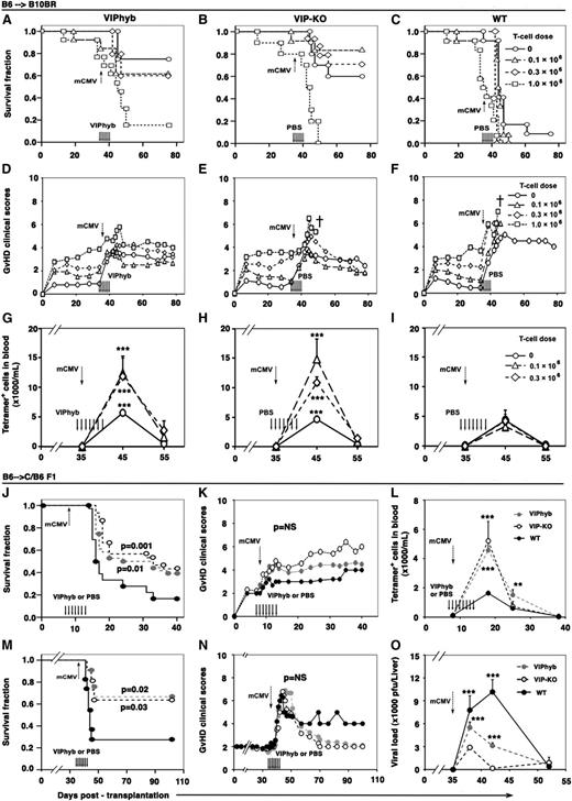
We first examined the effects of engrafting VIP-KO BM with graded doses of donor T-cells on GVHD in an established C57BL/6→B10BR MHC-mismatched allogeneic BMT (allo-BMT) model.12 We found similar survival rates and GVHD clinical scores comparing recipients of 1 × 10⁶, 3 × 10⁶, or 8 × 10⁶ splenocytes from WT versus VIP-KO donors (supplemental Figure 1). Next we used graded numbers of donor T-cells to test the effect of VIP-signaling blockade on survival, GVHD, and antigen-specific immune responses following mCMV infection after allo-BMT. Mice were transplanted with TCD-BM and T-cells from VIP-KO donors or from WT donors, treated with 7 daily injections of VIPhyb11 or phosphate-buffered saline (PBS), and infected with mCMV on day +35 posttransplant. Transplanting B6→B10BR mice with VIP-KO allografts or treating recipients of WT allografts with VIPhyb resulted in significantly better survival (60% to 85%) (Figure 1A-C) and low GVHD scores (Figure 1D-F) among recipients of no, low, or intermediate numbers of donor T-cells (0, 0.1 × 10⁶, or 0.3 × 10⁶) in comparison with 0% survival among corresponding PBS-treated groups that received WT allografts. All groups that received 1 × 10⁶ donor T-cells developed severe GVHD with nearly uniform mortality. Recipients of VIP-KO allografts and recipients of WT allografts treated with VIPhyb transplanted with low and intermediate T-cell doses (0.1 × 10⁶ or 0.3 × 10⁶) had significantly more mCMV-M45-peptide-MHC-tetramer+ CD8+ T-cells (tetramer+ CD8+ T-cells) in their blood than did PBS-treated recipients of WT allografts (Figure 1G-I).
Allo-BMT recipients treated with VIP antagonist or transplanted with VIP-KO donors had improved survival following mCMV infection. B10BR and CB6 F1 mice were transplanted with grafts from either VIP-KO or WT C57BL/6 mice. Transplant recipients were treated with 7 daily s.c. injections of 10 μg of VIPhyb or PBS starting the day prior to mCMV infection on day 8 or on day 35 posttransplant. Mice were examined daily for survival and clinical signs of GVHD. Peripheral blood was collected and tetramer+ CD8+ T-cells analyzed by flow cytometry. Viral loads in the liver from mice subjected to necropsy at predetermined time points were measured by plaque assay as previously described.8,14,15 (A-C) Survival of B6→B10BR recipients of 5 × 10⁶ TCD-BM alone or TCD-BM plus 0.1 × 10⁶, 0.3 × 10⁶, or 1 × 10⁶ splenic T-cells, treated with VIPhyb (A) or PBS (B, C). Mice were infected with 1 × 10⁵ mCMV PFU on day 35 posttransplant. Data are pooled from 2 replicate experiments with 12 total mice per group. (D-F) GVHD clinical scores for the B6→B10BR groups transplanted in panels A-C.23-25 (G-I) tetramer+ CD8+ T-cells in the B6→B10BR groups transplanted in panels A-C. (J) Survival of B6→CB6 F1 recipients of 5 × 10⁶ TCD-BM plus 3 × 10⁶ splenocytes following infection with 5 × 10³ mCMV PFU on day 8 posttransplantation. Data are from 3 independent experiments with a total of n = 22 mice for each group. (K) GVHD clinical scores in mCMV-infected B6→ CB6 F1 transplant recipients in panel J (SD were +/− 18% of mean values at respective time-points and omitted). (L) tetramer+ CD8+ T-cells in the B6→CB6 F1 groups transplanted in panel J. (M) Survival of B6→CB6 F1 recipients of 5 × 10⁶ TCD-BM plus 3 × 10⁶ splenocytes following 2 × 10⁴ mCMV PFU infection on day 35 posttransplant. Data are pooled from 3 independent experiments; n = 36 for both the WT group and VIP-KO group, and n = 30 for the VIPhyb group. (N) GVHD clinical scores of B6→CB6 F1 transplanted mice from M (SD were +/− 20% of mean values at respective time-points and omitted). (O) Viral load in the liver of B6→CB6 F1 recipients of 5 × 10⁶ TCD-BM plus 3 × 10⁶ splenocytes following infection with 2 × 10⁴ mCMV PFU on day 35 posttransplant. Data in panel O are mean values ± standard error of the mean from 2 replicate experiments with 10 mice per time point. **P < .01 and ***P < .001 signify significant differences between VIP-KO or VIPhyb-treated group and PBS-treated WT group. NS, not significant.
Allo-BMT recipients treated with VIP antagonist or transplanted with VIP-KO donors had improved survival following mCMV infection. B10BR and CB6 F1 mice were transplanted with grafts from either VIP-KO or WT C57BL/6 mice. Transplant recipients were treated with 7 daily s.c. injections of 10 μg of VIPhyb or PBS starting the day prior to mCMV infection on day 8 or on day 35 posttransplant. Mice were examined daily for survival and clinical signs of GVHD. Peripheral blood was collected and tetramer+ CD8+ T-cells analyzed by flow cytometry. Viral loads in the liver from mice subjected to necropsy at predetermined time points were measured by plaque assay as previously described.8,14,15 (A-C) Survival of B6→B10BR recipients of 5 × 10⁶ TCD-BM alone or TCD-BM plus 0.1 × 10⁶, 0.3 × 10⁶, or 1 × 10⁶ splenic T-cells, treated with VIPhyb (A) or PBS (B, C). Mice were infected with 1 × 10⁵ mCMV PFU on day 35 posttransplant. Data are pooled from 2 replicate experiments with 12 total mice per group. (D-F) GVHD clinical scores for the B6→B10BR groups transplanted in panels A-C.23-25 (G-I) tetramer+ CD8+ T-cells in the B6→B10BR groups transplanted in panels A-C. (J) Survival of B6→CB6 F1 recipients of 5 × 10⁶ TCD-BM plus 3 × 10⁶ splenocytes following infection with 5 × 10³ mCMV PFU on day 8 posttransplantation. Data are from 3 independent experiments with a total of n = 22 mice for each group. (K) GVHD clinical scores in mCMV-infected B6→ CB6 F1 transplant recipients in panel J (SD were +/− 18% of mean values at respective time-points and omitted). (L) tetramer+ CD8+ T-cells in the B6→CB6 F1 groups transplanted in panel J. (M) Survival of B6→CB6 F1 recipients of 5 × 10⁶ TCD-BM plus 3 × 10⁶ splenocytes following 2 × 10⁴ mCMV PFU infection on day 35 posttransplant. Data are pooled from 3 independent experiments; n = 36 for both the WT group and VIP-KO group, and n = 30 for the VIPhyb group. (N) GVHD clinical scores of B6→CB6 F1 transplanted mice from M (SD were +/− 20% of mean values at respective time-points and omitted). (O) Viral load in the liver of B6→CB6 F1 recipients of 5 × 10⁶ TCD-BM plus 3 × 10⁶ splenocytes following infection with 2 × 10⁴ mCMV PFU on day 35 posttransplant. Data in panel O are mean values ± standard error of the mean from 2 replicate experiments with 10 mice per time point. **P < .01 and ***P < .001 signify significant differences between VIP-KO or VIPhyb-treated group and PBS-treated WT group. NS, not significant.
Next, we repeated these experiments using the MHC mismatched B6→CB6F1 allo-BMT model,14 testing the effects of VIP signaling blockade on survival and immune responses after early (day +8) and late (day +35) posttransplant mCMV infection. Following early infection with low-dose mCMV (5 × 10³ PFU), recipients of VIP-KO grafts and VIPhyb-treated recipients of WT grafts had improved survival in comparison with PBS-treated recipients of WT grafts (Figure 1J), nonsignificantly increased with GVHD scores (Figure 1K). Mice treated with VIPhyb or transplanted with VIP-KO allografts had significantly more blood tetramer+ CD8+ T-cells (Figure 1L) expressing lower levels of PD-1 but equivalent levels of CD69 than did PBS-treated recipients of WT allografts (supplemental Figure 2). Following late, higher-dose mCMV infection (2 × 10⁴ PFU), recipients of VIP-KO grafts and VIPhyb-treated recipients of WT grafts had significantly better long-term survival (65%) than did PBS-treated recipients of WT allografts (25% survival) (Figure 1M). GVHD scores among groups with VIP signaling blockade were lower than in PBS-treated recipients of WT allografts (Figure 1N). Repeating the late mCMV infection experiment with a lower virus dose (1 × 10⁴ PFU, in which survival for all groups was >80%), we found a 75% to 90% reduction in the area under the curve (AUC) of liver mCMV PFU among recipients of VIP-KO grafts or VIPhyb-treated mice in comparison with PBS-treated recipients of WT allografts (Figure 1O). Finally, we noted that treatment with the VIP antagonist enhanced in vivo killing of CMV-peptide pulsed targets in comparison with PBS-treated recipients (supplemental Figure 3).
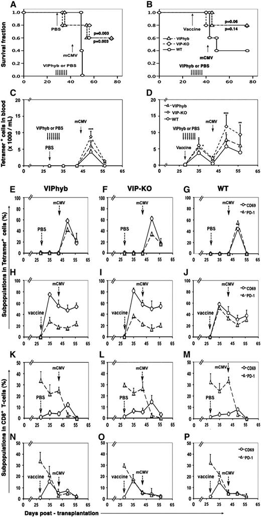
To explore VIP signaling blockade on immune responses to vaccination, we vaccinated B6→B10.BR allo-transplant recipients13 with Lm-MCMV (containing the immuno-dominant M45 mCMV peptide14 ) Recipients of WT allografts were treated with 7 days of PBS or VIPhyb starting 1 day before vaccination (day +28 posttransplant) followed by high-dose mCMV infection (1 × 10⁵ PFU) on day +42, 14 days postvaccination. Control groups were not vaccinated. Mice with VIP signaling blockade had better survival (Figure 2A-B) and more tetramer+ CD8+ T-cells following vaccination and mCMV infection (Figure 2C-D) than did PBS-treated mice engrafted with WT allografts. The magnitude of the effect of prior treatment with VIPhyb (ending 7 days before mCMV infection) on antigen-specific CD8+ T-cells was less than when VIPhyb was given concurrently with mCMV infection (compare Figures 1G and 2C). As expected, all groups of vaccinated mice had more blood tetramer+ CD8+ T-cells following subsequent mCMV infection than did nonvaccinated mice. Vaccinated mice with VIP signaling blockade had augmented cellular immune responses to subsequent mCMV infection in comparison with PBS-treated mice transplanted with WT allografts, with a higher frequency of antigen-specific CD8+ T-cells expressing CD69 and a lower fraction of antigen-specific T-cells expressing PD-1 (Figure 2E-J). Of note, the effect of VIP signaling blockade appeared to be antigen specific, because there were not substantive differences in the expression of PD-1 and CD69 in the total population of CD8+ T-cells comparing different treatment groups (Figure 2K-P).
Allo-BMT recipients treated with VIP antagonist or transplanted with VIP-KO donors had enhanced primary and secondary antigen-specific cellular immune responses to Lm-MCMV vaccine and mCMV infection. B10BR mice were transplanted with 5 × 10⁶ TCD-BM plus 0.3 × 10⁶ T-cells as described in Figure 1. Twenty-eight days posttransplant, mice were vaccinated with 1 × 10⁶ colony-forming units Lm-MCMV or PBS by i.p. injection and then infected with 1 × 10⁵ mCMV PFU on day +42 posttransplant. (A–B) Survival of transplant recipients treated on day 28 with either PBS (left) or VIPhyb (right). (C-D) Content of tetramer+ CD8+ T-cells in the blood of mice from panels A and B, respectively. (E-J) Percentage tetramer+ CD8+ T-cells expressing PD-1 or CD69. (K-P) Percentage of total CD8+ T-cells expressing CD69 or PD-1. Data are mean values ± standard deviation from 2 replicate experiments with 10 mice per time point. *P < .5, **P < .01, and ***P < .001 signify significant differences comparing mCMV-peptide MHC-tetramer+ CD8+ T-cell levels between VIP-KO or VIPhyb group and WT group.
Allo-BMT recipients treated with VIP antagonist or transplanted with VIP-KO donors had enhanced primary and secondary antigen-specific cellular immune responses to Lm-MCMV vaccine and mCMV infection. B10BR mice were transplanted with 5 × 10⁶ TCD-BM plus 0.3 × 10⁶ T-cells as described in Figure 1. Twenty-eight days posttransplant, mice were vaccinated with 1 × 10⁶ colony-forming units Lm-MCMV or PBS by i.p. injection and then infected with 1 × 10⁵ mCMV PFU on day +42 posttransplant. (A–B) Survival of transplant recipients treated on day 28 with either PBS (left) or VIPhyb (right). (C-D) Content of tetramer+ CD8+ T-cells in the blood of mice from panels A and B, respectively. (E-J) Percentage tetramer+ CD8+ T-cells expressing PD-1 or CD69. (K-P) Percentage of total CD8+ T-cells expressing CD69 or PD-1. Data are mean values ± standard deviation from 2 replicate experiments with 10 mice per time point. *P < .5, **P < .01, and ***P < .001 signify significant differences comparing mCMV-peptide MHC-tetramer+ CD8+ T-cell levels between VIP-KO or VIPhyb group and WT group.
We have previously shown that VIP-KO mice have more antiviral CD8 T-cells and enhanced antiviral cytolytic activity after mCMV infection.8 The current study demonstrates that transplanting VIP-KO allografts or treatment with a VIP antagonist enhances donor-derived cellular antiviral immunity and improves survival without increasing GVHD in allo-BMT. The mechanisms that VIP signaling blockade regulates cellular immunity may include inhibition of cyclic adenosine monophosphate (cAMP) induction leading to fewer Treg,17 fewer tolerogenic DC,18 and PD-1 expression,2,18 and counteracting immune-suppressive IL10-signaling derived from CMV-infected tissues and leukocytes.8,19 Acute20 and chronic21,22 viral infections increase PD-1 expression on antiviral effector CD8 T-cells, and PD-1 expression is associated with T-cell “exhaustion” in chronic viral infections.21,22 Of note, VIP-KO mice have decreased expression of PD-1 and PD-L1 on T-cells and DCs, respectively, after mCMV infection, and an abrogation of antiviral immunological “exhaustion.”8 The present study indicates that blocking VIP signaling may be a novel pharmacological strategy to enhance cellular immune responses to opportunistic infections in allo-BMT recipients by enhancing the numbers and activity of antigen-specific T-cells without increasing GVHD.
In conclusion, our current studies show that blockade of VIP signaling resulted in marked enhancement of cellular immune responses to mCMV infection and protection from lethal mCMV infections in allo-BMT. Pharmacological blockade of VIP signaling may be an attractive approach to evaluate in clinical trials enrolling recipients of allogeneic hematopoietic stem cell transplantation at high risk for CMV reactivation.
The online version of this article contains a data supplement.
The publication costs of this article were defrayed in part by page charge payment. Therefore, and solely to indicate this fact, this article is hereby marked “advertisement” in accordance with 18 USC section 1734.
Acknowledgments
The authors thank Drs. C. Giver and H. Yun for technical assistance and reviewing the manuscript.
This research was supported by National Institutes of Health grant R01 CA-74364-03 to E.K. Waller and a WES Foundation research grant to J.-M. Li.
Authorship
Contribution: J.L. designed and performed experiments and wrote the paper; M.S. and L.S. designed and performed experiments; E.K.W. designed experiments, coordinated the work, and wrote and edited the manuscript.
Conflict-of-interest disclosure: The authors declare no competing financial interests.
Correspondence: Edmund K. Waller, Department of Hematology/Oncology, Winship Cancer Institute, 1365B Clifton Road, Suite B5119, Emory University, Atlanta, GA 30322; e-mail: ewaller@emory.edu.


This feature is available to Subscribers Only
Sign In or Create an Account Close Modal