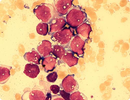
In March 2012, a 31-year-old male patient was diagnosed with alveolar rhabdomyosarcoma (ARMS) manifesting within the left sinus maxillaris with local lymphonodular metastases. No further metastases could be found by positron emission tomography–magnetic resonance imaging. He was treated by neo-adjuvant chemotherapy and consecutive radical endonasal resection and reconstruction in July 2012. In November 2012, he developed severe grade IV pancytopenia. Bone marrow aspiration revealed total bone marrow infiltration by cells with heavily vacuolated bluish cytoplasm similar to myelo-/lymphoblasts (magnification, ×100; May-Giemsa-Grünwald stain). Normal hematopoietic elements were absent. Immunohistochemistry in trephine biopsy showed a high expression of desmin. The patient received 5 cycles of palliative chemotherapy. The blood counts improved significantly with normal leukocyte and thrombocyte count. However, after 4 cycles of chemotherapy, bone marrow biopsy revealed 60% infiltration by the rhabdomyosarcoma.
Compared with embryonal rhabdomyosarcoma (ERMS), ARMS has a dismal prognosis and often presents with metastases at diagnosis. Explanations for the more aggressive course, different response to chemotherapy, and much shorter overall survival compared with ERMS might be that ARMS harbors a recurrent translocation involving either the PAX3 or PAX7 and FOXO1 genes leading to fusion transcripts, which likely plays a role in the more aggressive phenotype over ERMS.
For additional images, visit the ASH IMAGE BANK, a reference and teaching tool that is continually updated with new atlas and case study images. For more information visit http://imagebank.hematology.org.


This feature is available to Subscribers Only
Sign In or Create an Account Close Modal