Abstract
Recent studies indicate that acute myeloid leukemia (AML) cells, including leukemia-initiating cells, are highly dependent on oxidative phosphorylation (OXPHOS) for survival, while normal hematopoietic stem cells predominantly utilize glycolysis for energy homeostasis. We have reported development of a series of novel, highly potent mitochondrial complex I inhibitors, which in vitro inhibit complex I with IC50 values <10 nM (Marszalek et al., AACR 2014 Abstract #949). These inhibitors offer excellent therapeutic potential in the OXPHOS-dependent cancer models. IACS-1131 was selected as a preclinical tool compound from the series of more than 800 compounds across distinct structural classes. Here, we report the in vitro and in vivo efficacy of IACS-1131 in AML models.
Analysis of a panel of AML cell lines showed that a subset of leukemias are markedly dependent on OXPHOS for growth and survival; in this subset, IACS-1131 treatment caused steep decreases in viable cell number via induction of apoptosis. In sensitive cell lines (HL-60, OCI-AML3, KG-1, MV4;11, Kasumi-1), IACS-1131 induced pronounced apoptosis with EC50 between 10 and 100nM, consistent with the IC50 required to inhibit OXPHOS. MOLM13 and OCI-AML2 cells were less sensitive (EC50 250nM and 120nM, and a failure to induce cell death). In primary AML samples from patients with newly diagnosed or relapsed/refractory AML (n=12), the average EC50 for IACS-1131 was 14 ± 8nM in 9 samples, and exceeded 100nM in 3 samples. Consistent with the findings in AML cell lines, 10nM IACS-1131 resulted in partial responses, and 100-250nM resulted in profound loss of viability due to apoptosis induction. In contrast, this treatment caused only a moderate decrease in CD34+ cell numbers and <10% increase in apoptosis in 6 normal bone marrow samples.
The effects of IACS-1131 on the two major energy-generating pathways, mitochondrial OXPHOS and glycolysis, were investigated using the Seahorse Bioscience XF96 Analyzer. Treatment for 16 hrs caused a striking dose-dependent decrease in basal oxygen consumption rates (OCR), indicating OXPHOS inhibition; reduced ATP production; and decreased maximal respiratory capacity in OCI-AML3 cells and in primary AML blasts (n=9). We confirmed inhibition of complex I in AML cells using Seahorse mitochondrial electron flow assay. These changes preceded changes in viability or apoptotic markers; as such, loss of the mitochondrial membrane potential, annexin V positivity, and induction of mitochondrial reactive oxygen species were seen only at 72 hrs of exposure. Further time-course analysis demonstrated that 2 hrs of IACS-1131 exposure caused significant inhibition of OCR in both sensitive OCI-AML3 and resistant MOLM13 cells, but in MOLM13 cells there was a greater increase in extracellular acidification rates, suggesting compensation by glycolysis. In turn, inhibition of glycolysis with 2-DG, or blockade of pyruvate dehydrogenase kinase with dicholoroacetate (which forces entry into the TCA cycle) sensitized resistant cells to IACS-1131. The intracellular metabolome (polar fraction) of OCI-AML3 cells was characterized following 2, 4, 12 and 24 hrs of treatment with 100nm IACS-1131 using high-resolution magnetic resonance spectroscopy and high mass accuracy Orbitrap mass spectrometry. IACS-1131 modulated levels of the TCA intermediates, producing increased accumulation of citrate and fumarate and decreased succinate and malate, and increased glutathione, possibly because of the oxidative stress. Furthermore, the metabolic analysis indicated a strong effect on amino acid metabolism, whereby IACS-1131 reduced (between 25% and 62% of control) multiple anaplerotic amino acids (including arginine, leucine, isoleucine, valine, phenylalanine, asparagine, histidine, and glutamine, but not aspartate).
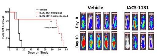
Finally, IACS-1131 at 60 mg/kg QD po demonstrated robust anti-leukemia activity in an orthotopic OCI-AML3 model. At this dose, IACS-1131 was well tolerated for >50 days and increased median survival duration by more than 5 times (Fig. 1). Studies exploring the anti-AML efficacy of single-agent IACS-1131 in primary AML xenografts are ongoing.
Taken together, these data strongly indicate that OXPHOS inhibition constitutes a novel therapeutic approach that targets a unique metabolic vulnerability of AML cells and indicate that further preclinical evaluation of OXPHOS inhibitors is warranted.
Treatment with IACS-1131 prolongs survival in a OCI-AML3 xenograft model. Luciferase-expressing OCI-AML3 cells were injected in the tail vein of NSG mice. On day 16 after injection, engraftment was confirmed and mice were randomized on the basis of IVIS-based imaging of luciferase activity after luciferin injection. For the next 58 days, mice received either vehicle or 60 mg/kg/day of IACS-1131 via oral gavage. Mice were sacrificed when body weight was reduced by >20% or for signs of morbidity. Right, bioluminescence imaging before (D0) and 10 days after 1 st dose; left survival.
Treatment with IACS-1131 prolongs survival in a OCI-AML3 xenograft model. Luciferase-expressing OCI-AML3 cells were injected in the tail vein of NSG mice. On day 16 after injection, engraftment was confirmed and mice were randomized on the basis of IVIS-based imaging of luciferase activity after luciferin injection. For the next 58 days, mice received either vehicle or 60 mg/kg/day of IACS-1131 via oral gavage. Mice were sacrificed when body weight was reduced by >20% or for signs of morbidity. Right, bioluminescence imaging before (D0) and 10 days after 1 st dose; left survival.
No relevant conflicts of interest to declare.
Author notes
Asterisk with author names denotes non-ASH members.


This feature is available to Subscribers Only
Sign In or Create an Account Close Modal