Abstract
Allogeneic stem cell transplant is a standard treatment for patients with high-risk and relapsed myeloid and lymphoid malignancies. However, donor T cells from the stem cell graft mediate graft-versus-host disease (GVHD), which is a common cause of morbidity and mortality for transplant recipients. Our group and others have shown that migration of donor T cells into secondary lymphoid tissue (SLT) and subsequent migration to target organs is critical to the pathogenesis of acute GVHD.
The Coronin family of proteins consists of actin-binding proteins, which regulate filament formation by interacting with the Arp2/3 complex. Coronin 1B, a ubiquitously expressed member of the Coronin family, is required for lamellipodial protrusion and effective cell migration. Previous work has not evaluated a role for this protein in the function of T lymphocytes or during acute GVHD.
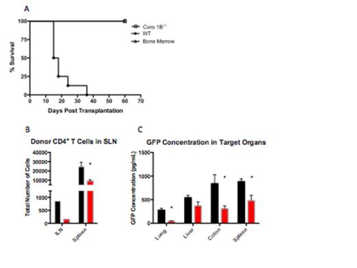
To evaluate the effect of Coronin 1B in acute GVHD pathogenesis, we transplanted B6 T cell depleted bone marrow cells with wild type or Coronin 1B-/- T cells to lethally irradiated B6D2 and BALB/c recipient mice and evaluated clinical score of GVHD and overall survival. B6D2 recipients of Coronin 1B-/- T cells demonstrated 100% survival (Figure 1A. p< .001 as determined by Log-rank (Mantel-Cox) test) and significantly decreased clinical scores after transplant. This was confirmed with improvement in survival in BALB/c recipients of Coronin 1B-/- T cells. Additionally, Coronin 1B-/- T cells were capable of eliminating P815 tumor cells, indicating that loss of Coronin 1B does not inhibit graft-versus-tumor activity. By day 12 post- transplant, all mice receiving bone marrow alone developed tumor compared to none of the mice receiving Coronin 1B-/- T Cells. However, protection was not complete as 40% of Coronin 1B-/- T cell recipients developed tumor by day 23.
To determine the effect of Coronin 1B on T cell migration during GVHD, B6D2 recipients were given GFP-expressing wild type or Coronin 1B-/- T cells along with T cell depleted bone marrow. Lymphoid tissue and target organs were harvested and analyzed by flow cytometry or GFP ELISA. We observed decreased accumulation of Coronin 1B-/- CD4+ (Figure 1B. p< .01 as determined by Student's t -test) and CD8+ T cells in the inguinal lymph node, mesenteric lymph node, and the spleen 4 days after transplant with no difference in accumulation in lymphoid tissue on days 7 and 14 after transplant. Additionally, we found decreased accumulation of Coronin 1B-/- donor T cells in the lung, colon and spleen 14 days after transplant (Figure 1C. p< .05 by Student's t -test). We also quantified the amount of cytokine in target organs by ELISA, and observed a decrease in IFN-γ and TNF-α in the colon 14 days after transplant.
Our data demonstrate that Coronin 1B-/- T cells elicit reduced GVHD compared to wild type T cells. This was correlated with decreased accumulation of Coronin 1B-/- T cells in SLT early after transplant. These data indicate that targeting the migration of T cells to SLT is a viable approach to prevent acute GVHD.
(A) Kaplan Meier curve comparing B6D2 recipients of Coronin 1B-/- T cells and wild type (WT) T Cells. (B) Decreased accumulation of Coronin 1B-/- T Cells 4 Days after transplant. For panels (B) and (C) black bars indicate recipients of WT T cells while red bars indicate recipients of Coronin 1B-/- T cells. Inguinal lymph nodes (ILN) were pooled from n=5 mice from each group. Spleens were analyzed individually. GFP expressing donor cells were analyzed by flow cytometry. Representative image of two experiments. (C) Coronin 1B-/- T cells express decreased accumulation in the lung, colon and spleen 14 days after transplant. Target organs were analyzed by GFP ELISA to detect GFP+ Donor Cells (n=5 in each group).
(A) Kaplan Meier curve comparing B6D2 recipients of Coronin 1B-/- T cells and wild type (WT) T Cells. (B) Decreased accumulation of Coronin 1B-/- T Cells 4 Days after transplant. For panels (B) and (C) black bars indicate recipients of WT T cells while red bars indicate recipients of Coronin 1B-/- T cells. Inguinal lymph nodes (ILN) were pooled from n=5 mice from each group. Spleens were analyzed individually. GFP expressing donor cells were analyzed by flow cytometry. Representative image of two experiments. (C) Coronin 1B-/- T cells express decreased accumulation in the lung, colon and spleen 14 days after transplant. Target organs were analyzed by GFP ELISA to detect GFP+ Donor Cells (n=5 in each group).
No relevant conflicts of interest to declare.
Author notes
Asterisk with author names denotes non-ASH members.


This feature is available to Subscribers Only
Sign In or Create an Account Close Modal