Abstract
Hyperbaric oxygen therapy (HBO) is being studied at our institution to improve hematopoietic stem cell homing and engraftment. Pre-clinical experiments have demonstrated that HBO therapy to the recipient prior to umbilical cord blood CD34+ cell infusion induces a low Erythropoietin (EPO) environment that results decreased erythroid differentiation and instead favored early bone marrow retention of CD34 cells with positive impact on engraftment in animal experiments1.
We hereby report a case series of 3 patients who received HBO on ongoing clinical trials and demonstrated unique patterns for lymphocyte recovery associated with unique clinical presentations. Patients were exposed to 100% oxygen at 2.5 atmosphere pressure for total of 2 hours prior to hematopoietic stem cell infusion.
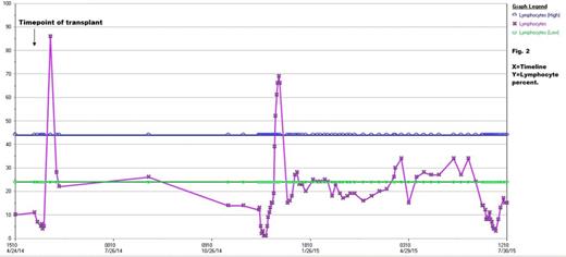
Case 1: A 64 year old male with IgG kappa multiple myeloma who relapsed post tandem cycles of high-dose Melphalan and autologous transplants. The patient received salvage therapy with Carfilzomib and Dexamethasone and achieved a partial response following which he underwent a preparative regimen of Carmustine, Etoposide, Cytarabine and Melphalan (BEAM) with subsequent autologous transplantation on an HBO clinical trial.
Post-transplant course was complicated by fever, rigors and extensive skin rash. These findings coincided with a rapid rise in white blood cell count predominantly composed of lymphocytes (Fig.2). Peripheral blood flow cytometry revealed atypical T-cell population with lymphocytes comprising 94% of total events. The majority were T cells (76%) with CD4 to CD8 ratio of 1:1 and co-expression of CD15, CD38 and HLA-DR. An associated infectious etiology was ruled out and the patient improved over a period of several days.
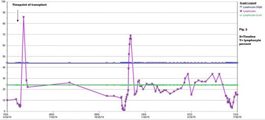
Case 2: A 33 year old male with refractory nodular sclerosis Hodgkin Lymphoma. He initially received Doxorubicin, Bleomycin, Vinblastine and Dacarbazine (ABVD) for 2 cycles with partial response. He was then switched to Bleomycin, Etoposide, Doxorubicin, Cyclophosphamide, Vincristine, Procarbazine and Prednisone (BEACOPP). After 2 cycles of BEACOPP, a repeat PET scan showed partial response. He was then salvaged with Brentuximab followed by BEAM and autologous transplant on HBO clinical trial.
The patient achieved neutrophil engraftment by day +11, of note his blood counts showed early increase in lymphocytes (86%) (Fig. 3). Post-transplant he was restarted on Brentuximab for two cycles with achievement with complete response, but his course was complicated by uveitis. An aqueous chamber fluid sample was sent for flow cytometry which showed Lymphocytes comprising 13% of total events. Flow cytometry of Cerebrospinal fluid revealed lymphocytes comprising 97% of total events. Both aqueous chamber and cerebrospinal fluids showed majority of T cells with normal CD4:CD8 ratio and antigen expression. His vision improved with topical steroid eye drops. Unfortunately, his disease progressed again off therapy.
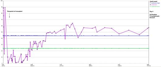
Case 3: A46 year old female with T cell large granular lymphoma who was initially treated with weekly methotrexate progressed to hepatosplenic gamma-delta T cell lymphoma and was treated with 4 cycles of hyper CVAD. Patient then received a reduced intensity preparative regimen of Fludarabine, Cytoxan, Total body irradiation and single unit umbilical cord blood transplantation on HBO clinical trial. The patient had an unremarkable post-transplant course and achieved neutrophil engraftment by day +25. Of note the patient demonstrated persistent peripheral lymphocytosis ranging between 42-64% of total white blood cell counts (Fig. 4). Bone marrow biopsies at multiple milestones showed her to be in complete remission and to be 100% donor by molecular tests.
Conclusions: Three unique patterns of lymphocyte recovery are seen in association with HBO therapy given prior to hematopoietic stem cell transplantation. We hypothesize that these patterns of lymphocyte recovery are secondary to engraftment kinetics caused by HBO and low EPO environment at the time of stem cell infusion. We suspect the early robust lymphocytosis may have played a role enhancing the inflammatory milieu resulting in fevers, rigors and extensive skin rash in case one, uveitis with CSF and aqueous chamber lymphocytosis in case two and persistent peripheral lymphocytosis in case three. In all three cases, the T cell exhibited normal antigen expression on flow cytometry.
No relevant conflicts of interest to declare.
Author notes
Asterisk with author names denotes non-ASH members.


This feature is available to Subscribers Only
Sign In or Create an Account Close Modal